Category: Obstetrics & Gynaecology

Reduced Antiepileptic Drug Effectiveness in Pregnancy Uncovered

Photo by Shvets Productions on Pexels

Blood levels of many commonly used antiepileptic drugs drop dramatically with the onset of pregnancy, which can result in ‘breakthrough seizures’ according to a study published in JAMA Neurology.

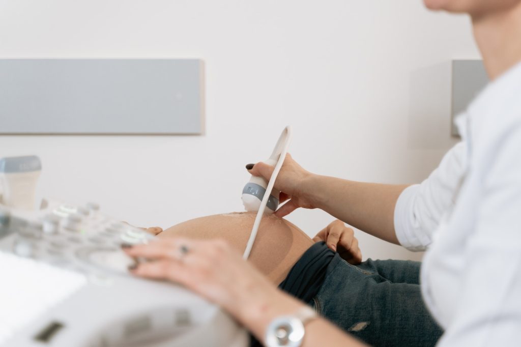
The findings, collected as part of the multicentre study Maternal Outcomes and Neurodevelopmental Effects of Antiepileptic Drugs (MONEAD), explain why many people with epilepsy start experiencing breakthrough seizures after conception, underscoring the need to increase antiseizure medication doses and closely monitor blood levels over the course of pregnancy.

A fine-tuned medication regime is critical in epilepsy. “Some people mistakenly believe that changes in the drugs’ blood concentration won’t occur until after 20 weeks of pregnancy, but our study shows how important it is to start monitoring and adjusting patients’ medication dosages early on,” said lead author Dr Page Pennelll. “Nearly half of all pregnancies in the United States are unplanned, so it is important to ensure that doctors have a clear picture of each patient’s baseline drug level even if they are not trying to conceive.”

A life-altering neurological condition, two-thirds of epilepsy cases do not have a known cause. In people with epilepsy, nerve cells in the brain are hyper-reactive, causing them to change the pattern of their electrical activity and become spontaneously active. That synchronous activation is manifested in seizures.

Epilepsy has a fraught history of diagnosis and management; people with epilepsy go undiagnosed or under-treated. First-generation drugs to control it had many dangerous side effects and were contraindicated for people who are trying to conceive.

Since then, safer medications have entered the U.S. market and become widely available, but clinicians started noticing a new problem – patients whose epilepsy was successfully managed with medications started having seizures soon after becoming pregnant.

“Identifying which antiseizure medications may have changes in concentrations and at what point in pregnancy those changes occur is important for determining which patients may need to be monitored more closely during pregnancy and after delivery,” said senior author Professor Angela Birnbaum at the University of Minnesota.

To solve the mystery, the researchers embarked on a study to analyse blood concentrations of 10 commonly used antiseizure drugs and compare them across different stages of pregnancy and after childbirth.

The study found that blood levels of seven out of 10 of the medications they examined dropped dramatically — from 29.7% for lacosamide, a commonly prescribed anticonvulsant, and up to 56.4% for lamotrigine.

In addition, the researchers noted that the drop in drug levels occurred mere days after conception.

Source: University of Pittsburgh

Teratogenic Drug Exposures Found in 1 in 16 Pregnancies

Source: Pixabay

Researchers have found, after reviewing a database containing 3 million pregnancies, that 1 in 16 women were exposed to teratogenic drugs.

The study, published in the American Journal of Obstetrics and Gynecology, highlights the need for women and their providers to carefully examine medications taken during pregnancy.

A teratogen is a substance that interferes with the normal development of a foetus. Hundreds of such drugs have been identified, including medications to treat seizures, migraines, obesity, acne, hypertension, bipolar disease and cancer.

University of Florida researchers investigated more than 200 teratogenic drugs and evaluated their exposure among 3.4 million pregnancies identified in a national private insurance database from 2006 to 2017. Prenatal exposure was defined by the mother taking at least one teratogenic drug during pregnancy.

The researchers divided drugs into two classes based upon their known teratogenic effect. About 140 drugs were known to have definite teratogenic effects, with another 65 identified as having potential teratogenic effects. The proportion of pregnancies with exposure to definite teratogens decreased slightly over the 12-year study period from 1.9% to 1.2%, while exposures for potential teratogens increased from 3.4% to 5.3%.

“While declining exposure rates among teratogenic drugs with definite risk are encouraging, the rising prenatal exposure to drugs with potential risk calls for more assessment,”  study author Professor Almut Winterstein, PhD, RPh. “To have 1 in 16 women and their unborn baby exposed to a teratogenic medication is simply too high, and we must identify strategies to improve pregnancy outcomes.”

The study also examined age and risk for prenatal exposure to teratogenic drugs and found teenagers and women in their 40s had the greatest risk. Both groups are known to have more unintended pregnancies and the drug exposure may have been accidental, which points to the need for more information about effective birth control and family planning when using teratogenic drugs.

The researchers were especially interested in prenatal exposure during more recent years, following the enactment of FDA requirements for risk mitigation strategies. Those are designed to reinforce safe medication-use behaviors, such as a pregnancy test before a teratogenic drug is started, and only a few medications require this extra safety precaution.

The 12 drugs with mitigation protocols in the study were found to be used infrequently and contributed to only a small portion of prenatal exposures. More research and regulatory action are needed to optimise the use of medications during pregnancy, the researchers concluded.

“There is much to do to address the evidence available regarding the risk-benefit of many drugs during pregnancy, and the availability of adequate risk-mitigation programs that ensure pregnancies are not unnecessarily exposed to teratogenic drugs,” Prof Winterstein said. “In the meantime, women and their providers must rely on the written information that is provided about the teratogenic risk for drugs during pregnancy.”

Source: University of Florida

A Small Risk of Increased Congenital Abnormalities from Opioids in Pregnancy

Photo by William-Fortunato on Pexels

In a new study in the Canadian Medical Association Journal, researchers drawing on a provincial database report a small increased risk of congenital abnormalities in infants exposed to opioid medications in the first trimester of pregnancy.

Prescribed opioid pain medications are capable of crossing the placenta and have the potential to cause harm. In a study comparing placental crossing rates for various opioids, oxycodone, a commonly prescribed opioid for pain relief, was the fastest. About 2%–4% of foetuses are exposed to these drugs. To determine the association between opioid pain medications in early pregnancy and congenital abnormalities in infants, investigators analysed administrative health data from Ontario on almost 600 000 birth parent–infant pairs. 

Among the infants included in the study, 2% (11 903) were exposed in utero to opioid analgesics, such as codeine, oxycodone, hydromorphone, tramadol, and morphine. Analysis showed a small increased risk of major anomalies with exposure to tramadol and morphine, and of minor anomalies with exposure to codeine, hydromorphone and oxycodone. Specific congenital anomalies observed included gastrointestinal and genital anomalies, neoplasms and tumours, and ankyloglossia.

This study adds to the evidence from previous studies in Sweden and Norway and also from a recent study of pregnant US Medicaid beneficiaries that suggested a small increased risk of congenital anomalies, an important finding for a pregnant person who may be prescribed opioids for pain relief.

“Both the potential for harm or distress to the pregnant person as a consequence of foregoing treatment and the subsequent risk to the infant must be considered for effective treatment,” the authors concluded. “These findings further quantify harms associated with prenatal exposure to opioid analgesics to inform treatment choices for pain in pregnancy.”

Source: EurekAlert!

Women in Labour Have Faster Gastric Emptying with an Epidural

Photo by Jonathan Borba on Unsplash

A study published in Anesthesiology finds gastric emptying is substantially slower during labour – but somewhat faster in women who receive an epidural for anaesthesia.

There is an ongoing debate as to whether it’s safe for women to eat solid food during labour. Physician anaesthesiologists prefer that labouring women have an empty stomach because of the lower risk for aspiration of food in case general anaesthesia for a caesarean section becomes necessary.

“These results suggest anaesthesiologists should remain cautious about permitting solid food during labour, especially when epidural analgesia is not used,” according to the report by Lionel Bouvet, MD, PhD, and colleagues of Hospices Civils de Lyon, France. 

Researchers assessed gastric emptying rates in four groups of women: 10 who were non-pregnant, 10 who were pregnant at full term (around 39 weeks) but not in labour, 10 in labour without an epidural, and 10 in labour who received an epidural for labour pain. On an empty stomach, each woman ate a light meal of yoghurt. Ultrasound scans were then used to compare the rate of stomach emptying among the four groups.

Stomach emptying was delayed for women in labour without epidural, in line with previous studies. The rate of stomach emptying from 15 to 90 minutes after eating was 52% in non-pregnant women and 45% in pregnant women at full term, compared to 31% for labouring women who received an epidural and 7% for women in labour without an epidural.

With epidural analgesia, gastric emptying occurred much faster during labour than during labour without epidural analgesia. After 90 minutes, the stomach was empty in 3 out of 10 labouring women who received an epidural, compared to 0 of 10 women in labour who had not received an epidural. By 2 hours, the stomach was empty in 6 of the women who received an epidural, compared to just 1 woman without an epidural.

Although clinical practice varies, current guidelines of the ASA and Society for Obstetric Anesthesia and Perinatology (SOAP) state that “Solid foods should be avoided in laboring patients,” reflecting a concern over the risk of aspiration in case anesthesia and surgery are needed. This new study is one of the first to systematically compare the extent of gastric emptying delay during late pregnancy and childbirth and with versus without epidural labor analgesia.

The results confirm a “statistically and clinically significant” longer time to an empty stomach among women in labour. However for those receiving epidural analgesia, stomach emptying appears to occur faster. Based on their findings, Dr Bouvet and co-authors suggest that a light solid meal “could probably be allowed” for women in labour who are receiving epidural analgesia and considered at a low risk of caesarean section within at least the next two hours.

“The report by Dr Bouvet and colleagues enables us to rethink our current practice of fasting during childbirth,” commented Anesthesiology editor Yandong Jiang, MD., PhD. “It is desirable that women giving birth with an epidural do not have the additional stress of hunger, but instead be allowed to eat a light meal.”

This contrasts with the ASA/SOAP recommendation that women in labour should consume only clear liquids to prevent aspiration, noted Mark Zakowski, MD, FASA, chair of ASA’s Committee on Obstetric Anesthesia. “This study clearly shows that stomach emptying is quite a bit slower for women in labor, and that if they eat even a light meal of about 4 ounces [about 120g] of yogurt, many will still have food in their stomach a few hours later,” Dr Zakowski said. “Since the need for emergency caesarean may arise at any time, the current ASA/SOAP guideline of clear liquids only during labour seems justified.”

Source: American Society of Anesthesiologists

Early Menopause and Oral Contraceptive Link

Photo by Reproductive Health Supplies Coalition on Unsplash

Long-term use of oral contraceptives, as well as certain methods of tubal ligation (TL), were linked to lower levels of antimüllerian hormone, a biomarker for ovarian aging, suggesting an increased risk for early menopause, according to preliminary research.

Using data from the Nurses’ Health Study II, researchers at the UMass Amherst School of Public Health and Health Sciences examined the association of oral contraceptive use and tubal ligation with antimüllerian hormone (AMH).

Published in the journal Menopause, the results were “intriguing,” according to lead author Christine Langton, PhD candidate.

“We’re one of the larger studies to have looked at both of these contraceptive methods at the same time,” says Langton, now a post-doctoral researcher at the National Institute of Environmental Health Sciences. “We feel we’re contributing to the story, and to the literature, though nothing we did was definitive. This is a piece of the puzzle.”

Early menopause, which occurs before 45, puts women at greater risk for a range of health conditions including cardiovascular disease, osteoporosis and dementia. The researchers noted that oral contraceptives change hormone levels and prevent ovulation; tubal ligation may affect blood supply to the ovaries, and certain methods of the procedure may damage the ovary and surrounding neural tissue. 

“Recently, AMH has become an established marker for the timing of menopause and was found to be strongly associated with the risk of early menopause,” the authors wrote. “Yet, the association of reproductive and lifestyle factors with AMH levels remains unclear.”

The team focused on a subset of 1420 premenopausal women in the Nurses’ Health Study prospective cohort who had provided a blood sample between 1996 and 1999. A history of their oral contraceptive use and tubal ligation began in 1989 and was updated every two years until their blood was collected.

“Women who reported that their [tubal ligation] procedure included the use of a clip, ring or band had significantly lower AMH levels compared to women who never had a TL procedure,” the researchers wrote.

One limitation is the small number of women reporting the type of tubal ligation, Langton added.

When it came to oral contraceptives, “we saw a significant inverse association – the longer the use of oral contraceptives, the lower the AMH levels were,” Langton said. “That particular finding was a little surprising to us because it didn’t completely align with what we saw when we looked at oral contraceptives and early menopause in the larger cohort” of more than 115,000 women.

Even after adjusting for factors including BMI, smoking, alcohol, number of pregnancies and breastfeeding, the inverse association between oral contraceptive use and AMH levels remained significant.

“We think further research is warranted,” Langton said.

Source: University of Massachusetts

Effects of Fathers’ Prenatal Alcohol Exposure Manifests in Offspring

Source: Pixabay

Researchers have discovered that males exposed to alcohol in utero later pass on those effects to their offspring during foetal development, through reduced placental efficiency. The study appears in FASEB Journal.

Dr Michael Golding, an associate professor at Texas A&M University has spent years investigating the father’s role, with regard to drugs and alcohol, in foetal development. Studies have shown that males pass down more than just their genetics, Dr Golding said, but exactly how that process works and the its consequences are still largely unknown.

“When you look at the data from throughout human history, there’s clear evidence that there’s something beyond just genetics being inherited from the male,” Dr Golding said. “So, if that data is solid, we’ve got to start looking more at male behaviour.

“Say you had a parent who was exposed to starvation – they could pass on what you might call a ‘thriftiness,’ where their kids can derive more nutrition from less food,” he said. “That could be a positive if they grow up in a similar environment, or they could grow up in a time when starvation isn’t an issue and they might be more prone to obesity or metabolic syndromes. That kind of data is clearly present in clinical data from humans.”

Epigenetics, which is Dr Golding’s area of study of how things beyond genes, such as behaviour and environment, affect development is called. One of the big questions in the search for answers on how male prenatal behaviour can impact foetal growth has been the way these epigenetic factors manifest.

The team has shown that prenatal exposure to alcohol in males can manifest in the placenta: in mice, offspring of fathers exposed to alcohol have a number of placenta-related difficulties, including increased foetal growth restriction, enlarged placentas, and decreased placental efficiency.

“The placenta supplies nutrients to the growing foetus, so foetal growth restriction can be attributed to a less efficient placenta. This is why placental efficiency is such an important metric; it tells us how many grams of foetus are produced per gram of placenta,” said Thomas, a graduate student at Texas A&M. “With paternal alcohol exposure, placentas become overgrown as they try to compensate for their inefficiency in delivering nutrients to the foetus.”

However,while these increases happened frequently in male offspring, the frequency varied greatly based on the mother; however, the same increases were far less frequent in female offspring. Dr Golding thinks that although information is passed from the father, the mother’s genetics and the offspring’s sex are also involved.

“This is a novel observation because it says that there’s some complexity here,” Dr Golding said. “Yes, men can pass things on to their offspring beyond just genetics, but the mom’s genetics can interpret those epigenetic factors differently, and that ultimately changes the way that the placenta behaves.”

These results don’t draw a clear line in how drinking in human males prior to conception impacts foetal development, but they continue to at least point to it being a question that needs to be explored. 

Dr Golding is hoping that more questions will be asked about male prenatal behaviour so that there’s more data from which to work.

“The thing that I want to ultimately change is this stigma surrounding the development of birth defects,” Dr Golding said. “There’s information coming through in sperm that is going to impact the offspring but is not tied to the genetic code; it’s in your epigenetic code, and this is highly susceptible to environmental exposures, so the birth defects that we see might not be the mother’s fault; they might be the father’s or both, equally.”

Source: Texas A&M University

Study Confirms COVID Vaccination does not Affect Fertility in IVF

Photo by Shvets Productions on Pexels

Vaccination against COVID did not affect fertility outcomes in patients undergoing in-vitro fertilisation (IVF), according to a new study. The findings, which were published in Obstetrics & Gynecology, add to the growing body of evidence providing reassurance that COVID vaccination does not affect fertility.

Investigators compared rates of fertilisation, pregnancy, and early miscarriage in IVF patients who had received two doses of vaccines manufactured by Pfizer or Moderna with the same outcomes in unvaccinated patients.

“The study found no significant differences in response to ovarian stimulation, egg quality, embryo development, or pregnancy outcomes between the vaccinated compared to unvaccinated patients.” said first author Devora Aharon, MD.

The study involved patients whose eggs were frozen and then thawed for in vitro fertilisation and womb transfer, and patients who underwent medical treatment to stimulate the development of eggs. The two groups of patients who underwent frozen-thawed embryo transfer (214 vaccinated and 733 unvaccinated) had similar rates of pregnancy and early pregnancy loss. The two groups of patients who underwent ovarian stimulation (222 vaccinated and 983 unvaccinated) had similar rates of eggs retrieved, fertilisation, and embryos with normal numbers of chromosomes, among several other measures.

The authors of the study anticipate that the findings will ease the anxiety of people considering pregnancy. 

Patients undergoing IVF treatment are closely tracked, enabling the researchers to capture early data on the implantation of embryos in addition to pregnancy losses that might be undercounted in other studies.

Previous studies have found that COVID vaccination helped protect pregnant persons (already at greater risk from severe illness and death from COVID) from severe illness, conferred antibodies to their infants, and did not raise the risk of preterm birth or foetal growth problems.

Source: EurekAlert!

Assisted Reproductive Technology Doubled Birth Rates in Middle-aged Women

Photo by Christian Bowen on Unsplash

A study found birth rates among middle-aged women in Northern European increased thanks to assisted reproductive technologies.

The study examined the total population of women aged 40–49 years between 2008–2018 in Denmark, Norway, and Sweden, birth rates increased in Denmark and Sweden, and births resulting from assisted reproductive technology doubled in all three countries. The findings were published in Acta Obstetricia et Gynecologica Scandinavica.

The study found that use of hormonal contraception increased among women aged 40–44 years in Denmark from 24% to 31%, in Sweden from 27% to 30%, and in Norway from 22% to 24%. Additionally, birth rates among women 40–44 years increased from 9.5 to 12 per 1000 women in Denmark and from 11.7 to 14.3 per 1,00 in Sweden, while they remained stable in Norway at approximately 11 per 1000 women.

In women aged 40–49 years, there was a doubling of assisted conceptions in Denmark from 0.71 to 1.71 per 1000 women; in Sweden from 0.43 to 0.81 per 1000; and in Norway from 0.25 to 0.53 per 1000. The study also found that Sweden had the highest induced abortion rate (7.7 to 8.1 per 1000 women) in women aged 40–49 years during the study period.

“This study confirms the trend of postponing childbirth observed for most of Europe and demonstrates the important role of assisted reproductive technology on birth rates in this age group,” said lead author Ingela Lindh, MD, of Sahlgrenska University Hospital. “The study provides valuable information to improve women’s knowledge about their fertility.”

Source: Wiley

Synthetic Progestogen in Utero Leads to Doubled Cancer Rate in Offspring

Photo by Shvets Productions on Pexels

In utero exposure to a synthetic progestogen used to prevent miscarriage can lead to an increased risk of developing cancer, according to a new study.

The study by researchers at The University of Texas Health Science Center at Houston (UTHealth Houston) was published in the American Journal of Obstetrics and Gynecology.

The drug, 17α-hydroxyprogesterone caproate (17-OHPC), is a synthetic progestogen frequently used by women in the 1950s and 1960s, and is still prescribed today to women to help prevent preterm birth. Progesterone helps the uterus grow during pregnancy and prevents early contractions that may lead to miscarriage.

“Children who were born to women who received the drug during pregnancy have double the rate of cancer across their lifetime compared to children born to women who did not take this drug,” said the study’s lead author, Caitlin C. Murphy, PhD, MPH, associate professor in the Department of Health Promotion and Behavioral Sciences at UTHealth School of Public Health in Houston. “We have seen cancers like colorectal cancer, pancreatic cancer, thyroid cancer, and many others increasing in people born in and after the 1960s, and no one really knows why.”

Researchers reviewed data from the Kaiser Foundation Health Plan on women who received prenatal care between June 1959 and June 1967, and the California Cancer Registry, which traced cancer in offspring through 2019.

Out of more than 18 751 live births, researchers discovered 1008 cancer diagnoses were made in offspring ages 0 to 58 years. Additionally, a total of 234 offspring were exposed to 17-OHPC during pregnancy. Offspring exposed in utero had cancer detected in adulthood at more than twice the rate of of those unexposed: 65% of cancers occurred in adults younger than 50.

“Our findings suggest taking this drug during pregnancy can disrupt early development, which may increase risk of cancer decades later,” Murphy said “With this drug, we are seeing the effects of a synthetic hormone. Things that happened to us in the womb, or exposures in utero, are important risk factors for developing cancer many decades after we’re born.”

A new randomised trial shows there is no benefit of taking 17-OHPC, and that it does not reduce the risk of preterm birth, according to Murphy.

The U.S. Food and Drug Administration proposed in October 2020 that this particular drug be withdrawn from the market.

Source: University of Texas Health Science Center at Houston

Thyroid Conditions may Affect Egg Reserves in Ovaries

Photo by Mart Production on Pexels

In an analysis published in Reproductive Medicine and Biology, researchers found evidence that thyroid conditions in women could negatively impact egg reserves in their ovaries.

Hypothyroidism is a disease that is prevalent in women, even in those of reproductive age. Thyroid hormones are involved in the control of the menstrual cycle. Oocytes express cell surface receptors for thyroid hormones that affect the actions of follicle-stimulating hormone and luteinising hormone through steroid biosynthesis. As such, thyroid dysfunction disturbs menstrual regularity and ovulation. However, the effects of low thyroid hormone levels (hypothyroidism) and thyroid autoimmune diseases on the functioning of a woman’s ovaries are not fully understood. This analysis aimed to investigate whether hypothyroidism and/or thyroid autoimmunity  affect the ovarian reserve, evaluated using levels of anti-Mullerian hormone (AMH).

The researchers found nine relevant trials, and their findings indicated that hypothyroidism and thyroid autoimmunity can negatively impact a woman’s ovarian reserve, or the total number of healthy immature eggs in the ovaries.

“Our age-stratified analysis demonstrated that thyroid autoimmunity and hypothyroidism possibly have different impacts on the ovarian reserve. It provides an important clue in determining how these conditions affect the development of ovarian follicles,” said senior author Akira Iwase, MD, PhD, of the Gunma University Graduate School of Medicine, in Japan.

Source: Wiley