A quarter of people with diabetes develop foot ulcers, which are slow to heal due to hypoxic conditions in the wound from impaired blood vessels and increased inflammation. These wounds can become chronic, leading to poor quality of life and possibly amputation.
Jianjun Guan, professor of mechanical engineering and materials science at the McKelvey School of Engineering at Washington University in St. Louis, has developed a hydrogel that delivers oxygen to a wound and decreases inflammation, helps to remodel tissue and speeds up healing. The results are published in Science Advances.
Prof Guan’s new hydrogel uses microspheres to gradually release oxygen to interact with the cells by means of an enzyme coating that converts the microsphere’s contents into oxygen. In this way, the hydrogel delivers oxygen over two weeks, reducing inflammation and promoting healing. “The oxygen has two roles: one, to improve skin cell survival under the low-oxygen condition of the diabetic wound; and two, oxygen can stimulate the skin cells to produce growth factors necessary for wound repair,” Prof Guan said.
In a unique pilot project in Sweden, drones were used to deliver defibrillators to real-life alerts of suspected cardiac arrest. The drones were dispatched in more than a fifth of the emergencies and arrived on target and ahead of the ambulance in most cases.
”This is the first time in the world that a research group can report results from a study where drones flew defibrillators to location of real-life alerts of suspected cardiac arrest,” says lead researcher Andreas Claesson, associate professor at the Center for Resuscitation Science at the Department of Clinical Science and Education, Södersjukhuset, Karolinska Institutet.
With sudden cardiac arrest, every minute counts. Currently, the odds of surviving an out-of-hospital cardiac arrest are 10 percent. However, with early CPR and a shock from an automated external defibrillator (AED), the chances of survival could reach 50-70 percent but response time needs to improve. In 2019 the median response time from alert to ambulance arrival for out-of-hospital cardiac arrest (OHCA) in Sweden was 11 minutes. To try and reach cardiac arrest victims sooner, researchers investigated using the rapid dispatch of drone-carried defibrillators in parallel with ambulances. Drones are already used in some countries to dispatch medicines and medical supplies to remote rural regions, The study, conducted in mid-2020 in western Sweden, describes an integrated method where emergency operators, drone pilots and air traffic control worked together to facilitate the dual response.
The drones took off in response to 12 out of 53 alerts of suspected cardiac arrest over a four-month period, successfully delivering an AED to the site in 11 of those cases. In seven of those cases, the drones arrived before the ambulance, with a median time benefit of 1 minute and 52 seconds. However, no drone-delivered defibrillators were attached to the patients before ambulances arrived.
“Even if none of the AEDs were used this time, our study shows that it is possible to use drones to transport defibrillators in a safe way and with target precision during real-life emergencies,” said first-author Sofia Schierbeck, PhD student at the Center for Resuscitation Science at the Department of Clinical Science and Education, Södersjukhuset, Karolinska Institutet. “A precondition for their future use is that the dispatcher takes initiative and instructs people on site to quickly collect and attach the AED in order to help the person with cardiac arrest.”
More work is needed to increase the dispatch rate and time benefits. For instance, in 2020 the drones were grounded if it was dark, rainy or the winds were too strong. The software system was also configured to avoid routes above densely populated areas, meaning that some alerts were too far out of range.
“Since this study was completed, we have identified several areas of improvement,” Andreas Claesson said. “In April this year, we began a follow-up study with a more optimised system. In that study, we want to test if we can use the drones in more alerts and reduce the response time further and thereby increase the time benefit as compared to the ambulance. Every minute without treatment in the early stages reduces the chance of survival by around 10 percent, and that is why we believe this new method of delivery has the potential to save lives.”
Researchers believe that antibacterial properties of sugars in human breast milk could be harnessed for new antimicrobial therapies.
Group B Streptococcus (GBS) bacteria are a common cause of blood infections, meningitis and stillbirth in newborns, and are becoming resistant to antibiotics. Researchers have now discovered that human milk oligosaccharides (HMOs), short strings of sugar molecules abundant in breast milk, can help prevent GBS infections in human cells and tissues and in mice. This might yield new antibiotic treatments, the researchers believe.
“Our lab has previously shown that mixtures of HMOs isolated from the milk of several different donor mothers have antimicrobial and antibiofilm activity against GBS,” says Rebecca Moore, who is presenting the work at a meeting of the American Chemical Society (ACS). “We wanted to jump from these in vitro studies to see whether HMOs could prevent infections in cells and tissues from a pregnant woman, and in pregnant mice.” Moore is a graduate student in the labs of Steven Townsend, PhD, at Vanderbilt University and Jennifer Gaddy, PhD, at Vanderbilt University Medical Center.
According to the US Centers for Disease Control and Prevention, about 2000 babies in the U.S. get GBS each year, with 4-6% of them dying from it. The bacteria are often transferred from mother to baby during labour and delivery. An expectant mother who tests positive for GBS is usually given intravenous antibiotics during labor to help prevent early-onset infections, which occur during the first week of life. Notably, late-onset infections (which happen from one week to three months after birth) are more common in formula-fed than breastfed infants, suggesting breast milk has factors which could help protect against GBS. If so, the sugars could be a replacement for current antibiotics which are steadily becoming less effective.
The researchers studied the effects of combined HMOs from several mothers on GBS infection of placental macrophages and of the gestational membrane. “We found that HMOs were able to completely inhibit bacterial growth in both the macrophages and the membranes, so we very quickly turned to looking at a mouse model,” Moore says. They examined whether HMOs could prevent a GBS infection from spreading through the reproductive tract of pregnant mice. “In five different parts of the reproductive tract, we saw significantly decreased GBS infection with HMO treatment,” Moore notes.
To determine which HMOs and other oligosaccharides have these antimicrobial effects and why, the researchers made an artificial two-species microbiome with GBS and the beneficial Streptococcus salivarius species growing in a tissue culture plate, separated by a semi-permeable membrane. Then, the researchers added oligosaccharides that are commonly added to infant formula, called galacto-oligosaccharides (GOS), which are derived from plants. In the absence of the sugar, GBS suppressed the growth of the “good” bacteria, but GOS helped this beneficial species grow. “We concluded that GBS is producing lactic acid that inhibits growth, and then when we add the oligosaccharide, the beneficial species can use it as a food source to overcome this suppression,” Moore explained. The first HMOs tested did not have this effect, but Townsend says it’s likely that one or more of the over 200 unique sugars in human milk will show activity in the artificial microbiome assay. There are likely two reasons why HMOs can treat and prevent GBS infection: they prevent pathogens from sticking to tissue surfaces and forming a biofilm, and they could also act as a prebiotic by promoting good bacteria growth.
“HMOs have been around as long as humans have, and bacteria have not figured them out. Presumably, that’s because there are so many in milk, and they’re constantly changing during a baby’s development,” Townsend said. “But if we could learn more about how they work, it’s possible that we could treat different types of infections with mixtures of HMOs, and maybe one day this could be a substitute for antibiotics in adults, as well as babies.”
Researchers at Empa have developed a patch that stably seals two sutured pieces of intestine and thus prevents dangerous leaks.
A burst appendix or a life-threatening intestinal volvulus are emergencies that need to be treated by surgeons immediately. However, operations carry risks: highly acidic digestive juices and intestinal bacteria can leak out, causing peritonitis and sepsis.
Sealing sutured tissue with a plaster has already been tried, but the first were not well tolerated or were even toxic. Currently, these plasters are made of biodegradable proteins, which have variable clinical results. These is because they are mainly intended to support the healing process, and dissolve too quickly when in contact with digestive juices and don’t always hold tight. “Leaks after abdominal surgery are still one of the most feared complications today,” explained Empa researcher Inge Herrmann, who is also professor for nanoparticulate systems at ETH Zurich.
Searching for a material that could reliably seal intestinal injuries and surgical wounds, Hermann’s team found a synthetic composite material made up of four acrylic substances that, together, form a chemically stable hydrogel. Additionally, the patch actively cross-links with the intestinal tissue until it is fluid-tight. The quadriga of acrylic acid, methyl acylate, acrylamide and bis-acrylamide works in perfect synergy, as each component conveys a specific feature to the final product: a stable bond to the mucosa, the formation of networks, resistance to digestive juices and hydrophobicity. This new technology is detailed in Advanced Functional Materials.
In lab experiments, the researchers found the polymer system met their expectations. “Adhesion is up to ten times higher than with conventional adhesive materials,” said researcher Alexandre Anthis from Empa’s Particles-Biology Interactions lab in St. Gallen. “Further analysis also showed that our hydrogel can withstand five times the maximum pressure load in the intestine.” The material’s design uses its tailored effect: The rubbery composite selectively reacts with digestive juices that might leak through intestinal wounds, expands and closes all the more tightly. The inexpensive, biocompatible super glue, could thus shorten hospital stays and save healthcare costs, and Anthis is making plans to bring it to market.
A new electromedical device provides important data about possible cardiovascular and pulmonary risks before an operation.
Before any operation, it is important to properly assess the individual risk: Are there perhaps circulatory or pulmonary problems that need special consideration? To what extent can special risks be taken into account when planning the anaesthesia? Previously, clinicians have had to rely on rather subjective empirical values or carry out more elaborate examinations when in doubt. To address this, a novel device has been developed by TU Wien and MedUni Wien to objectively measure the cardiovascular and pulmonary system fitness of patients.
Pre-op interviews are important—but subjective Complications often occur after surgical interventions. In addition to blood loss and sepsis, perioperative cardiovascular and pulmonary problems are among the most common causes of death in the first 30 days after surgery.
To minimise this risk, anesthesiologists routinely talk to patients before surgery, in addition to measuring their blood pressure, performing an electrocardiogram, or conducting more laborious examinations. But assessing responses can be highly individualised. “There are also objectively measurable parameters by which one could easily identify possible risks,” said Prof Eugenijus Kaniusas (TU Wien, Faculty of Electrical Engineering and Information Technology). “So far, however, they have not been routinely measured.”
Just hold your breath This new device uses multiple sensors to determine key metrics in a completely non-invasive way. All the patient has to do is hold their breath for a short time to slightly outbalance their body, which responds reflexively with various biosignals. “Holding your breath is a mild stress for the body, but that is already enough to observe changes in the regulatory cardiovascular and pulmonary systems,” explained Eugenijus Kaniusas. “Oxygen saturation in the blood, heart rate variability, certain characteristics of the pulse waveform—these are dynamic parameters that we can measure in a simple way, and from them we could ideally infer individual fitness in general, especially before surgery.”
Since the device is non-invasive, medical training is not needed to operate it, and has no side effects. The result is easy to read: A rough assessment according to the three-color traffic light system or a score between 0 and 100 is displayed. The measurement can also be carried out at the bedside without any problems for people with limited mobility.
“Our laboratory prototype is being tested at MedUni Wien in cooperation with Prof. Klaus Klein from the University Department of Anesthesia, General Intensive Care Medicine and Pain Therapy. We hope to bring the device to market in the next 5 years with the help of research and transfer support,” said Eugenijus Kaniusas.
There is an urgent need for more standardised and detailed reporting of research on mammalian cells, and for greater control over and measurement of the environmental conditions of cell cultures, according to a recent study. This will improve the precision of human physiology models and contribute to the reproducibility of research.
Researchers analysed 810 randomly selected papers on mammalian cell lines. Fewer than 700 of those, involving 1749 individual cell culture experiments, included relevant data on the environmental conditions of the media in which the cells were cultured. The analysis suggests that the relevance and reproducibility of this type of research needs significant improvement.
“Mammalian cell cultures are fundamental to manufacturing viral vaccines and other biotechnologies,” explained marine scientist, Shannon Klein. “They are used to study basic cell biology, replicate disease mechanisms and investigate the toxicity of novel drug compounds before they are tested on animals and humans.”
Though cells are cultured in controlled incubators in line with standard protocols, cells grow and ‘breathe’ over time and exchange gases with their surrounding environment. This impacts their immediate environment, and even these small changes can affect parameters like culture acidity and dissolved oxygen and carbon dioxide. These changes in turn can affect cell function, causing different conditions to that found in a living human body.
The researchers found that around half of the papers analysed failed to report the temperature and carbon dioxide settings of their cell cultures. Less than 10 percent reported the atmospheric oxygen levels in the incubator and less than 0.01 percent reported the medium’s acidity. No papers reported the dissolved oxygen or carbon dioxide in their media.
“We were very surprised that researchers largely overlooked the maintenance of environmental factors, like culture acidity, at levels relevant to the physiological body over the full course of the cell cultures, despite it being well known that this is important for cell function,” said Ph.D. student Samhan Alsolami.
The team, led by KAUST’s marine ecologist Carlos Duarte and stem cell biologist Mo Li in collaboration with developmental biologist Juan Carlos Izpisua Belmonte from the Salk Institute, who is currently a visiting professor at KAUST, recommends that biomedical scientists develop standard reporting and control and measuring procedures, in addition to employing specialised instruments for controlling the culture environments of different cell types. Additionally, scientific journals should establish reporting standards and require adequate monitoring and control of culture medium acidity and dissolved oxygen and carbon dioxide.
“Better reporting, measurement and control of the environmental conditions of cell cultures should improve how well scientists can repeat and reproduce experimental results,” said Alsolami. “More careful attention could drive new discoveries and increase the relevance of preclinical research to the human body.”
Researchers have created biocompatible generators which harvest body motion to produce electrical impulses for medical applications such as wound healing.
Piezoelectric materials such as ceramics and crystals can generate an electrical charge when mechanically stressed, and are used in many devices such as ultrasound transducers, vibration sensors, and cell phones. In medicine, electrostimulation using piezoelectric devices has been shown to be beneficial for accelerating wound and bone fracture healing, maintaining muscle tone in stroke victims, and chronic pain reduction. However, lack of biocompatibility has stalled progress in the field.
Now bioengineers at the University of Wisconsin’s Department of Materials Science and Engineering, led by Professor Xudong Wang, have developed implantable piezoelectric therapeutic devices. These thin, flexible devices make use of the piezoelectric properties of non-rigid, nontoxic biological materials such as silk, collagen, and amino acids. The team came up with a method for self-assembly of small patch-like constructs that use the amino acid lysine as the piezoelectric generator. The self-assembly process incorporates a biocompatible polymer shell that surrounds the lysine as the polymer/lysine solution evaporates. Chemical interactions between the inner layer of lysine and the polymer coating orient the lysine into the crystal structure necessary for it to produce electric current when flexed.
“This work is an outstanding example of using the chemical properties of the materials to create a self-assembling product,” explained David Rampulla, director of the Division of Discovery Science and Technology at the National Institute of Biomedical Imaging and Bioengineering. “The process used is rapid and inexpensive, making production of such wafers for therapeutic applications feasible. That the wafers are biodegradable opens the possibility for creating electrotherapies that could be used to accelerate healing of an injured bone or muscle, for example, and then degrade and disappear from the body.”
In one of a number of tests, wafers were placed in the leg and chest of rats, movements of which compressed the piezoelectric wafers enough to create an electrical output. Blood tests performed after the transplanted wafer dissolved showed normal levels of blood cells and other metabolites, indicating no harmful effects from the dissolved device.
Prof Wang emphasises the simplicity of the elegant work. “We believe the technology opens a vast array of possibilities including real-time sensing, accelerated healing of wounds and other types of injuries, and electrical stimulation to treat pain and other neurological disorders. Importantly, our rapid self-assembling technology dramatically reduces the cost of such devices, which has the potential to greatly expand the use of this very promising form of medical intervention.”
A small study has shown that a doctor’s presence during a blood pressure measurement skews the results, according to researchers who studied the effect by measuring nerve activity.
The phenomenon known as ‘white coat hypertension‘ is where the mere presence of a medical professional can raise blood pressure. Known about for decades, it occurs in about a third of patients.
In a small study published in the journal Hypertension, researchers probed the effect by measuring blood pressure, heart rate and nerve traffic in the skin and muscles with and without a doctor present.
The researchers found a “drastic reduction” in the body’s alarm response when a doctor was not present, said co-lead author Dr Guido Grassi, professor of internal medicine at the University of Milano-Bicocca.
Blood pressure and heart rate increases in response to a perceived threat, said Dr Meena Madhur, associate professor of medicine in the divisions of clinical pharmacology and cardiology at Vanderbilt University.
“If you’re out in the wild and a bear was charging after you, you’d want your blood vessels in your skin, for example, to constrict and the blood vessels in your muscles to dilate to provide more blood flow to those organs so that you can run really fast,” said Prof Madhur, who was not involved in the new research.
The study included 18 people, 14 of them men, with untreated mild to moderate hypertension. Each participant was examined in a lab, where an electrode measured nerve activity in the skin and muscles. Readings were taken twice in the presence of a doctor and twice without.
Both blood pressure and heart rate rose when the doctor was present, with nerve traffic patterns to the skin and skeletal muscle suggesting a classic fight or flight reaction.
Without the doctor’s presence, cardiovascular and neural responses were “strikingly different,” the researchers wrote. Fight or flight response indications were “entirely absent”.
Peak systolic blood pressure was an average of 14 points lower when the participant was alone than when a doctor was present, and peak heart rate was lowered by nearly 11 beats per minute.
This was the first study to actually measure sympathetic nervous system responses to doctors supervising a blood pressure measurement, the researchers wrote.
The study’s findings illustrated the complexity of blood pressure measurement and how it is affected by involuntary nervous system reactions, Grassi said. “Measurements without the doctor’s presence may better reflect true blood pressure values.”
White coat hypertension is not a new concept, Prof Madhur said, “this just drives home the fact that we should be more conscious of how the blood pressure is taken in the clinic.”
Last year, the American Medical Association and AHA issued a joint report endorsing more blood pressure measurement at home.
Limitations included the small study size due to the complexity of the measurements, the researchers said. Subsequent research would need to examine blood pressure medication as they could affect the fight or flight response, said Orof Madhur.
The work needs to be repeated with more women to examine possible sex differences. And she’d be interested in seeing whether people have the same response to nurses and other medical professionals as they did to doctors in this study.
Previous work shows that when nurses take blood pressure measurements, the white coat effect is reduced.
This latest research emphasises the need for people to handle blood pressure measurements with care, Prof Madhur said.
“I always tell my patients that we really can’t rely on a single office blood pressure measurement, because that’s just a random point in time,” she said.
Prof Madhur said that to take an accurate reading at home, a patient should sit still, with their back straight and supported and feet on the floor, waiting at least a few minutes before recording blood pressure. They should take multiple readings at the same time of day over the course of a week, and bring that log to their doctor’s appointment. Those at-home readings should be the ones used for planning treatment, she said.
“But,” Prof Madhur added, “if we are going to do an office blood pressure reading, it should be taken with the doctor not in the room.”
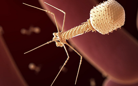
A group of newly discovered bacteriophages named after the UK village of Colney could help combat C. difficile infections.
Clostridioides difficile, or C. diff, is a species of bacteria that infects the human gut. It can become a major problem when our normal gut microbes are impaired, most commonly during a course of antibiotics. This leads to an overgrowth of C. diff, with toxins it produces causing diarrhoea and severe inflammation.
Treatment involves further courses of antibiotics, but relapse and recurrent infections are common. The strains are becoming more resistant to antibiotics and causing more severe illness.
This prompted researchers in Norwich to look for the bacteria’s natural enemy, bacteriophages. They screened 27 different C. diff strains for any bacteriophages, finding one, which they called ΦCD27 (phiCD27). Genome sequencing confirmed this phage had not been discovered before. In fact, the members of the International Committee on Taxonomy of Viruses (ICTV) decided it was genetically distinct enough to form a new group, or genus of phages.
The ICTV decided to name the new genus Colneyvirus, the Colney parish address of the Institute of Food Research (IFR, now part of Quadram Institute), where it was first discovered.
Like normal viruses, phages reproduce by injecting their genetic material into bacteria, making viral copies using the host’s own machinery. Using enzymes called endolysins, they destroy the bacterial cell wall and escape.
The researchers extracted the gene for ΦCD27’s endolysin and put it into another bacterium, E. coli so that they could produce and purify the endolysin. It was proven active against 30 different C. diff strains, including hypervirulent strains behind the current epidemic. It also didn’t affect other common bacterial species in the human gut microbiome.
”This phage and the endolysin encoded by its genome can provide a targeted approach to combat C. diff infections, in contrast to use of broad spectrum antibiotics that cause collateral damage by inhibiting other members of the gut bacterial population” said Professor Arjan Narbad, Group Leader at the Quadram Institute.
However, to be effective the endolysins need to be delivered into the gut, so the team also put the gene into a strain of lactic acid bacteria that has previously been used to deliver proteins and vaccines to the gut.
The research team believes this could serve as the basis for future new treatments C. diff. The system needs more work, but in the battle against this bacterial pandemic, the colneyvirus could be a vital ally.
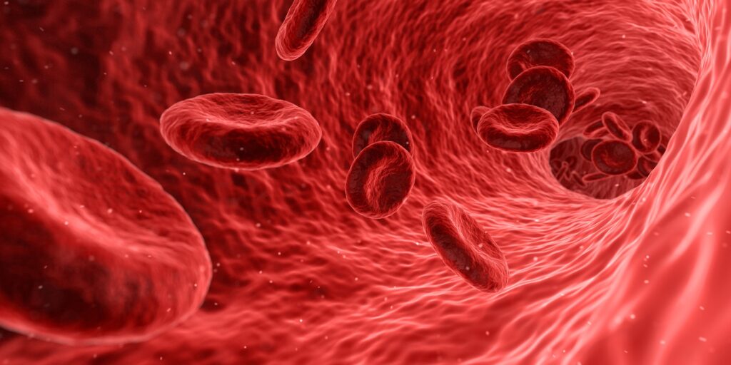
After 60 years of fruitless searches by scientists, researchers from the University of Virginia have finally determined the location of our bodies’ natural blood-pressure sensors.
These cellular sensors monitor blood pressure and adjust hormone levels to keep it in check. Scientists have long suspected that these ‘baroreceptors’, may exist in or around specialised kidney cells called renin cells, but no one has been able to locate the baroreceptors within the cell until now.
The new findings, from UVA Health’s Dr Maria Luisa S Sequeira-Lopez and colleagues, finally reveal where the barometers are located, how they work and how they help prevent hypertension or hypotension. The study was published in Circulation Research.
“It was exhilarating to find that the elusive pressure-sensing mechanism, the baroreceptor, was intrinsic to the renin cell, which has the ability to sense and react, both within the same cell,” said Dr Sequeira-Lopez. “So the renin cells are sensors and responders.”
Back in 1957, it was first proposed that a pressure sensor existed inside renin cells because the cells had to know when to release renin, a hormone that helps regulate blood pressure. Though the baroreceptors had to exist, scientists couldn’t tell what it was and whether it was located in renin cells or surrounding cells.
To tackle this decades-old mystery, the study’s researchers used a combination of innovative lab models and determined that the baroreceptor was a ‘mechanotransducer’ inside renin cells. This mechanotransducer detects pressure changes outside the cell, then transmits these mechanical signals to the cell nucleus, akin to how the cochlea turns sound vibrations into nerve impulses.
Through in vitro tests, the researchers found that applying pressure to renin cells triggered changes within the cells and decreased activity of the renin gene, Ren1. The scientists also compared differences in gene activity in kidneys exposed to lower pressure and those exposed to higher pressure.
Ultimately, when the baroreceptors detect excess pressure outside the renin cell, renin production is cut back, while low blood pressure prompts more renin production.
Dr Sequeira-Lopez said she is looking forward to the work to “unravel the signaling and controlling mechanisms of this mechanotransducer and how we can use the information to develop therapies for hypertension.”