Anti-rheumatic drugs used for rheumatoid arthritis (RA) might prevent the development of autoimmune thyroid disease, according to a new observational study by researchers from Karolinska Institutet which is published in the Journal of Internal Medicine.
Patients with RA at increased risk of autoimmune thyroid diseases such as Hashimoto’s disease and Graves’ disease. While patients with RA are usually treated with immunomodulatory drugs that affect the immune system, such drugs are rarely used in autoimmune thyroid diseases. Instead, such patients are treated with thyroid hormones such as levothyroxine to compensate for the changes in normal thyroid function that accompany autoimmune thyroid disease.
In this study, the researchers wanted to investigate whether immunomodulatory drugs that reduce inflammation in the joints of patients with RA might also reduce the risk of these patients developing autoimmune thyroid disease. Previous studies in mice suggest that so-called DMARDs, a type of immune-modulatory drugs used to treat rheumatoid arthritis, can reduce inflammation in the thyroid gland. Still, knowledge of whether this effect also applies to humans is limited, according to the research team.
The researchers used data between 2006 and 2018 on over 13 000 patients with rheumatoid arthritis and their treatment, as well as data from over 63 000 individuals in a matched control group without rheumatoid arthritis.
The researchers found that the risk of developing an autoimmune thyroid disease among RA patients was lower after their onset of the rheumatic disease than before diagnosis.
The most greatest risk reduction was seen in RA patients treated with immunomodulatory drugs or ‘biological DMARDs’. In these patients, the risk of autoimmune thyroid disease was 46% lower than in the control group without rheumatoid arthritis.
“These results support the hypothesis that certain types of immunomodulatory drugs could have a preventive effect on autoimmune thyroid disease,” says Kristin Waldenlind, researcher at the Department of Medicine, Solna, Division of Clinical Epidemiology, Karolinska Institutet, specialist in rheumatology at Karolinska University Hospital and first author of the study. She continues:
“Our results do not prove that it is the treatment with immunomodulatory drugs that led to the reduced risk of autoimmune thyroid disease, but provide support for this hypothesis. The results, if they can be replicated in further studies, open up the possibility of studying more directly in clinical trials whether the immunomodulatory drugs currently used for rheumatoid arthritis could also be used for the early treatment of autoimmune thyroid disease, ie for new areas of use of these drugs, known as drug repurposing.”
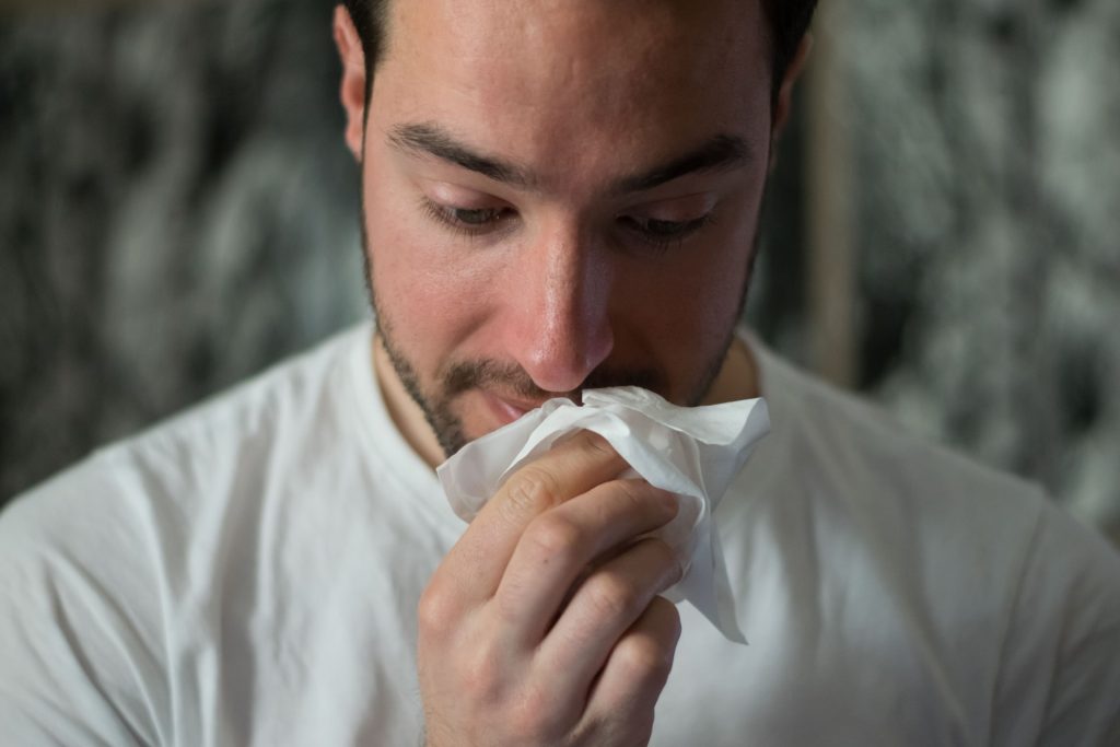
A study of firefighters on a punishing training course has revealed clues as to why extreme exercise temporarily weakens the immune system – a phenomenon seen in elite athletes. The findings, published in Military Medical Research, may lead to better ways to support the health of people who undergo extreme exertion, such as firefighters tackling wildfires.
Thirteen firefighters volunteered for the study, average age 25 and male. They went through a rigorous training exercise, carrying 9 to 20kg of gear over hilly terrain during a 45-minute training exercise in the California sun. Gloves, helmets, flashlights, goggles, and more weighted them down as they sprinted through the countryside wearing fire-resistant clothing to show they were ready to serve as wildland firefighters.
After the training, they immediately gave samples of their blood, saliva, and urine for analysis. Two were excluded, one being unable to finish the course and the other arriving to late to provide a sample. The 11 participants who completed the course lost an average of 2.2% of their initial weight.
Then, the scientists from the Department of Energy’s Pacific Northwest National Laboratory (PNNL) analysed more than 4700 molecules, consisting of proteins, lipids, and metabolites, from each of the firefighters, looking to understand what happens when the body undergoes intense physical exercise. Measuring and interpreting the data from thousands of such measurements is a specialty of PNNL scientists who explore issues related to climate science and human health by analysing millions of sensitive measurements using mass spectrometry each year.
The researchers’ aim was to increase safety for first responders and others.
“Heat stress can be life threatening,” said Kristin Burnum-Johnson, a corresponding author of the study. “We wanted to take an in-depth look at what’s happening in the body and see if we’re able to detect danger from exhaustion in its earliest stages. Perhaps we can reduce the risk of strenuous exercise for first responders, athletes, and members of the military.”
As expected, the team detected hundreds of molecular changes in the firefighters. The differences before and after exercise underscored the body’s efforts at tissue damage and repair, maintenance of fluid balance, efforts to keep up with increased energy and oxygen demand, and the body’s attempts to repair and regenerate its proteins and other important substances.
But in the saliva, the team found some unexpected results. There was a change in the microbial mix of the mouth – the oral microbiome – showing that the body was increasingly on the lookout for bacterial invaders. Scientists also saw a decrease in signaling molecules important for inflammation and for fighting off viral infections.
A decrease in inflammation makes sense for people exercising vigorously; less inflammation allows people to breathe in air more quickly, meeting the body’s eager demand for more oxygen. Having fewer inflammatory signals in the respiratory system helps the body improve respiration and blood flow.
Less inflammation, more inhalation
But less inflammation leaves the body more vulnerable to viral respiratory infection, which other studies observed in elite athletes and others who exercise vigorously. Some studies have shown that a person is up to twice as likely to come down with a viral respiratory infection in the days after an especially energetic workout.
“People who are very fit might be more prone to viral respiratory infection immediately after vigorous exercise. Having less inflammatory activity to fight off an infection could be one cause,” said Ernesto Nakayasu, a corresponding author of the paper. He notes that the work provides a molecular basis for what clinicians have noticed in their patients who do strenuous workouts.
The team hopes that the findings will help explain why come people are more vulnerable to respiratory infection after a workout.
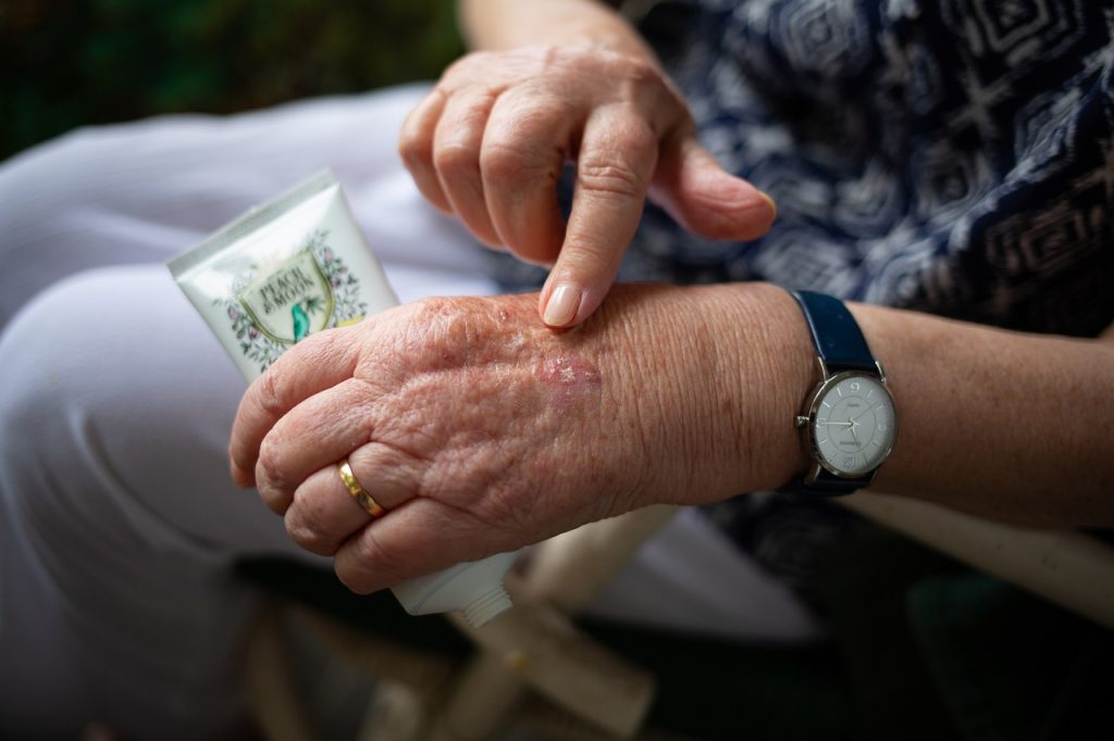
Photo by Towfiqu barbhuiya: https://www.pexels.com/photo/person-feeling-pain-in-the-knee-11349880/
New research reported in the journal Nature could lead to new targeted treatments for rheumatoid arthritis (RA). The findings showed that guesswork could be taken out of selecting treatments for each patient, and this might one day also be extended to other autoimmune conditions.
The study was led by University of Colorado School of Medicine faculty members Fan Zhang, PhD, and Anna Helena Jonsson, MD, PhD. The Accelerating Medicines Partnership: Rheumatoid Arthritis and Systemic Lupus Erythematosus (AMP: RA/SLE) Network collected inflamed tissue from 70 patients with RA from across the country and the United Kingdom. Jonsson supervised the team of scientists who processed these samples for analysis, and Zhang led the computation analysis of the data. These efforts yielded a cell atlas encompassing more than 300 000 cells from synovial tissue. Further analysis revealed that there are six different subgroups of RA based on their cellular makeup.
“We hope the data will help us discover new treatment targets,” says Jonsson, assistant professor of rheumatology. “We wanted to make it public so that researchers across the country and across the world can continue working on new treatment ideas for rheumatoid arthritis going forward.”
No more guess-and-check
Jonsson, a practicing rheumatologist as well as a researcher, knows that RA patients respond differently to different treatments. Until now, she says, rheumatologists used a “guess and check” method to find a treatment that works for an individual patient.
With the new data and powerful computational classification methods developed by Zhang and the computational analysis team, the researchers were able to quantitatively classify RA types into what they call ‘cell-type abundance phenotypes’, or CTAPs. Developed methods, together with the new cell atlas, can start to identify which patients will respond to which treatments.
“Even when you classify rheumatoid arthritis inflammation using these simple markers – T cell markers, B cells, macrophages and other myeloid cells, fibroblasts, endothelial cells – what we found is that each of those categories is associated with very specific kinds of pathogenic cell types we’ve already discovered,” Jonsson says. “Previous rheumatoid arthritis research found that T cell populations called peripheral helper T cells are relevant in rheumatoid arthritis, as are B cells called antibody-producing B cells, and other specific cell types. What we found is that they’re usually not found all together.
“For example, the peripheral helper cells are found with the B cells in only one category of RA, and the pathogenic macrophage populations tend to exist in a different category. Because of this, we can start asking questions about how these specific partners work together.”
Scratching an itch can be a relief, but for many patients it can get out of control, becoming a serious health problem. So what normally stops this progression?
A paper published in Science Immunology reports a breakthrough that could transform how doctors treat conditions from atopic dermatitis to allergies, they have discovered a feedback loop centred on a single immune protein called IL-31 that both causes the urge to itch and dials back nearby inflammation.
The findings, by Scientists at UC San Francisco, lay the groundwork for a new generation of drugs that interact more intelligently with the body’s innate ability to self-regulate.
Previous approaches suggested that IL-31 signals itch and promotes skin inflammation. But the UCSF team discovered that nerve cells, or neurons, that respond to IL-31, triggering a scratch, also prevent immune cells from overreacting and causing more widespread irritation.
“We tend to think that immune proteins like IL-31 help immune cells talk to one another, but here, when IL-31 talks to neurons, the neurons talk right back,” said Marlys Fassett, MD, PhD, UCSF professor of dermatology and lead author of the study. “It’s the first time we’ve seen the nervous system directly tamp down an allergic response.”
The discovery could eventually change how asthma, Crohn’s and other inflammatory diseases are treated, due to IL-31’s presence throughout the body.
“IL-31 causes itch in the skin, but it’s also in the lung and in the gut,” said Mark Ansel, Ph.D., UCSF professor of immunology and senior author of the study. “We now have a new lead for fighting the many diseases involving both the immune and nervous systems.”
More than an itch
IL-31 is one of several “itch cytokines” because of its ability to instigate itch in animals and people. Fassett, a dermatologist and a researcher, has wanted to know why since she arrived at UCSF in 2012, a few years after its discovery. She reached out to Ansel, a former colleague and asthma expert who welcomed her into his lab.
First, Fassett removed the IL-31 gene from mice and exposed them to the house dust mite, a common, itchy allergen.
“We wanted to mimic what was actually happening in people who are chronically exposed to environmental allergens,” Fassett said. “As we expected, the dust mite didn’t cause itching in the absence of IL-31, but we were surprised to see that inflammation went up.”
Why was there inflammation but no itching? Fassett and Ansel found that a cadre of immune cells had been called into action in the absence of the itch cytokine. Without IL-31, the body was blindly waging an immunological war.
IL-31 brings balance to the forces
Ansel and Fassett then homed in on the nerve cells in the skin that received the IL-31 signal. They saw that the same nerve cells that spurred a scratch also dampened any subsequent immune response. These nerve cells were integral to keeping inflammation in check, but without IL-31, they let the immune system run wild.
The findings squared well with what dermatologists were increasingly seeing with a new drug, nemolizumab, which blocked IL-31 and was developed to treat eczema. While clinical trial patients found that the dry, patchy skin of their eczema receded on the drug, other skin irritation, and even inflammation in the lungs, would sometimes flare up.
“When you give a drug that blocks the IL-31 receptor throughout the whole body, now you’re changing that feedback system, releasing the brakes on allergic reactions everywhere,” Ansel said.
Fassett and Ansel also found that these neurons released their own signal, called CGRP, in response to the itch signal, which could be responsible for dampening the immune response.
“The idea that our nerves contribute to allergy in different tissues is game changing,” Fassett said. “If we can develop drugs that work around these systems, we can really help those patients that get worse flares after treatment for itch.”
Fassett recently founded her own lab at UCSF to tease apart these paradoxes in biology that complicate good outcomes in the clinic. And Ansel is now interested in what this itch cytokine is doing beyond the skin.
“You don’t itch in your lungs, so the question is, what is IL-31 doing there, or in the gut?” Ansel asked. “But it does seem to have an effect on allergic inflammation in the lung. There’s a lot of science ahead for us, with immense potential to improve therapies.”
Over the last few decades, there has been growing popularity for the ‘hygiene hypothesis’, which suggests that some level of microbial exposure helps protects against developing allergy. Now, an article published in Science Immunology by researchers from Karolinska Institutet challenges this hypothesis by showing that mice with high infectious exposures from birth have the same, if not an even greater ability to develop allergic immune responses than ‘clean’ laboratory mice.
Studies have suggested that certain infections might reduce the production of inflammatory antibodies to allergens and alter the behaviour of T cells involved in allergies. It has also been suggested that ‘good’ intestinal bacteria could shut off inflammation elsewhere in the body.
Robust allergic responses
Researchers have now compared the allergic immune response in ‘dirty’ wildling mice to those of typical clean laboratory mice. They found very little evidence that the antibody response was altered or that the function of T cells changed in a meaningful way. Nor did anti-inflammatory responses evoked by good gut bacteria appear to be capable of switching off the allergic immune response. On the contrary, wildling mice developed robust signs of pathological inflammation and allergic responses when exposed to allergens.
“This was a little unexpected but suggests that it’s not as simple as saying, ‘dirty lifestyles will stop allergies while clean lifestyles may set them off’. There are probably very specific contexts where this is true, but it is perhaps not a general rule,” says Jonathan Coquet, co-author of the study and Associate Professor at Karolinska Institutet.
More like the human immune system
The wildling mice are genetically identical to clean laboratory mice but are housed under ‘semi-natural’ conditions and have rich microbial exposures from birth.
“The immune systems of wildling mice better represent the human immune system and so we hope that they can bring us closer to the truth of how microbes act upon the body,” says Jonathan Coquet.
The findings contribute to the general understanding of how allergies may arise and may also have clinical implications. Using experimental infections to treat patients suffering from inflammatory diseases has also been attempted in recent clinical trials. For example, infecting people with worms or performing faecal transplantations has been proposed as a tool to combat inflammatory diseases. Newborns delivered through C-section, have had maternal faecal transplantation and bacterial supplementation with the aim of promoting good bacteria in the baby’s gut and the child’s future health.
Beneficial effects of exposure not clear as we’d like
“This field of research can provide important insights into how infections and microbes can be used to facilitate health, but it is still in its infancy. Our study is a reminder that general and broad exposures to microbes may not have the clear beneficial effects that we wish them to have,” says Susanne Nylén, co-author of the study and Associate Professor at the Department of Microbiology, Tumor and Cell Biology at Karolinska Institutet.
Adults with atopic dermatitis (AD) have a 34% increased risk of developing new-onset inflammatory bowel disease (IBD) compared to those without the skin condition, according to a new recently published in JAMA Dermatology. The study also shows for children, the risk increase is 44%. Additionally, as the severity of AD increased, the risk of developing IBD rose.
These findings clear up ambiguity from previous research, especially among populations of children and between the different types of IBD: ulcerative colitis and Crohn’s disease. While IBD is located in the gut and AD affects the skin, both diseases are driven by the immune system and are categorised by severe inflammation. Insight offered from this study from the Perelman School of Medicine at the University of Pennsylvania could lead to new treatments for both IBD and AD.
“It is imperative for clinicians to understand atopic dermatitis and the trajectory of our patients with it in order to provide the best standard of care,” said senior author Joel M Gelfand, MD, dermatology professor at Penn. “There are new and better treatments for AD today, and there will likely continue to be more. But providers have to understand how those treatments could impact other autoimmune diseases. For patients with AD and another autoimmune disease, some currently available medications can exacerbate symptoms of their other disease or can help treat two immune diseases at the same time.”
While this is not the first study to explore AD and IBD, its size, with one million adult and child participants with AD drawn from a UK medical database, and its separation between ulcerative colitis and Crohn’s disease advances previous research.
When looking at ulcerative colitis and Crohn’s disease separately, AD was not linked to higher ulcerative colitis in children unless the kids had severe AD. Children with AD, however, had a 54–97% increased relative risk of Crohn’s disease, and among children with severe AD, their risk was roughly five times higher. Results among adults were more straightforward. Adults with AD had a 32% increased relative risk of ulcerative colitis and a 36% increased relative risk of Crohn’s disease. Gelfand notes that the absolute extra risk of developing IBD in individuals with AD is still quite small, but the association is meaningful in better understanding health outcomes in AD. Moreover, since millions of people have AD, this small increase in risk spread among many people is likely important from a public health perspective.
Although Penn researchers did not look at the root cause of IBD linked to AD, they have strong hypotheses about the links.
“AD and IBD can cause changes in the microbiome, chronic inflammation, and the dysfunction in the skin and gut barrier respectively,” said Gelfand, who is also the director of the Center for Clinical Sciences in Dermatology at Penn. “There are also specific cytokines, certain kinds of proteins, that play a role in immune system activity and that seem to be related to AD and IBD. For example, we think dysfunction of types of T cells common to both AD and IBD, could be the culprits. Those need to be explored further to uncover both what’s happening at a microscopic level and what proteins or structures could be targeted to treat one or both conditions.”
As a leading expert on psoriasis, a disease known to be tied to IBD genetically, Gelfand is well aware of how closely skin health can affect other parts of the body. He and his colleagues are also studying AD’s relationship to infections, neurologic and psychiatric disorders, and cardiovascular disease.
“Investigating the relationship between skin diseases and other diseases doesn’t just offer new insight into how these diseases can affect a patient with both, but these studies are especially powerful because they also highlight unique characteristics of each disease and how they behave individually,” Gelfand shared.
Signalling from inflammatory cytokines activates several protein kinases in a chain – at the end of this process MKK6 (yellow) ‘switches on’ p38α (green) by binding it and adding phosphates. The combination of computer simulations, SAXS, and cryo-EM has shown how the two proteins interact and bind to each other to transmit the signal. Credits: Ella Marushchenko, Isabel Romero Calvo/EMBL
Cytokine storms, where inflammatory cascades during an infection that can spiral out of control and lead to severe disease and even death, were recently highlighted during the COVID-19 pandemic. In a new paper published in Science, researchers report their discovery of a cellular ‘switch’ which may lead to new drugs to prevent deadly cytokine storms.
EMBL Grenoble and University of Geneva researchers provide essential insights on a protein called p38α which is an important cellular ‘switch’ triggering the inflammatory response. They have obtained the first structure of p38α being activated by another regulatory protein kinase, MKK6, opening up new directions to develop drugs to stop cytokine storms.
The final switch: a drug target
Matthew Bowler, a researcher at EMBL Grenoble, has been studying kinases for more than a decade. This group of enzymes plays an important role in regulating complex processes in the cell by acting as a ‘switch’ to transmit signals and activate gene expression. They do so by phosphorylation, in which phosphate is added to other molecules to modulate their function.
Bowler’s work particularly focuses on belonging to the Mitogen Activated Protein (MAP) family of kinases, key players involved in the inflammatory response. Inflammation is switched on via a series of kinases, which activate each other in a cascade of reactions, the final kinase in the series being responsible for activating gene transcription required for inflammation. This process releases cytokines, pro-inflammatory signalling molecules, which, in case of overactivation, can lead to cytokine storms.
This kinase chain reaction is well regulated and is similar to a logic circuit: the inflammation response requires specific buttons to be switched on, ultimately activating p38α – the convergence point for all the signals and the last switch of the inflammatory process.
Because the kinase chain reaction can come from different ‘branches’ of the logic circuit, this last switch is a particularly relevant drug target. The inflammatory response is regulated by p38α and is highly activated during a cytokine storm. Inactivating it could prevent inflammation from occurring, instead of trying to treat it while it is already underway.
Protein kinases, including p38α, have therefore been heavily studied. The first protein kinase structure was solved 30 years ago and many more structures have followed, with over 7000 structures now available in the Protein Data Bank.
However, important parts of the puzzles are still missing. “Structural biologists have obtained detailed information on the structure and functions of protein kinases, but mostly in isolation. So we don’t really know how these enzymes are activated along the chain reaction,” explained Bowler.
Without this essential information about how the activation is triggered, drugs have mostly targeted the kinases’ nucleotide-binding site – a common and well-known pocket present in all kinases, where the phosphate transfer occurs. The lack of drug specificity due to a common binding site across kinases means that a drug designed to stop one kinase from signalling could also stop others. This presents a problematic side effect, considering the essential role of kinases as key regulators in cellular processes.
“There are many molecules that have been designed to target p38α, especially its nucleotide-binding site, but none have yet made it past clinical trials due to this lack of specificity,” added Bowler.
Cracking the activation mechanism
Since 2009, Bowler and a former PhD student in his lab, Erika Pellegrini, have therefore been investigating the interactions between p38α and MKK6, the kinase which activates it. But studying the interaction between kinases proves to be extremely complex. “These enzymes are very dynamic molecules; they transmit important signals and need to act quickly. In the case of p38α, it has to go into the nucleus and activate lots of other different proteins,” said Bowler.
They were hampered by the fact that the interactions of the MKK6-p38α complex cannot be determined by macromolecular crystallography, a structural biology technique often employed to investigate proteins but that is particularly challenging to apply in the case of such dynamic proteins.
Recent developments in cryo-electron microscopy (cryo-EM), particularly during the last decade, raised new hopes. In 2016, Bowler and new PhD student and first author of the paper, Pauline Juyoux, decided to switch to this technique – even though the protein complex was at the time considered too small for cryo-EM analysis. They were supported by Pellegrini, who had acquired expertise in this technique.
Using cryo-EM and complementary techniques, such as X-ray crystallography and small-angle X-ray scattering (SAXS), the team managed to obtain the 3D structure of the complex and identify a previously unknown docking site where the two enzymes interact – crucial information for understanding how p38? is activated. “This could be an interesting target for inhibitors that block this specific interaction, and consequently the signal triggering the inflammatory response,” explained Juyoux.
A collaboration with the Gervasio Lab from the University of Geneva, which uses molecular dynamics simulations, provided further insight into how the two kinases interact. “They showed that the model we had generated was compatible with the enzymatic activity and that the phosphorylation site was at the right distance from the active site,” explained Juyoux. “They also classified the different types of conformations of the complex to show how they assemble.”
Crucially, by comparing these simulations with the SAXS data they were able to model how the two proteins interact prior to catalysis. “The beauty of combining the state-of-the-art simulations with SAXS and cryo-EM data through advanced statistical approaches is that we can ‘see’ the dance of the two kinases approaching one another, while knowing that what we see in the computer is fully supported by all the experimental data available,” explained Francesco Gervasio. “The simulations required several months of supercomputing time generously allocated by the Swiss National Supercomputing Centre,” he continued, “But it was well worth it, given the relevance of the final results.”
These results provide an alternative drug target site to explore and also open the door to studying similar processes in two other families of MAP kinases: ERK kinases, which are involved in cancer, and JNK kinases, which are also involved in inflammation, especially in Alzheimer’s disease.
“Kinases are very similar to one another in terms of sequence and structure, but we don’t know how and why they respond or send a specific signal,” said Juyoux, whose current research project as a postdoctoral fellow at Institut de Biologie Structurale in Grenoble focuses on JNK kinases. “Comparing these different families of kinases could help explain the specificity of interactions and lead the way to new therapeutic approaches.”
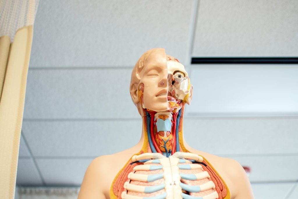
Researchers have identified stem cells in the human thymus for the first time. These cells represent a potential new target to understand immune diseases and cancer and how to boost the immune system. Their reported their discovery in the journal Developmental Cell.
The thymus is a gland located in the front part of the chest, the place where thymocytes (the cells in the thymus) mature into T cells, specialised immune cells crucial to fighting disease. The thymus has a unique and complex 3D structure, including an epithelium (a lining of cells able to guide T cell maturation) that forms a mesh throughout the whole organ and around the thymocytes.
Owing to its relatively inaccessible location, comparatively recent discovery and the fact that it shrinks with age, the thymus has only been investigated for a short period of time compared to other organs. Until now, scientists believed it didn’t contain ‘true’ epithelial stem cells, but only progenitors arising in foetal development.
However, these findings from researchers at the Francis Crick Institute, show for the first time the presence of self-renewing stem cells, which give rise to the thymic epithelial cells instructing thymocytes to become T cells. This suggests the thymus plays an important, regenerative role beyond childhood, which could be exploited to boost the immune system.
In the course of their experiments, the researchers examined these stem cells based on the expression of specific proteins in the human thymus. They identified stem-cell niches (areas where stem cells are clustered) in two locations in the thymus: underneath the organ capsule, or outer layer, and around blood vessels in the medulla, the central part.
They demonstrated that thymic stem cells contribute to the environment by producing proteins of the extracellular matrix, which functions as their own support system.
By using state-of-the-art techniques to map gene expression in single cells and tissue sections, they found that these stem cells, named Polykeratin cells, express a variety of genes allowing them to give rise to many cell types not previously considered to have a common origin. They can develop into epithelial as well as muscle and neuroendocrine cells, highlighting the importance of the thymus in hormonal regulation.
The researchers isolated Polykeratin stem cells in a dish and were able to show that thymus stem cells can be extensively expanded. They demonstrated that all the complex cells in the thymus epithelium could be produced from a single stem cell, highlighting a remarkable and yet untapped regenerative potential.
Roberta Ragazzini, postdoctoral research associate at the Crick and UCL, and first author, said: “It’s paradoxical that stem cells in the thymus – an organ which reduces in size as we get older – regenerate just as much as those in the skin – an organ which replaces itself every three weeks. The fact that the stem cells give rise to so many different cell types hints at more fundamental functions of the thymus into adulthood.”
It’s understood that the thymus’ activity is tightly regulated in adults, providing enough immune support to fight infections but not overshooting to the degree of attacking the body’s own cells.
However, in certain people, the thymus isn’t working properly, or their immune system has reduced capacity. Today’s findings suggest it could be helpful in these cases to stimulate the stem cells to regrow the thymus and rejuvenate their immune system.
Paola Bonfanti, senior group leader of the Epithelial Stem Cell Biology and Regenerative Medicine Laboratory at the Crick, said: “This research is a pivotal shift in our understanding of why we have a thymus capable of regeneration. There are so many important implications of stimulating the thymus to produce more T cells, like helping the immune system respond to vaccinations in the elderly or improving the immune response to cancer.”
The researchers will next study thymic stem cell properties throughout life and how to manipulate them for potential therapies.
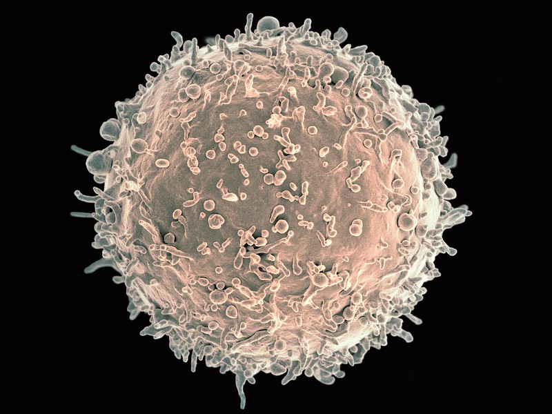
Scanning electron micrograph of a B cell. Credit: NIH
When the immune system is compromised due to various conditions and medicines, patients can experience opportunistic infection. Now, researchers reporting in Cell Reports have uncovered a sex-based variance in the trained immune memory response to infection in mice that might translate to humans.
The researchers, from the University of Missouri School of Medicine, found that female mice were more vulnerable to opportunistic infection from a bacterial pathogen to which they had previously been exposed when progesterone levels were naturally elevated as part of their reproductive cycle.
“Differences in immune response in males and females have been observed before. For instance, males had increased morbidity and severity of COVID-19 from SARS-CoV-2 infections,” said Dr Adam Schrum, associate professor in the Department of Molecular Microbiology and Immunology. “But females are known to suffer other infections worse than males. Our research found that female mice were far more vulnerable to opportunistic bacterial infection than male mice because of a sex-based difference in their trained immunity.”
To understand why the immune systems of female and male mice responded differently to a bacterial pathogen, the researchers examined whether the reproductive cycle affected immune training. They found that elevated progesterone levels correlated with lower trained immune responses. To test this more fully, the researchers gave the female mice progesterone blockers and found that their trained immune response was subsequently enhanced.
“The female mice had significantly restored trained immune response when progesterone was blocked, reaching comparable levels to those of male mice,” said Schrum. “Sex hormone-based modulation of immune function needs more study to be fully understood, but as a first step we can conclude that immune training is influenced by a progesterone-dependent mechanism that results in a sex bias in mice.”
In addition to further study to understand how and why progesterone specifically influences trained immune responses in mice, the researchers pointed out that because mice have shorter estrous cycles than the human menstrual cycle, further research is needed to understand how sex hormones might affect human immune training.
People often say whether they feel like their immune system is ‘down’ – but could there be some truth to this? A recent study showed that when freshly vaccinated people self-assessed the strength of their immune response, their estimates correlated well to their measured antibody levels. They were even more accurate when their immune response was weak. The results were published in the journal Biological Psychology.
At the University of Konstanz, Stephanie psychologist Dimitroff researches the connection between our brain and our immune system. “Listen to your body,” she concludes from her study. “The field of medicine is moving towards greater patient orientation. Our findings support the idea that patients’ self-perceptions provide valuable clues about their state of health. Physicians should listen to them more.”
Communication between the immune and nervous systems
One part of our brain, the insula, receives information from the body and gives us a basic impression of its condition, which until now was assumed to be quite general in nature. Stephanie Dimitroff’s study now suggests that our brain can perceive the body’s condition more specifically than previously thought. Is it possible that our brain can assess the state of our immune system?
“Of course, our brain does not count antibodies. But our immune system is intrinsically connected to the central nervous system,” Dimitroff explains. “The immune system is regulated via this connection. And our brain also receives information from the immune system.”
This communication between the immune system and the central nervous system is key for our sense of well-being or illness. “It is important to know here: When we feel ill, for example, we have a cold, this feeling is caused quite significantly by the immune system’s communication with the central nervous system,” says Dimitroff. “The brain receives signals that something is wrong with the body and causes the feeling of illness as a result.”
The same flow of information between the immune and nervous systems can generally also take place when the body is not ill. This means it could be possible that this communication process gives us an impression of our immune system even when we are healthy. Stephanie Dimitroff’s study investigates whether this is actually the case.
Results of the study
The study looked at people who had received the COVID-19 vaccine. This group of participants was chosen because a particularly large number of people received the vaccine in the summer of 2021, when the study was conducted. 166 people between the ages of 18 and 59 participated in the study.
After vaccination, the participants in the study were able to assess surprisingly well how strongly their immune system was positioned to fight the respective illness. This was especially true for people who had developed only a few antibodies. In fact, 71% of participants who did not feel well protected after vaccination also had a below-average immune response. “Our most notable finding is that those who felt they had not produced high levels of antibodies after vaccination were often correct in their assessment.”
By contrast, participants who assessed their immune response as good were not always right. However, all of those who had a particularly strong immune response also reported feeling well protected.
Alternative interpretations
For Stephanie Dimitroff, however, it is still too early to draw any final conclusions. The psychologist is considering other possible causes, including the placebo effect. This is because communication between the brain and the immune system runs in both directions. The signals from our brain can therefore also influence our immune system. People who firmly believe in vaccination or are basically optimistic could thus actually develop a better immune defence (placebo effect) and also feel better protected. It is therefore possible that belief in the effectiveness of a vaccine is what improves its efficacy, and this could also explain the high accuracy of the self-assessments.
“Our results suggest that it is quite likely that people have a real ability to assess their own health. However, I cannot rule out that there is a combination of effects at play, including the placebo effect and/or feelings of optimism,” Dimitroff says. In her view, it would make sense to repeat the study in order to confirm the results and rule out alternative causes.