Category: Gender

For Women, Lymphoma Chemotherapy more Effective in Afternoon

Photo by Malvestida on Unsplash

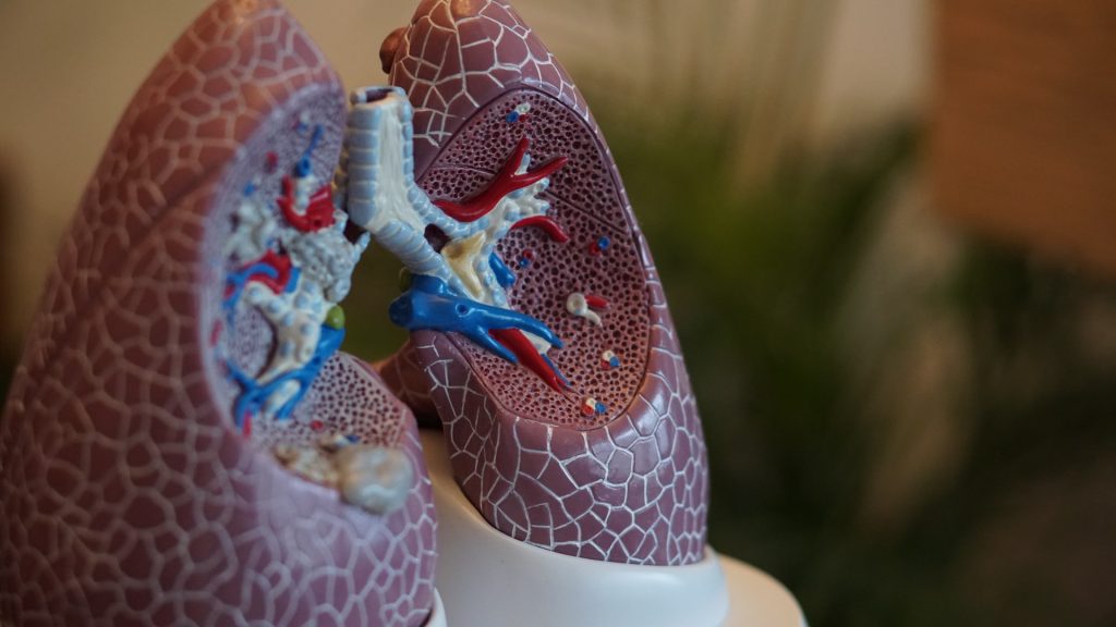
While chemotherapy is highly effective at killing cancer cells, it also kills healthy cells, something which medical research is trying minimise. Recently, ‘chronochemotherapy’ has garnered growing attention in the research community. As the name suggests, the aim is timing the delivery of the drug when the body is least vulnerable to the harmful effects of the drug, while the cancer cells are at their most vulnerable.

Chronochemotherapy exploits the fact that human physiological processes, including cell proliferation and differentiation, are regulated by the circadian clock. However, it is not yet widely exploited in real-world clinical settings because there at present is no systematic method to find the optimal chemotherapy delivery time.

This problem was tackled by an interdisciplinary team of researchers from the Institute for Basic Science (IBS), South Korea. They were led by the principal investigators, mathematician Kim Jae Kyoung at IBS and oncologist Koh Youngil at Seoul National University Hospital. The researchers studied a group of patients suffering from diffuse large B-cell lymphoma (DLBCL), which accounts for about 30 to 40% of non-Hodgkin’s lymphoma. Their findings are published in the journal JCI Insights.

The research team noticed that DLBCL patients at Seoul National University Hospital received chemotherapy at two different schedules, with some patients receiving morning treatment (8:30), while others taking the drugs in the afternoon (14:30). All patients received R-CHOP, which is a combination of targeted therapy and chemotherapy, 4 to 6 times in the morning or afternoon at intervals of about 3 weeks.

Figure 1. Chemotherapy in the afternoon can improve treatment outcomes
The daily fluctuation of proliferative activity of bone marrow is larger in females than in males, and it becomes higher in the morning (left). Thus, chemotherapy in the morning strongly inhibits proliferative activity in female lymphoma patients, resulting in a higher incidence of adverse events such as neutropenia and infections. This forces the clinicians to reduce the dose intensity (center). Consequently, female patients undergoing the morning treatment show a lower survival probability than those undergoing the afternoon treatment (right). Specifically, only ~13% of female patients treated in the afternoon had a worse outcome and ~2% of them died while ~37% of female patients treated in the morning had a worse outcome and ~25% of them died. Male patients did not show any difference in treatment outcomes depending on the chemotherapy delivery time.

They analysed 210 patients to investigate differences between morning and afternoon treatment. They found that female patients who received afternoon treatment had 12.5 times reduced mortality rate (25% to 2%), while the cancer recurrence after 60 months was decreased by 2.8 times (37% to 13%). In addition, chemotherapy side effects such as neutropenia were more common in female patients who received morning treatment.

Surprisingly, there was no difference in treatment efficiency depending on the treatment schedule in the case of male patients.

To understand the cause of gender differences, the research team analysed ~14 000 blood samples from the Seoul National University Hospital Health Examination Center. It was found that in females, white blood cell count tends to decrease in the morning and increase in the afternoon. This indicates that the bone marrow proliferation rate is higher in the morning than in the afternoon because there is a ~12hr delay between the bone marrow proliferation and blood cell production.

This means that if a female patient receives chemotherapy in the morning when bone marrow is actively producing blood cells, the possibility of adverse side effects becomes greater. These results are consistent with the findings from recent randomised clinical trials that showed female colorectal cancer patients treated with irinotecan in the morning suffered from higher drug toxicities.

One confounding variable was the drug dose. Since the morning female patients suffered from greater adverse side effects, oftentimes the dose had to be reduced in these patients. On average, the drug dose was reduced by ~10% compared to the dose intensity given to female patients receiving the afternoon treatment.

Unlike female patients, it was found that male patients did not show a significant difference in white blood cell count and bone marrow cell proliferation activity throughout the day, which is the reason why the timing of the treatment had no impact.

Professor Koh Young-il said, “We plan to verify the conclusions of this study again with a large-scale follow-up study that completely controls confounding variables, and to confirm whether chemotherapy has similar effects in other cancers.”

Ci Kim Jae Kyung said, “Because the time of the internal circadian clock can vary greatly depending on the individual’s sleep-wake patterns, we are currently developing a technology to estimate the time of the circadian clock from the patient’s sleep pattern. We hope that it can be used to develop an individualised anti-cancer chronotherapy.”

Source: Institute for Basic Science

Why Many Lung Diseases are Sex-biased

Anatomical model of lungs
Photo by Robina Weermeijer on Unsplash

By studying sex differences in expression across more than 2500 genes in mouse lung cells, researchers may have found an explanation for sex biases in the prevalence and severity of lung diseases, such as the greater severity of COVID for males. In particular, very high numbers of X-linked genes escape transcriptional silencing in lung alveolar type 2 (AT2s) cells, according to their study published in Stem Cell Reports.

“Our study is the first to compare male and female AT2 cells for gene expression, and our findings suggest that there are likely sex differences in lung repair following viral-induced injury,” says co-senior author Montserrat Anguera, an associate professor at the University of Pennsylvania.

Sex differences exist for many lung diseases, but the mechanistic basis for this remains unclear. “We started this project during the beginning of the pandemic, because we were curious about the sex bias with COVID disease, where more older men have increased morbidity, and wondered whether X-chromosome inactivation (XCI) might contribute to this sex bias,” Anguera says. “We realised that the SARS-CoV2 virus first encounters AT2 cells in the lung, and that the virus enters cells through the angiotensin-converting enzyme 2 (Ace2) receptor, which is located on the X chromosome.”

XCI is a process by which one of the copies of the X chromosome is inactivated in female mammals. The inactive X chromosome is silenced by being packaged into a transcriptionally inactive structure called heterochromatin. XCI prevents female mammals from having twice as many X-chromosome gene products as males, who only possess a single copy of the X chromosome.

In the new study, Anguera and co-senior author Andrew Vaughan, an assistant professor at the University of Pennsylvania, investigated XCI maintenance and sex-specific gene expression profiles using male and female AT2s. The results showed that approximately 68% of expressed X-linked genes in mouse AT2s escape XCI. These genes include Ace2, which serves as the entry point into cells for SARS-CoV-2, but is also involved in lung repair.

There were genome-wide expression differences between male and female AT2s, possibly contributing to sex differences in lung injury and repair in multiple settings. Taken together, the findings demonstrate that AT2 cells have the highest levels of XCI escape for mouse cells reported to date and support a renewed focus on AT2s as a potential contributor to sex-biased differences in lung disease.

In addition, the results showed that AT2 cells, similar to immune cells, do not strictly follow the classic rules of XCI. “We were surprised to find that female AT2 cells lack canonical epigenetic modifications that are typically enriched on the inactive X as a result of XCI. These include the long noncoding RNA Xist and heterochromatic histone modifications H3K27me3 and H2AK119-ubiquitin,” Anguera says. “Because the inactive X in female AT2 cells has fewer epigenetic marks, this enables more gene expression chromosome wide, including the Ace2 gene.”

For now, it remains an open question whether ACE2 escapes XCI in human AT2 cells. to The authors say this is a likely scenario because there are significantly higher numbers of XCI escape genes in human cells compared to mouse cells.

Moving forward, the authors plan to investigate how expression from the inactive X in AT2 cells is affected by SARS-CoV2 infections. They also will continue to study how expression from the X chromosome is regulated in other cell types that do not exhibit conventional XCI maintenance. “Our findings open the door to future work investigating the genetic and epigenetic basis, residing within the X chromosome, of sex differences in immune responses to inhaled viruses,” Anguera says.

Source: Science Daily

Why Obesity’s Health Impacts are Worse for Males

Toilet sign male and female
Photo by Tim Mossholder on Unsplash

A newly published study in iScience sheds light on the biological underpinnings in sex differences in obesity-related disease, with researchers observing “striking” differences in the cells that build blood vessels in the fatty tissue of male versus female mice.

Men are more likely than women to develop conditions associated with obesity such as cardiovascular disease, insulin resistance and diabetes, says study leader Professor Tara Haas at York University.

“People have used rodent models to study obesity, and the diseases that are associated with obesity – like diabetes – but they’ve typically always studied male rodents, because females are resistant to developing the same kinds of diseases,” says Haas. “We were really interested in exploring that difference because, to us, it spoke of something really fascinating happening in females that protects them.”

In earlier work, Haas and her team saw that when mice become obese, females grow a lot of new blood vessels to supply the expanding fat tissue with oxygen and nutrients, whereas males grow a lot less. For this study, Haas and her co-authors focused on differences in the endothelial cells that make up the building blocks of these blood vessels in fat tissue.

The team used software to help sift through thousands of genes to zero in on the ones that would be associated with blood vessel growth. They discovered that processes associated with the proliferation of new blood vessels were high in the female mice, whereas the males had a high level of processes associated with inflammation.

“It was very striking the extent of inflammation-associated processes that were prevalent in the males,” Haas recalls. “Other studies have shown that when endothelial cells have that kind of inflammatory response, they’re very dysfunctional, and they don’t respond to stimuli properly.”

York PhD student Alexandra Pislaru, who works in Haas’ lab and is a co-first author of the study, participated in this project as part of her dissertation.

“It is exciting to observe the continuing resilience that female endothelial cells display even when stressed by a long-term high-fat diet,” Pislaru says. “The findings from our study can help researchers to get a better understanding of why obesity manifests differently in men and women.”

The researchers also examined the behaviour of the endothelial cells when they were taken out of the body and studied in petri dishes.

“Even when we take them out of the body where they don’t have the circulating sex hormones or other kinds of factors, male and female endothelial cells still behave very differently from each other,” Haas explains.

Female endothelial cells replicated faster, while male endothelial cells displayed greater sensitivity to an inflammatory stimulus. By comparing with previously published data sets, the researchers found endothelial cells from aged male mice also displayed a more inflammatory profile compared to female cells.

“You can’t make the assumption that both sexes are going to respond to the same series of events the same way,” says Haas. “This isn’t just an obesity related issue – I think it’s a much broader conceptual problem that also encompasses healthy aging. One implication of our findings is that there will be situations where the treatment that is ideal for men is not going to be ideal for women and vice-versa.”

While humans and mice have different genes that may be turned up or down, Haas believes the general findings would likely apply and is interested studying the same cells in humans in future research.

Source: York University

Teachers Grade Girls Higher, Study Finds

Photo by Mary Taylor on Pexels

Girls are routinely being given more generous grades than boys with the same academic competences, according to a new study of tens of thousands of Italian pupils and their teachers. This reflects other European studies reflecting higher teacher grading for girls.

This bias against boys could mean the difference between a pass and a fail in subjects such as maths. The researchers warn that it could also have wider consequences in areas such as university admission, job choice and earnings.

Their study, published in British Journal of Sociology of Education, is the first to demonstrate that the problem is systemic – it is present across a variety of educational environments, regardless of teachers’ characteristics.

Gender-related gaps in educational achievement are common worldwide.  However, the nature of the gap differs with different ways of measuring achievement.

When the results of standardised tests, which have a standard scoring system, are used, girls typically outperform boys in humanities, languages and reading skills, while boys do better in maths.

In contrast, when grades are awarded by teachers, females do better than males in all subjects.

To find out how teachers’ evaluations tend to favour females, the University of Trento researchers began by comparing the scores students received in standardised tests of language and maths with the grades they achieved in their classroom exams.

The 38 957 students were around 15-16 years old.  The standardised tests were set nationally and marked anonymously, while the classroom exams were set in the classroom and marked non-anonymously by their teachers.

In line with previous studies, the girls performed better than the boys in the standardised tests of language, while the boys were ahead at maths.

The teachers however, put the girls in front in both subjects. The girls’ average grade in language was 6.6 (out of 10), compared to 6.2 for the boys.  In maths, the average grade for the girls was 6.3, while the boys averaged 5.9 – which is below the pass mark of 6.

The analysis also showed that when a boy and a girl were similarly competent at a subject, the girl would typically receive a higher grade.

The researchers then looked whether factors, such as the type of school and the size and gender make-up of classes, were driving the gender grade gap.

They also investigated whether characteristics of teachers themselves, such as how senior or experienced they were and whether they were male or female, helped explain girls’ more generous grades.

Only two factors were found to have an effect – and only in maths.  The gender gap in maths grades was greater when classes were bigger.  Girls were also graded as being further ahead than boys in technical and academic schools than they were in vocational schools.

None of the other factors had any significant effect in reducing the gender grading gap.

Taken overall, the results show for the first time that higher grading of girls is systemic – rather than stemming from one particular failing, it is embedded in the whole school system.

The study’s authors say it’s possible that, in reading, teachers unconsciously reward students exhibiting traditionally female behaviour, such as quietness and neatness, which make teaching easier for the teachers.  Another theory is that inflated grades in mathematics are a way of trying to encourage girls, who are often seen as weaker in this subject.

The study’s authors conclude that bias against boys in Italian schools is considerable and could have long-term consequences.

“There is a strong correlation between having higher grades and desirable educational outcomes, such as gaining admission to good colleges or having a lower probability of dropping out of school,” says researcher Ilaria Lievore, a PhD candidate in Sociology.

“Consequently, higher grades are also correlated with other outcomes, such as having higher earnings, a better job or even higher life satisfaction.”

She adds that although other European countries also grade girls more generously than boys, the reasons for this could differ from place to place and won’t necessarily mirror those in Italy.

The study’s limitations include using grades which were awarded part-way through the school year,  which may have differed from differed from the students’ final grades.

Source: Taylor & Francis

X Chromosome is Shut Down in Some Male Cancers

Source: NIH

Researchers report in Cell Systems that they have discovered another difference between cancer cells and normal cells besides mutations: the X chromosome, typically only inactivated in XX female cells, can also be inactivated across different male-derived cancers.

“To balance the expression of genes between the sexes, in normal development, one copy of the female X chromosome is inactivated at random across the human body. We wanted to know if this process that occurs in normal development goes awry in genetically unstable male or female cancer cells,” says senior author Srinivas Viswanathan, a cancer geneticist and medical oncologist at the Dana-Farber Cancer Institute.

By using publicly available datasets comprising of thousands of DNA samples from cancer patients around the world, the team of researchers stumbled upon the high expression of XIST – the gene responsible for shutting down gene expression on the X chromosome – in about 4% of the male cancer samples analysed.

While XIST may be expressed in very early development in all sexes, X inactivation is thought to be a female-specific process later in development. It was previously shown that some female cancer cells may lose the ability to turn off one of the X chromosomes, leading to increased X-linked gene expression, but this ability of X inactivation had still only been studied primarily in female cells.

Within the 4% of anomalous male cancer samples identified, 74% were from reproductive cancers already shown to inactivate the X chromosome, but that left 26% of samples from other cancer types. These included liver, brain, skin, heart, lung, and thyroid cancers.

“We were very surprised by this result since XIST is a transcript typically used to classify female cancers, and so we wanted to ensure that this was not merely a result of mis-annotation. Yet, we do in fact see that some male cancers of diverse subtypes activate XIST and display features of X inactivation,” says Viswanathan.

“We have to be aware of the caveats of working with these types of datasets. These samples have been in many people’s hands, and there is more room for human error,” said co-corresponding author Cheng-Zhong Zhang, cancer biologist at the Dana-Farber Cancer Institute. “This is the biggest source of uncertainty for us; we have to be creative in how we look at the data and find controls.”

One possible explanation for why this phenomenon is occurring is genetic instability. Cancers often have multiple copies of chromosomes, and if two X chromosomes happen to be in one cell, then it may be necessary to inactivate one of them by activating XIST, regardless of whether that cell is in a female or male individual.

“Another possibility is there are some important genes on the X chromosome that, when silenced, enable the cancer to grow. We will investigate this in future studies,” says Viswanathan.

“In some ways, sex is the ultimate biomarker in that it subdivides the human population, but we often don’t think about how genetic differences between the sexes may inform cancer prognosis or response to therapy,” says Viswanathan.

Source: Cell Press

Why Women Have the Edge in Recovery from Kidney Injury

Anatomic model of a kidney
Photo by Robina Weermeijer on Unsplash

Women have a better ability to recover from kidney injury than men, but the reasons are not well understood. A study in Cell Reports may provide answers, as researchers found that females have an advantage at the molecular level that protects them from a newly discovered form of cell death that occurs in injured kidneys. This protection could be exploited as a potential therapeutic.

“Kidney disease afflicts more than 850 million people worldwide every year, so it’s important to understand why female kidneys are more protected from these acute and chronic injuries,” said Tomokazu Souma, MD, PhD, assistant professor at Duke University School of Medicine. “Our study is a step toward identifying the causes and suggests that this female resilience could be therapeutically harnessed to improve kidney repair in both sexes.”

Souma and colleagues conducted studies in mice focusing on a form of cell death called ferroptosis, which was only recently discovered. This form of cell death is dependent on iron and oxidative stress. It has been identified as a key player in kidney diseases.

Using genetic and single-cell RNA transcriptomic analysis in mice, the researchers found that being female confers striking protection against ferroptosis through a particular pathway called nuclear factor erythroid 2-related factor 2, or NRF2.

In females, NRF2 is highly active, keeping cell death in check. In males, however, the sex hormone testosterone reduces the activity of NRF2, thus promoting ferroptosis and undermining cell resiliency in kidney injury.

Further experiments showed that chemically activating NRF2 protected male kidney cells from ferroptosis, demonstrating that NRF2 could be a potential therapeutic target to prevent failed renal repair after acute kidney injury.

“By identifying the mechanism in which the female hormonal environment protects and the male hormonal environment aggravates acute and chronic kidney injuries, we believe there is strong potential to boost the resilience of kidneys,” Souma said.

Source: Duke University Medical Center

Women with Autism have a Greater Mental Illness Risk

Mirror symbolising schizophrenia
Source: Vince Fleming on Unsplash

Autistic young men and women are more affected by psychiatric conditions and have an increased risk of hospitalisation as a result of their mental illness. Autistic women are particularly vulnerable, as shown in a study published in JAMA Psychiatry.

Autistic people have an increased risk of suffering from mental illness. Current data indicates that autistic women are more vulnerable than autistic men, but few studies have been able to establish that there are sex differences.

The researchers, from Karolinska Institutet, conducted a register-based cohort study with more than 1.3 million people in Sweden who were followed from the age of 16 to 24 between 2001 and 2013. Just over 20 000 of these were diagnosed with autism.

“We saw an increased risk of eleven different psychiatric conditions, including depression, anxiety disorders, self-harm and difficulty sleeping,” says Miriam Martini, a doctoral student in psychiatric epidemiology at Karolinska Institutet and first author of the study.  

High hospitalisation rates 

Something that Miriam Martini finds particularly worrying is that 32 out of 100 autistic women had been hospitalised as a result of their mental illness, compared with 19 out of 100 autistic men. For non-autistic people, the corresponding figure was less than five out of 100.   

The study focuses on young adults who are at a crucial time in their life when many mental health problems increase, while the transition to adulthood often means poorer access to care, says Miriam Martini.   

“Healthcare for young adults needs to be expanded, especially for autistic women, so that mental illness can be detected in time to avoid worsening of symptoms resulting in hospitalization,” says Miriam Martini.  

The reason why autistic women are more affected by mental illness than autistic men is not clear, but in the study, the researchers point to several possible factors. Previous research has shown that autistic women to a greater extent use compensatory behaviours to camouflage their autism, which may be due to the fact that women generally tend to adapt to the expectations of those around them. This delays diagnosis and the provision of assistance, which can negatively affect their mental health.  

Overlooked by the healthcare system 

Another possible explanation may be that it could be difficult to detect autism in women using diagnostic criteria.  

“It may be that autism manifests differently in women than in men, which means that women are not detected using today’s diagnostic criteria. This is something we need to do more research on,” says Miriam Martini.  

Source: Karolinska Institutet

Selection for Metabolism and Immunity Explains Fatty Liver Sex Differences

Anatomy of the gut
Source: Pixabay CC0

A new way of understanding sex-biased diseases has been developed by scientists at UC San Francisco (UCSF). Their evolutionary biology-based theory, published in in Science, is that males and females took opposing paths in a trade-off between immunity and metabolism that happens in the liver. This helped males fight bacterial infections from wounds received in dominance fights, while helping females store subcutaneous fat to survive when food is scarce.

Working with mice, the scientists described the activity of a signalling pathway that regulates lipids, storing fat in the liver in males and releasing it into the bloodstream in females. This pathway also responds to growth hormone.

This phenomenon may have shaped male biology in ways that increase risk from modern, high-calorie diets. The findings are particularly important for fatty liver, which affects a quarter of the US population. It is seen predominantly in men until women reach menopause.

“Scientists have only recently started to understand there are these profound differences between males and females,” said Holly Ingraham, PhD, UCSF professor and co-senior author of the study. “Understanding these differences is going to be the key to unlocking therapeutics for sex-biased diseases. Fatty liver is one example.”

The experiments found that male mice were three times more likely than females to survive infection with E. coli. The females developed hyperlipidaemia, a condition that is also seen in humans with severe sepsis. Lowering their lipid levels helped them to survive.

The investigators then examined how males and females respond to the contemporary environmental challenge of overeating by feeding the mice high-fat chow. males developed fatty liver and glucose intolerance, but females did not. This was true even when males and females gained a similar amount of weight.

Searching the literature for something that could explain this, the team identified a transcription factor called BCL6, which prevents the breakdown of fat in the liver and is much more present in male mice.

Deleting the gene for this protein eliminated liver fat in the males, and also their ability to survive the infection.

“The host defence programs in the liver are the predisposing factors that drive fatty liver in males,” said Joni Nikkanen, PhD, a postdoctoral fellow and one of the study researchers.

“We have an evolutionary perspective on why such programs have developed — because they protect males against bacterial infections,” he said. “But in another context, these same programs are not good for you anymore, and you will develop more severe fatty liver.”

The team also examined how the presence of BCL6 affected gene expression in the liver. This process begins at puberty when males produce more testosterone, and their pituitary glands start to secrete growth hormone in sharp peaks and valleys.

These intermittent bursts, likely testosterone-regulated, are important. When researchers infused male mice continuously with growth hormone the way it is secreted in females, BCL6 disappeared from their livers, and they lost the ability to fight E. coli infection.

The results point to growth hormone as a potential therapy for adults with fatty liver disease, an idea that is currently being tested. Its effects are already well established in children whose pituitaries don’t produce enough growth hormone. Male children especially tend to develop fatty liver, but it goes away when they are given growth hormone to treat their short stature.

The work also expands the scientific view of how the body fights infection to include organs like the liver.

“The fight is still between the infection and the immune system,” said Omer Gokcumen, PhD, an evolutionary anthropologist at the University at Buffalo and a co-author of the study. “But the liver is determining the battlefield.”

Source: University of California – San Francisco

Cardiovascular Disease Risk Factors the Same for Men and Women

Photo by Ketut Subiyanto on Pexels

For men and women, the risk factors for cardiovascular disease are largely the same, an extensive global study involving over 155 000 participants shows.

The study, published in The Lancet, includes participants from 21 countries with differing GDP. Cardiovascular disease is more widespread in low- and middle-income countries.

The 21-country study used data were taken from the Prospective Urban Rural Epidemiological (PURE) Study and comprised 155 724 participants aged 35–70 years with no history of cardiovascular disease when they joined the study. All cases of fatal cardiovascular disease, heart attack, stroke, and heart failure during the follow-up period, which averaged ten years, were registered.

The risk factors studied were metabolic (such as high blood pressure, obesity, and diabetes), behavioral (tobacco smoking and diet), and psychosocial (economic status and depression).

No clear gender or income divide

Metabolic risk factors were found to be similar in both sexes, except for high values of low-density lipoprotein (LDL, often known as bad cholesterol), where the association with cardiovascular disease was stronger in men. In the researchers’ opinion, however, this finding needs confirmation in more studies.

Depressive symptoms were another risk factor for cardiovascular disease that proved to be more significant among the men than the women. On the other hand, the link between a poor diet and cardiovascular disease was closer in women; and smoking, though markedly more frequent among men, was just as injurious a risk factor for women.

Overall, the researchers found broadly similar risk factors for cardiovascular disease for the male and female participants, irrespective of their countries’ income level. This highlights the importance of disease prevention strategies, too, being the same for both sexes.

Similarities greater than differences

The women’s lower overall risk of cardiovascular disease, especially heart attack (myocardial infarction), may be explained by the younger women’s higher tolerance to risk factors. Their estrogen makes vessel walls more compliant and affects the liver’s capacity to get rid of LDL.

Among the 90 934 women in the study, 5.0 cases of stroke, heart attack, and/or cardiovascular disease were registered per 1000 persons per year. The corresponding number in the group of men (64 790 individuals) was 8.2 cases.

Annika Rosengren, Professor of Medicine at Sahlgrenska Academy, University of Gothenburg, is the second author of the study, in charge of the Swedish part of the PURE population study of 4 000 individuals in Gothenburg and Skaraborg.

“When it comes to cardiovascular disease in men and women, the similarities in terms of risk factors are considerably greater than the differences. But men are more vulnerable to high levels of LDL, the bad cholesterol, and we know from other studies that they develop pathological changes in the coronary arteries at a lower age than women, and tend to start developing myocardial infarction quite a lot earlier. With respect to early stroke, though, the sex differences are less pronounced, as we’ve also seen in other studies,” Prof Rosengren said.

Source: University of Gothenburg

Sex Differences in Post-stroke Biomarkers

Credit: American Heart Association

Although males and females are equally affected by stroke Since oestrogen and progesterone have known neuroprotective effects, it is important to compare the size, severity and biochemical composition of the brain tissue following stroke in female and male animal models. In a paper published in IBRO Neuroscience Reports, researchers have discovered that certain biomarkers were different in the brains of male and female mice.

Stroke neurosurgeon and research coordinator Prof Nicole Sylvain and her colleagues are looking at treatments for post-stroke recovery that help supplement these energy losses. Using the Canadian Light Source (CLS) at the University of Saskatchewan (USask), the team was able to identify energy biomarkers in the brain, which could eventually inform clinicians about the effects of potential stroke treatments on brain recovery after a stroke.

The group’s recent study examined post-stroke differences between male and female mice, and found that female mice have higher amounts of glycogen in their brains. Glycogen is a sugar-like substance that circulates in our blood and nourishes our cells. When the supply of glycogen is disrupted by stroke, the brain is severely impacted.

Most pre-clinical stroke research has been performed using male lab animals, with results usually generalised to both sexes. In clinical stoke cases, females have a higher incidence of ischaemic stroke and poorer outcomes, compared to males.

“We found that, for the most part, male data can be generalised for females, however, some of the metabolic markers we measured were actually different,” Sylvain said. “It’s really important to do the research on both sexes.”

It would be impossible for the team to detect the biomarkers without to the Mid-IR beamline.

“The only way to detect them in such an accurate way across the brain is with infrared imaging, so the CLS has been absolutely vital to our research.”

Source: University of Saskatchewan