Category: Allergies

Gluten Free Diet Reduces Coeliac Symptoms – and ‘Good’ Gut Bacteria

Photo by Mariana Kurnyk: https://www.pexels.com/photo/two-baked-breads-1756062/

A research team led by the University of Nottingham has used magnetic resonance imaging (MRI) to better understand the impact a gluten free diet has on people with coeliac disease, which could be the first step towards finding new ways of treating the condition.

The MARCO study – MAgnetic Resonance Imaging in COliac disease is published in Clinical Gastroenterology and Hepatology (CGH) (link connects to BioRxiv copy).

Coeliac disease is a chronic condition affecting around one person in every 100 in the general population. When people with coeliac disease eat gluten, which is found in pasta and bread, their immune system produces an abnormal reaction that inflames and damages the gut tissue and causes symptoms such as abdominal pain and bloating.

The only treatment is a life- long commitment to a gluten free diet, which helps recovery of the gut tissue but still leaves many patients with gastrointestinal symptoms.

Luca Marciani, Professor of Gastrointestinal Imaging at the University, led the study. He said: “Despite being a common chronic condition, we still don’t precisely know how coeliac disease affects the basic physiological functioning of the gut and how the gluten free diet treatment may further change this.

“We launched the MARCO study to try and address this issue, by using MRI along with gut microbiome analysis to give us new insights into how a gluten-free diet affects people with coeliac disease.”

The team recruited 36 people who had just been diagnosed with coeliac disease and 36 healthy volunteers to participate in the study. Images were taken of their guts with MRI, along with blood and stool samples. The patients then followed a gluten free diet for one year and came back to repeat the study. The healthy participants came back one year later too and repeated the study, but they did not follow any diet treatment.

The study found that the newly diagnosed patients with coeliac disease had more gut symptoms, more fluid in the small bowel and that the transit of food in the bowel was slower than in the healthy controls.

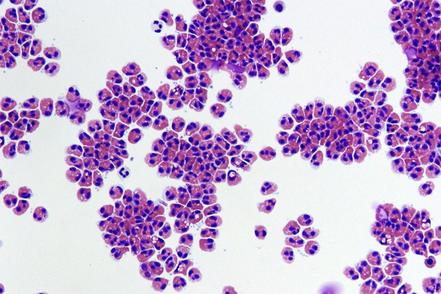
The microbiota (the ‘bugs’ living in the colon) of the patients showed higher levels of ‘bad bugs’ such as E.coli. After one year of a gluten free diet, gut symptoms, bowel water and gut transit improved in the patients, but without returning to normal values. But the gluten free diet also reduced some of the ‘good bugs’ in the microbiota, such as Bifidobacteria associated with reduced intake of starch and wheat nutrients, due to the different diet.

The patient study was conducted by Radiographer Dr Carolyn Costigan, from Nottingham University Hospitals, as part of her PhD studies at the University of Nottingham.

It was particularly interesting to see how the imaging results on gut function correlated with changes in the ‘bugs’ in the colon microbiota. The findings increase our understanding of gut function and physiology in coeliac disease and open the possibility of developing prebiotic treatments to reverse the negative impact of the gluten free diet on the microbiome.”

Luca Marciani, Professor of Gastrointestinal Imaging

Dr Frederick Warren from the Quadram Institute, which contributed to the research, said: “This study is the result of an exciting and innovative research collaboration bringing together medical imaging technology and gut microbiome analysis. We provide important insights which pave the way for future studies which may identify novel approaches to alleviate long-term symptoms in coeliac patients.”

Source: University of Nottingham

Can Adrenaline Auto-injectors Prevent Fatal Anaphylaxis?

Photo by Mat Napo o Unsplash

Individuals at risk of anaphylaxis are often prescribed adrenaline (epinephrine) autoinjectors such as EpiPens. A recent review published in Clinical & Experimental Allergy finds that these autoinjectors, which people use to self-administer adrenaline into the muscle, can deliver high doses of adrenaline into the blood, but these levels are short-lived and may not be sufficient to save lives in cases of fatal anaphylaxis.

Anaphylaxis is an acute systemic hypersensitivity reaction to an allergen or trigger, typically associated with skin reactions, nausea/vomiting, difficulty breathing, and shock.

Investigators noted that data from animal and human studies suggest that intravenous adrenaline infusions delivered directly into the blood can prevent fatal anaphylaxis, but adrenaline autoinjectors may have little impact in such deadly cases.

“For effective management of the most severe allergic reactions, adrenaline given by continuous intravenous infusion, with appropriate fluid resuscitation, is likely to be required—how this is safely achieved in the pre-hospital setting remains to be determined,” the authors wrote. This challenge stems from the fact that fatal anaphylaxis is unpredictable and fast. Fortunately, fatality is rare, with a population incidence of 0.03–0.51 per million per year.

Source: Wiley

Better Allergy Diagnosis with Mast Cell Activation Testing

Photo by Corleto on Unsplash

Researchers at the University of Bern and Bern University Hospital have developed a test to simplify the diagnosis of allergies by testing mast cells. Its effectiveness has now been confirmed in clinical samples from children and adolescents suffering from a peanut allergy. The results, recently published in the European Journal for Allergy and Clinical Immunology (Allergy), could fundamentally improve the clinical diagnosis of allergies in future.

Food allergies are a major health problem worldwide. In some countries, up to 10% of the population is affected, mainly young children. Peanut allergy, in particular, is one of the most common diseases and often manifests itself in severe, potentially life-threatening reactions. The stress of food allergies not only affects the individuals concerned, but also has far-reaching consequences for their families, the health system and the food industry. The oral food challenge test, in which people consume the allergen (such as peanut extract) under supervision to test the allergic reaction, is still considered the gold standard in diagnosis. However, the method is complex and carries health risks. The allergen skin prick test and blood test are often not very accurate, which can lead to misdiagnoses and unnecessary food avoidance. 

A team of researchers led by Prof Dr Alexander Eggel and Prof Dr Thomas Kaufmann from the University of Bern, developed an alternative test in 2022. It mimics the allergic reaction in a test tube and thus offers an attractive alternative to standard tests. The Bern researchers have now investigated the effectiveness of the test on samples from children and adolescents with confirmed peanut allergy and a healthy control group in a clinical study in collaboration with partners from the Hospital for Sick Kids in Toronto, Canada. They were able to show that the new test has a higher diagnostic accuracy than the methods used so far.

Mast cell activation test as appropriate alternative

“The most common food allergies are type I allergies. They develop when the body produces immunoglobulin E (IgE) antibodies in response to substances that are actually harmless (allergens),” explains Alexander Eggel. These antibodies bind to specific receptors on the mast cells, which are immune cells that play an important role in allergic reactions and inflammation. They are mainly located in the tissue, for example, in the intestinal mucosa, and are prepared for and sensitised to the allergen by binding to the antibodies. Upon renewed contact with the allergen, it binds directly to the mast cells loaded with antibodies, activating them and triggering an allergic reaction.

“In the Hoxb8 mast cell activation test (Hoxb8 MAT), which we developed, mast cells grown in the laboratory are brought into contact with blood serum from allergic patients. The mast cells bind the IgE antibodies from the serum and are sensitised by them. We can then stimulate the mast cells with different amounts of the allergens to be tested,” says Eggel. Quantifying the activated mast cells suggests how allergic a patient is to the allergen tested without needing to consume the food. 

Higher diagnostic accuracy than standard tests

The study used serum samples from a total of 112 children and adolescents who had already participated in a study in Canada and for whom clear diagnostic data on their peanut allergy status were available. The mast cells cultured in the laboratory were sensitised with their serum and then stimulated with peanut extract. “The cell-based test was easy to carry out and worked perfectly. All samples were measured within two days, which was very fast,” says Thomas Kaufmann. The results showed that a large number of sera from allergic patients exhibited allergen dose-dependent activation, while almost all samples from the non-allergic control subjects did not activate the mast cells. “An exceptionally high diagnostic accuracy of 95% could be calculated from these data,” Eggel adds.

In addition, the data measured in the study were analysed in direct comparison with other diagnostic methods established at the hospital. It was found that the Hoxb8 MAT test had significantly higher accuracy than the standard measurement of allergen-specific IgE antibodies in the blood or the frequently used skin test. “Comparison with other clinical tests was crucial to determine which of them reflected the patients’ allergic reaction best. The new mast cell activation test has the advantage that it is functional and therefore incorporates many parameters that are important for triggering the allergy,” says Thomas Kaufmann, adding: “The new test is also based on stable blood serum, which can be drawn using simple blood sampling and then stored in the freezer. This eliminates the challenging logistical obstacles that arise with other methods.” The study also showed that the Hoxb8 MAT test leads to less false negative results. 

“What has been shown in this study on the diagnosis of peanut allergies can also be applied to other allergies in a simple way. The technology is a perfect example of how basic research from the University of Bern can be brought to the clinical practice, and might ultimately simplify life for patients and physicians,” concludes Eggel.

Source: University of Bern

Scientists Discover that Mast Cells Gobble up Other Immune Cells

This scanning electron microscopy image captures the moment where degranulating mast cells (pseudo-colored in sepia) attract and start to incorporate living neutrophils (pseudo-colored in cyan), forming cell-in-cell structures where mast cells trap living neutrophils inside them. © Marcus Frank & Karoline Schul11z, Universitätsmedizin Rostock, Germany

When it comes to allergies, mast cells are key immune system players, releasing pro-inflammatory substances in response to allergens. Now, scientists in Germany have discovered something weird: other immune cells nested inside them like Russian dolls. But how exactly did these cells wind up there?

As reported in the journal Cell, the researchers observed mast cells observed capturing and making use of neutrophils. This surprising discovery sheds new light on how our immune system works, particularly during allergic reactions.

Mast cells, residing in tissues and critical for initiating inflammation, are filled with granules containing pro-inflammatory substances. These granules are released upon encountering potential dangers, including allergens, causing allergic reactions – which for some includes innocuous materials like pollens. But despite how common allergies are, the interaction between mast cells and other immune cells at sites of allergic responses has been largely unexplored.

The research group at the Max Planck Institute of Immunobiology and Epigenetics in Freiburg and the University of Münster used specialised microscopy to visualise the real-time dynamics of activated mast cells and other cell types during allergic reactions in living mouse tissues. The team discovered a surprising interaction: neutrophils were found inside mast cells.

“We could hardly believe our eyes: living neutrophils were sitting inside living mast cells. This phenomenon was completely unexpected and probably would not have been discovered in experiments outside a living organism and highlights the power of intravital microscopy,” says Tim Lämmermann, research leader and Director at the Institute of Medical Biochemistry at the University of Münster.

Pulling a neutrophil trick to trap neutrophils

Neutrophils are frontline immune system defenders, responding quickly and broadly to potential threats. They circulate in the blood and quickly exit blood vessels at sites of inflammation. They are well-equipped to combat pathogens by engulfing the invaders, releasing antimicrobial substances, or forming web-like traps known as ‘neutrophil extracellular traps’. Additionally, neutrophils can communicate with each other and form cell swarms to combine their individual functions for the protection of healthy tissue. While much is known about neutrophils’ role in infections and sterile injuries, their role in inflammation caused by allergic reactions is less understood.

“It quickly became clear that the double-pack immune cells were no mere coincidence. We wanted to understand how mast cells trap their colleagues and why they do it,” explains Michael Mihlan, first and co-corresponding author of the study. Once the team was able to mimic the neutrophil trapping observed in living tissue in cell culture, they we were able to identify the molecular pathways involved in this process. The researchers found that mast cells release leukotriene B4, a substance commonly used by neutrophils to initiate their own swarming behaviour.

By secreting this substance, mast cells attract neutrophils. Once the neutrophils are close enough, mast cells engulf them into a vacuole, forming a cell-in-cell structure that the researchers refer to as ‘mast cell intracellular trap’ (MIT). “It is ironic that neutrophils, which create web-like traps made of DNA and histones to capture microbes during infections, are now trapped themselves by mast cells under allergic conditions,” says Tim Lämmermann.

Recycled neutrophils to boost mast cell function

With the help of an international team, the researchers confirmed the formation of MITs in human samples and investigated the fate of the two cell types involved after trapping. They found that trapped neutrophils eventually die, and their remains get stored inside mast cells. “This is where the story takes an unexpected turn. Mast cells can recycle the material from the neutrophils to boost their own function and metabolism. In addition, mast cells can release the newly acquired neutrophil components in a delayed manner, triggering additional immune responses and helping to sustain inflammation and immune defense”, says Michael Mihlan.

“This new understanding of how mast cells and neutrophils work together adds a whole new layer to our knowledge of allergic reactions and inflammation. It shows that mast cells can use neutrophils to boost their own capabilities – an aspect that could have implications for chronic allergic conditions where inflammation occurs repeatedly,” says Tim Lämmermann. The researchers have already begun investigating this interaction in mast cell-mediated inflammatory diseases in humans, exploring whether this discovery could lead to new approaches to treating allergies and inflammatory diseases.

Source: Max Planck Institute of Immunobiology and Epigenetics

New Guidance Available for Peanut Desensitisation Therapy

Photo by Corleto on Unsplash

Based on focus groups with children and young people with peanut allergy, experts have published guidance for clinicians working in the UK’s National Health Service (NHS) to help them safely and equitably implement Palforzia® peanut oral immunotherapy. Their recommendations are published in Clinical & Experimental Allergy.

In 2022, the National Institute for Health and Care Excellence in the UK recommended the use of Palforzia® – which has defatted peanut powder as its active ingredient – for desensitising children and young people with peanut allergy in the NHS.

The new consensus guidance will inform and support healthcare professionals as they implement Palforzia® for desensitisation and as they gradually increase peanut dosing in patients.

“It is great we can now offer an actual treatment for peanut allergy, rather than just recommend avoidance and educate patients on how to recognise and manage reactions, but the challenge in our current NHS is how we can provide this to eligible patients equitably, regardless of where they live and their backgrounds,” said corresponding author Tom Marrs, PhD, of Guy’s and St Thomas’ NHS Foundation Trust. “This guidance outlines what NHS services need to be able to offer this treatment at scale and to advocate for patients so that we can develop best-practice models.”

Source: Wiley

Allergies: Advances in Understanding Eosinophils

Human eosinophils, a type of white blood cell, are shown isolated from blood. In patients with eosinophilic oesophagitis, these cells store and release packages of inflammatory proteins (red) that can damage the throat and pesophagus. Credit: Julie Caldwell, Cincinnati Children’s Hospital Medical Center

Eosinophils, specialised immune cells, are identified by their distinctive granules that stain red when treated with an acidic reagent, eosin, which gave them their name. Eosinophils are typically rare in blood and tissues, accounting for about 3% of white blood cells. Their biological roles are poorly understood, but recent studies suggest that eosinophils are involved in regulating fat metabolism, repairing certain tissues, and helping fight different infections and cancers. Now, a new study has found that the cytokine Interleukin-5 (IL-5) acts differently on the production of eosinophils than previously believed.

In common diseases such as allergic asthma and rhinosinusitis, eosinophils are abnormally numerous in the blood and tissues, a condition known as eosinophilia. Eosinophilia is a clinical sign that aids in diagnosing these “eosinophil-associated” diseases and guides their treatment. It is known that eosinophilia is driven by increased production of eosinophils by the bone marrow. Since the 1990s, it has also been known that IL-5 is essential for eosinophilia. This led to the development and market introduction of precision therapies targeting IL-5 with monoclonal antibodies to treat severe forms of eosinophilic diseases. However, the effects of IL-5-blocking treatments on eosinophils remain poorly described.

The Laboratory of Cellular and Molecular Immunology (LCMI) of the University of Liege, under the direction of Fabrice Bureau and Christophe Desmet, aimed to better understand the origin of eosinophils and eosinophilia, and the effects of treatments targeting eosinophils. As Christophe Desmet explains, “these questions previously suffered from a too rudimentary definition of the eosinophil development pathway in our bone marrow.” Two doctoral students from the laboratory, Joseph Jorssen and Glenn Van Hulst, combined their talents in bioinformatics and flow cytometry with the help of the Genomics and Flow Cytometry platforms of the GIGA Institute to finely characterise, using different approaches to analyse the surface protein and messenger RNA composition of eosinophils at various stages of their development. Although the mouse remains a reference model, collaboration with the Hematology Department of Liege University Hospital and GIGA also allowed for a very detailed and updated mapping of eosinophil development in human bone marrow, and to observe its conservation through evolution.

This detailed characterisation work, published in the journal Immunity, provides the community with simple-to-use methods and freely accessible bioinformatics data that will greatly facilitate future studies of eosinophils. Using these resources, the same study showed that IL-5 does not act as previously believed by researchers and clinicians. Most thought that IL-5 promoted the maturation of cells destined to become eosinophils and that IL-5-targeting treatments blocked this maturation. “Our study actually supports the opposite hypothesis,” explains Christophe Desmet: IL-5 slows down the maturation of developing eosinophils, allowing them to multiply longer. By stimulating this “transit amplification,” IL-5 promotes eosinophilia, and by inhibiting this process, IL-5-targeting treatments reduce it.

The researchers also showed that interferon response factor-8, considered an essential promoter of leukocyte production, was not intrinsically required for eosinophil production.

This study thus provides resources, methods, and perspectives to understand the origin of eosinophils, the effects of current precision therapies, and the regulation of eosinophil development and numbers in normal and disease conditions.

Source: University of Liège

Celiac Disease: New Findings on the Effects of Gluten

Photo by Mariana Kurnyk

May 16 is International Celiac Day. Celiac disease is a chronic autoimmune condition that occurs in around 1% of the world’s population. It is triggered by the consumption of gluten proteins from wheat, barley, rye and some oats. A gluten-free diet protects celiac patients from severe intestinal damage. Together with colleagues, chemist Dr Veronica Dodero from Bielefeld University was able to determine new details on how certain gluten-derived molecules trigger leaky gut syndrome in celiac disease.

The key finding of the study: a particular protein fragment formed in active celiac disease forms nanosized structures, the so-called oligomers, and accumulates in a gut epithelial cell model. The technical name of the molecule is 33-mer deamidated gliadin peptide (DGP). The study team has now discovered that the presence of DGP oligomers may open the tightly closed gut lining, leading to the leaky gut syndrome. The study has now been published in the journal Angewandte Chemie.

Wheat peptides causing leaky gut

Gluten proteins cannot be completely broken down by the gut. This can lead to the formation of large gluten fragments (peptides) in our gut. In cases of active coeliac disease, researchers discovered that the enzyme tissue transglutaminase 2 (tTG2) present in humans modifies a specific gluten peptide, resulting in the formation of the 33-mer DGP. This usually happens in a part of our gut called the lamina propria. However, recent research has shown that this process can also occur in the gut lining.

‘Our interdisciplinary team characterized the formation of 33-mer DGP oligomers through high-resolution microscopy and biophysical techniques. We discovered the increased permeability in a gut cell model when DGP accumulates, reports Dr. Maria Georgina Herrera, the first author of the study. She is researcher at the University of Buenos Aires in Argentina and was a postdoctoral fellow at Bielefeld.

When the intestinal barrier is weakened

Leaky gut syndrome occurs when the lining of the intestine becomes permeable, allowing harmful substances to enter the bloodstream, leading to inflammatory responses and different diseases. In celiac disease, there’s debate about the early stages of increased permeability. The mainstream theory suggests that chronic inflammation in coeliac disease leads to a leaky gut. However, there is a second theory that proposes that gluten’s effects on gut lining cells are the primary cause. In this view, gluten directly damages the cells of the intestinal lining, making them permeable, which triggers chronic inflammation and potentially leads to celiac disease in predisposed people.

However, since gluten is consumed daily, what molecular triggers lead to the leaky gut in celiac disease patients? If 33-merDGP oligomers are formed, they may damage the epithelial cell network, allowing gluten peptides, bacteria, and other toxins to pass massively into the bloodstream, leading to inflammation and, in celiac disease, autoimmunity.

‘Our findings reinforce the medical hypothesis that impairment of the epithelial barrier promoted by gluten peptides is a cause and not a result of the immune response in celiac patients,’ says the lead author of the study, Dr Veronica Dodero from the Bielefeld Faculty of Chemistry.

The relationship between 33-mer DGP and Celiac Disease

Human leukocyte antigens (HLAs) are proteins found on the surface of cells in the body. They play a crucial role in the immune system by helping it distinguish between self (the body’s own cells) and non-self (foreign substances like bacteria or viruses). In celiac disease, two specific HLA proteins, namely HLA-DQ2 and HLA-DQ8, are strongly associated with the condition. The 33-mer DGP fits perfectly with HLA-DQ2 or HLA-DQ8 and triggers an immune response, leading to inflammation and small intestine villous atrophy. This strong interaction turns the DGP into what scientists call a superantigen. For those affected, a gluten-free diet is the only lifelong therapy.

Source: Bielefeld University

Certain Gut Bacteria Assist in Immunotherapy for Milk Allergy

Gut Microbiome. Credit Darryl Leja National Human Genome Research Institute National Institutes Of Health

Researchers in Japan have discovered a link between gut bacteria and the success of milk-allergy oral immunotherapy. Published in the scientific journal Allergology International, the study found that Bifidobacterium – a genus of beneficial bacteria in the gut – was associated with a higher chance of successful treatment. The finding may help in the development of more effective oral immunotherapies, perhaps by combining them with probiotic supplements.

Many children have allergic reactions to certain milk proteins. Most grow out of it but some have to contend with lifelong allergy. Milk allergy can be improved by oral immunotherapy – taking small, increasing doses of milk. Unfortunately, while allergic reactions are controlled during treatment, in most cases, tolerance disappears soon after the treatment ends.

Gut bacteria are thought to help reduce allergic reactions to some foods, but little is known about the link between these bacteria and oral immunotherapy for milk allergy. Hiroshi Ohno led a team at the RIKEN Center for Integrative Medical Sciences to find out why. The researchers examined 32 children with cow’s milk allergy who received oral immunotherapy, with the first month being conducted in a hospital. “Oral immunotherapy is not without risk,” explains Ohno. “We closely monitored the children in the hospital, and in fact 4 children had such severe reactions to the milk that we could not allow them to continue the treatment.”

The remaining 28 children then completed an additional 12 months of treatment at home. Next, they avoided milk for two weeks, and were then tested on a double-blind, placebo-controlled food challenge to see if they could still tolerate milk without any allergic reactions. During the food challenge, children were initially given a tiny amount of placebo or milk (only 0.01mL) which was gradually increased every 20 minutes until they had an allergic reaction or until they could drink the final 30mL without a reaction.

The researchers focused their analyses on immunological and bacterial changes during the treatment and the relationship between gut bacteria and successful treatment—which was defined as showing milk tolerance that lasted beyond the treatment period by passing the food challenge. They found that during treatment, immunological markers for cow’s milk allergy improved, and bacteria in the gut changed. Nevertheless, after two weeks of avoiding milk, only 7 of the 28 children passed the food challenge, even though they had been able to drink milk safely at the end of the treatment.

To understand why the treatment worked for these seven children but not the others, the team looked for the clinical factors and types of gut bacteria that were related to successful treatment. Of the clinical factors, unsuccessful treatment was more likely in children who were being treated for eczema or asthma and in children who initially had higher levels of milk-protein antibodies. Among the gut bacteria, the presence of Bifidobacterium, a genus of beneficial bacteria in the Bifidobacteriaceae family was related to a higher chance of successful treatment. In fact, only children who passed the final food challenge showed an increasing trend in these bacteria over the course of treatment. When considering ways to improve oral immunotherapy, this is good news because while the first two factors are difficult to change, the types of bacteria in one’s gut are not set in stone.

“With this study, we have identified gut environmental factors that help establish immune tolerance against cow’s milk allergy via oral immunotherapy,” says Ohno. “The next step is to examine the mechanisms underlying this phenomenon and to develop ways to improve the effectiveness of oral immunotherapy, such as the addition of probiotic supplements.”

Source: RIKEN

Respiratory Allergies: Newly Discovered Inflammatory-triggering Molecule

Photo by Andrea Piacquadio on Unsplash

One of the molecules responsible for triggering the inflammation that causes allergic respiratory diseases, such as asthma and allergic rhinitis, has just been discovered by scientists in France. This molecule, from the alarmin family, represents a therapeutic target of major interest for the treatment of allergic diseases. The study is published in the Journal of Experimental Medicine on 10 April.

The inflammation process plays a crucial role in allergic respiratory diseases, such as asthma and allergic rhinitis. Although the pulmonary epithelium is recognised as a major player in the respiratory inflammation that causes these diseases, the underlying mechanisms are still poorly understood.

The research team, from the CNRS, Inserm and the Université Toulouse III – Paul Sabatier and co-directed by Corinne Cayrol and Jean-Philippe Girard, has identified one of the molecules responsible for triggering these allergic reactions,. This molecule from the alarmin family, named TL1A, is released by lung epithelium cells a few minutes after exposure to a mould-type allergen.

It cooperates with another alarmin, interleukin-33, to alert the immune system.

This double alarm signal stimulates the activity of immune cells, triggering a cascade of reactions responsible for allergic inflammation.

Alarmins, therefore, constitute major therapeutic targets for the treatment of respiratory allergic diseases. In a few years’ time, treatments based on antibodies blocking the TL1A alarmin could benefit patients suffering from severe asthma or other allergic diseases.

Source: CNRS

Sublingual Immunotherapy for Food Allergies Safe and Effective for High-risk Children

Photo by Corleto on Unsplash

New research from the University of British Columbia reveals a safe path to overcoming food allergies for older children and others who can’t risk consuming allergens orally to build up their resistance. Sublingual immunotherapy (SLIT) involves placing smaller amounts of food allergens under the tongue.

A study conducted by UBC clinical professor and paediatric allergist Dr Edmond Chan and his team at BC Children’s Hospital Research Institute found SLIT to be as safe and effective for high-risk older children and adolescents as oral immunotherapy is for preschoolers.

The study was published in The Journal of Allergy and Clinical Immunology.

“Our work confirms the safety and effectiveness of SLIT for older children and adolescents with multiple food allergies at higher risk of severe reaction,” said Dr Chan. “These are patients for whom oral immunotherapy would typically be denied because it’s felt to be too risky, so this could be the best approach for that population.”

Previously published research from Dr Chan’s team has shown that preschool oral immunotherapy is safe and effective in the real world. The protocol involves a “build-up phase” of several months, when patients visit a clinic every two weeks to ingest a higher dose of an allergen under medical supervision before continuing the same daily dose between visits. When they reach a certain dose – usually around 300mg of protein – they enter a “maintenance phase” during which they take that target daily dose at home. After a year of maintenance doses, approximately four out of five patients are able to pass an oral challenge test in which they tolerate a much higher dose of 4000mg of protein.

However, the build-up phase is risky for older children and those with a history of severe reactions. Dr Chan’s group has been looking for a safer way to get this at-risk group of patients to the maintenance phase.

They recruited about 180 such patients between the ages of four and 18, most with multiple food allergies. The SLIT protocol (started when COVID-19 pandemic restrictions were in place) required patients to have virtually supervised appointments 3-5 times over several months to build up to a small dose – in most cases, just 2mg of protein – which is absorbed through the membranes under the tongue rather than swallowed and ingested.

The patients’ caregivers learned how to mix and administer these doses at home using novel recipes based on products you can buy at the grocery store, developed with the team’s research dietitian. A wide variety of allergens were treated, including peanut, other legumes, tree nuts, sesame, other seeds, egg, cow’s milk, fish, wheat, shrimp, and other allergens. Patients took these doses daily for 1–2 years.

“It takes up to twice as long as oral immunotherapy, but we wouldn’t have had it any other way, because we needed the superior safety of SLIT for these older kids that are felt to be more severe,” said Dr. Chan.

While most patients had mild symptoms during the build-up phase, none had severe reactions during either build-up or maintenance. Seventy per cent of those tested at the end of the protocol could tolerate 300mg of their allergen – a success rate nearly as high as that for oral immunotherapy.

The results were encouraging for a therapy that any family can undertake at home with guidance from professionals.

“Besides safety considerations in older children, allergists are often quite burdened by the oral immunotherapy build-up phase, where a patient may require 11 or more visits to the clinic. They just don’t feel they have the capacity to offer that many visits in their office,” said Dr Chan. “In our clinic, we are starting to do more home-based approaches because the demand for medical appointments that would allow supervision far outstrips the supply. We are trying to develop an approach, based on data, that matches a patient’s risk level with the appropriate amount of supervision. Our SLIT data suggests that home-based SLIT build-up is safe.”

Ultimately, the trial highlights an alternative that allergists should now consider for patients who cannot safely undertake oral immunotherapy. The trade-off for greater safety is simply a longer timeline, but it comes with the benefit of keeping clinics free for those who need them most.

Source: University of British Columbia