The cellular slime mold Dictyostelium discoideum is a soil microbe that produces diverse natural products with potential antibiotic activity. Previously, three chlorinated compounds had been detected in Dictyostelium, but only the most abundant compound (CDF-1) was identified and shown to be almost as effective an antimicrobial as ampicillin. In research published in FEBS Open Bio, investigators optimised lab culture conditions of Dictyostelium cells to boost the levels of low-abundance chlorinated compounds and to characterise their antimicrobial properties.
The optimized culture conditions took advantage of propionic acid and zinc supplementation to increase the yield of the chlorinated compounds, leading to the identification of CDF-2 and CDF-3 in addition to CDF-1. The molecular structure of CDF-2 and CDF-3 was similar to that of CDF-1, aside from the length of a molecular structure called an acyl side chain. When their antibacterial activity was tested, similarly to CDF-1, CDF-2 and CDF-3 exhibited stronger activity against Gram-positive bacteria than ampicillin but limited activity against Gram-negative bacteria.
Because these compounds are conserved across distantly related Dictyostelium species, CDFs may fulfill a critical role in protecting against harmful bacteria.
“Soil presents both opportunities and dangers for the Dictyostelium amoeba, and we believe this amoeba responds by producing specialised chemicals to attract, repel, or eliminate friends, prey, and predators. We are just starting to discover these chemicals, including this new, potent antibiotic,” said corresponding author Tamao Saito, PhD, of Sophia University, in Japan.
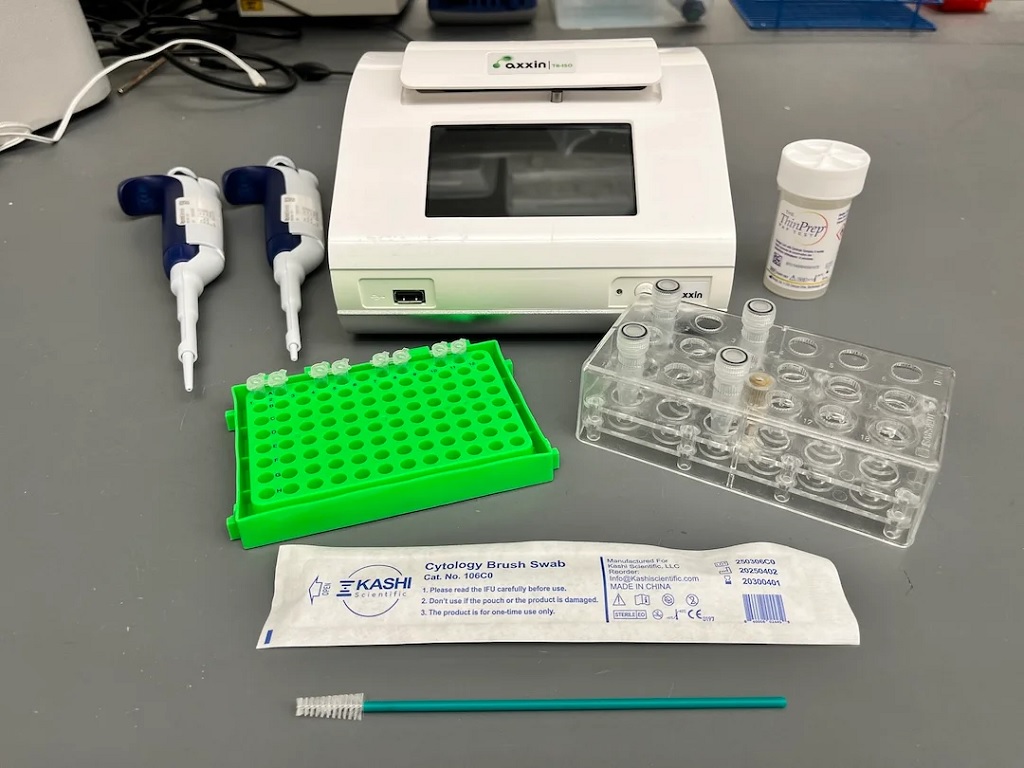
Materials used to run the HPV LAMP assay. A cytology brush is used to collect a cervicovaginal swab sample into ThinPrep buffer. Samples are lysed in screw-on tubes and lysate is added to LAMP reagents in PCR tubes. The assay is run on the Axxin T8-ISO heater/fluorimeter.
A team of researchers led by Rice University, in collaboration with colleagues in Mozambique and the US, has developed a simple, affordable human papillomavirus (HPV) test that delivers results in less than an hour with no specialised laboratory required. The breakthrough could provide an option for women in low-resource settings to be screened and treated for cervical cancer in a single clinic visit, a step that global health experts say could save countless lives. The research was recently published in Nature Communications.
Cervical cancer is considered easily preventable, yet it remains one of the deadliest cancers for women worldwide. According to the World Health Organization (WHO), each year more than 350 000 women die from the disease, and nearly 90% of those deaths occur in low- and middle-income countries where access to regular cervical cancer screening is limited. Persistent infection with high-risk types of HPV causes nearly all cases of cervical cancer. While vaccines are helping reduce HPV infections globally, most women at risk today are adults who did not get the vaccine in childhood. For them, regular and reliable screening is the only path to early detection and lifesaving treatment.
“Cervical cancer is almost entirely preventable, yet it still claims hundreds of thousands of lives each year,” said first author Maria Barra, a bioengineering graduate student at Rice. “Our goal was to build a test accurate enough to guide treatment, fast enough to use during a clinic visit and inexpensive enough to scale. This assay meets all three goals.”
The WHO recommends HPV DNA testing as the gold standard for cervical cancer screening, but existing HPV DNA tests often require expensive lab equipment and trained laboratory technicians – barriers that make widespread use in low-resource settings unattainable. As a result, many women are not screened for cervical cancer. Even where screening programs exist, results may take days or weeks to return. Patients leave to await results. However, where care facilities are remote, few in number and difficult to access, patients are often unable to return for treatment, leaving precancerous lesions to progress unchecked. A faster test without reliance on a lab could provide results and prompt treatment during the same patient visit.
“This is the kind of pragmatic innovation we focus on when engineering for global health – fewer steps, lower cost, higher impact,” said Rebecca Richards-Kortum, Professor of Bioengineering and co-director of the Rice360 Institute for Global Health Technologies at Rice. “Our data show you can bring lab-grade molecular screening to almost any setting without sacrificing reliability. Providing accurate results quickly enables clinicians to start treatment without delay.”
The new test uses a method called loop-mediated isothermal amplification (LAMP), which simplifies DNA detection by running at a single temperature. Instead of requiring DNA extraction – a complicated step in many existing tests – this process is extraction-free. A swab sample is chemically lysed, added directly to the LAMP reagents and incubated for about 45 minutes in a portable heater then read by fluorescence.
The test detects three of the most dangerous HPV types (HPV16, HPV18 and HPV45), which together cause about 75% of all cervical cancers. It also includes a built-in cellular control to ensure that the sample was collected properly.
In clinical studies, the test showed 100% agreement with the reference standard in 38 samples from Houston and 93% agreement in 191 samples from Maputo, Mozambique. The cost of the test is projected to be less than $8 each, and the portable device it runs on is battery-operated, making it ideal for clinics without consistent electricity.
“High mortality rates from cancer are closely associated with delays in diagnoses and limited access to early treatment,” said Cesaltina Lorenzoni, head of the National Cancer Control Program at the Mozambican Ministry of Health, director of science and teaching at Maputo Central Hospital and professor of pathology at the Eduardo Mondlane University Faculty of Medicine. “Point-of-care technologies that can aid clinicians in identifying cancer and guide treatment options in a single patient visit could be lifesaving in clinical settings in Maputo. This assay performed very well in our clinical setting and holds promise of delivering the kind of rapid, specific, cost-effective cancer detection that would meaningfully improve outcomes for women in our country.”
The WHO has set ambitious targets to screen 70% of women worldwide by 2030 as part of its public health campaign to eliminate cervical cancer. Meeting that goal will require screening millions of women in various global settings that lack advanced lab equipment or resources.
By cutting out expensive instruments, minimising sample handling and delivering rapid, accurate results, the LAMP assay represents a significant step toward realistically achieving the WHO goal. Critically, it opens the door to “screen-and-treat” strategies, where if a positive result is found, the patient can be treated on the same medical visit, reducing treatment delays and loss to follow-ups.
The team is currently working to expand the test to cover additional high-risk HPV types and is also working on lyophilised (freeze-dried) reagents that don’t require refrigeration, further increasing the test’s usability in rural or resource-limited areas. The team also plans to conduct usability studies with frontline health workers to refine the design before larger clinical rollouts.
“Our goal is a complete, field-ready kit that community clinics can use anywhere,” Richards-Kortum said. “If we can help health systems move to same-day screen-and-treat, we can move towards a future where cervical cancer can be eliminated globally.”
A research team at the LKS Faculty of Medicine of the University of Hong Kong (HKUMed) discovered that certain dietary fatty acids can supercharge the human immune system’s ability to fight cancer. The team found that a healthy fatty acid found in olive oil and nuts, called oleic acid (OA), enhances the power of immune γδ-T cells, specialised cells known for their cancer-fighting properties.
Conversely, they found that another fatty acid, called palmitic acid (PA), commonly found in palm oil and fatty meats, diminishes the ability of these immune cells to attack tumours. This groundbreaking study, published in the academic journal Signal Transduction and Targeted Therapy, offers an innovative approach using dietary OA supplementation to strengthen the antitumour immunity of γδ-T cells.
Dietary fatty acids and cancer immunotherapy
Dietary fatty acids are essential for health, helping with growth and body functions. They may also play a role in cancer prevention and treatment, but understanding how they affect cancer is challenging because of the complexity of people’s diets and the lack of detailed studies. Recently, scientists have learned that fatty acids can influence the immune system, especially in how it fights cancer. Specialised immune cells, called γδ-T cells, are particularly good at attacking tumours. These cells, once activated, have helped some lung and liver cancer patients live longer. However, this therapy is not effective for all patients, partly because the variation of the metabolic status, such as fatty acid metabolism, can influence its efficacy in the patients.
Oleic acid may improve cancer treatment outcomes
The research team identified a correlation between PA and OA levels and the efficacy of cancer therapies. ‘Our research suggests that dietary fatty acid supplementation, particularly with foods rich in OA, such as olive oil and avocados, could enhance γδ-T cell immunosurveillance, leading to more effective cancer treatments,’ said Professor Tu Wenwei from the Department of Paediatrics and Adolescent Medicine, School of Clinical Medicine, HKUMed, who led the study.
The team also discovered that another fatty acid, called PA, can weaken these immune cells and how OA can counteract this. ‘The results indicate that cancer patients should avoid PA and consider OA supplementation in their diets to improve clinical outcomes of γδ-T cell-based cancer therapies,’ added Professor Tu.
Significant impact from simple dietary changes
Professor Tu said, ‘This study is the first to show that the fatty acids we eat can directly affect how well our immune cells fight cancer.’ It reveals how PA can harm these cells and how OA helps them through a specific process involving a protein called IFNγ. By analysing blood samples, the researchers confirmed that the levels of these fatty acids are linked to the outcome of cancer immunotherapy.
‘For cancer patients, this discovery suggests simple changes, like eating more foods rich in OA (such as olive oil, avocados and nuts) and cutting back on PA (found in processed foods, palm oil and fatty meats), could improve the effectiveness of cancer treatments. The study also points to novel strategies, like combining dietary changes with specific drugs to further boost the immune system,’ added Professor Tu.
This study demonstrates that personalised nutrition may serve as an effective strategy to enhance immune function and support cancer treatment. It also suggests that new drugs targeting the processes affected by these fatty acids could enhance the power of γδ-T cell therapies. By integrating nutritional interventions with immunotherapy, this discovery could help more cancer patients achieve better outcomes.
Both Leathan (L) and Godfrey (R) have aplastic anaemia, which can treated with a stem cell donation. Leathan received stem cells from his twin sister, who is a perfect match. But Godfrey must travels from KwaMhlanga to Pretoria for life-sustaining blood transfusions.
When aplastic anaemia struck two young South Africans, their fates diverged dramatically. While one received a life-saving stem cell transplant, the other continues to fight every day. The rare blood disease affects fewer than six people per million, but for Leathan and Godfrey, the statistics became deeply personal.
Understanding Aplastic Anaemia: When Hope Meets Science
Aplastic anaemia is a devastating condition where the bone marrow fails to produce sufficient blood cells, leaving patients vulnerable to infections, bleeding, and severe anaemia. Given this rare disease’s high mortality rates, prompt recognition and immediate action are critical for survival. “The challenge with aplastic anaemia is that early symptoms can be subtle,” explains Dr Gugulethu Jali, a Clinical Haematologist and Haematopathologist at the Department of Health Kwa-Zulu Natal. “However, advances in treatment, particularly hematopoietic stem cell transplantation (HSCT), have transformed the prognosis, with survival rates now exceeding 80% when matched donors are found.”
Leathan’s Journey: From Crisis to Recovery
Seventeen-year-old Leathan had his whole life mapped out. The passionate soccer player dreamed of becoming a criminal lawyer, balancing his love for the game with serious academic ambitions. But subtle symptoms began to appear, including weight loss and nosebleeds that seemed minor at first.
When he suddenly collapsed at home, his family rushed him to hospital where doctors discovered his blood levels were critically low. Tests revealed that his bone marrow had completely stopped producing blood cells. Without immediate intervention, he would need blood transfusions and platelets for the rest of his life.
But Leathan had something that changes everything in aplastic anaemia cases: a perfect genetic match. His twin sister, without hesitation, donated her stem cells , giving her brother the ultimate gift of life.
Today, Leathan represents the success story that medical advances have made possible. Since the transplant, he has not needed further transfusions, and his blood counts are steadily stabilising. However, he may still need additional stem cell support to fully restore his health.
Currently, he’s on the path back to his soccer dreams and law school aspirations, a living example of what’s achievable when the right match is found.
Godfrey’s Battle: The Same Disease, Different Circumstances
While Leathan’s recovery shows what’s possible, eleven-year-old Godfrey from KwaMhlanga, Mpumalanga, is still living with the daily reality of aplastic anaemia. Like Leathan, Godfrey was once full of energy and loved soccer.
Then the familiar pattern began to emerge: Godfrey started moving more slowly, struggling with everyday tasks that once came easily. When uncontrollable bleeding began, his family knew something was seriously wrong. After a long diagnostic journey that began in 2019, Godfrey received the same diagnosis Leathan had faced: aplastic anaemia.
Unlike Leathan, Godfrey doesn’t have a twin sister who’s a perfect match. Instead, every month, he travels from KwaMhlanga to Pretoria for life-sustaining blood transfusions. The physical and emotional toll has been devastating. He was unable to pass Grade 5 last year, not because he lacks ability, but because fighting for your life leaves little energy for schoolwork.
Your Role in Changing Godfrey’s Story
For Godfrey to follow the same path as Leathan, he needs his genetic match. That person could be you.
Compatible donors are often found within similar ethnic backgrounds, making diversity in donor registries crucial for patients like Godfrey. If you’re between 17 and 55 and in good health, registering as a stem cell donor takes minutes and costs nothing. Register today at https://www.dkms-africa.org/save-lives.
The Hospital Association of South Africa (HASA) has announced the appointment of a new Board of Directors following its Annual General Meeting held on Monday, 6 October 2025, in Sandton.
Gale Shabangu from Mediclinic Southern Africa has been elected Chairperson, succeeding Melanie Da Costa from Netcare. Mark Bishop from Lenmed will serve as Deputy Chairperson.
Shabangu is widely recognised for her leadership in advancing inclusive, values-driven corporate cultures across South Africa’s private sector.
The newly elected Board represents a broad cross-section of the private hospital industry, from day hospitals, large hospital groups and smaller hospital operators, bringing together strategic insight, operational experience to strengthen HASA’s role in advancing the country’s healthcare priorities.
HASA Chief Executive Officer, Dr Dumisani Bomela, welcomed the new Board and extended appreciation to the outgoing Chairperson and Board members, and said: “I am pleased to share that HASA has elected a new Board of Directors for 2025/2026 to help steer the Association through the next phase of its journey. We also wish to extend our sincere gratitude to Melanie Da Costa, our outgoing Chairperson, for her dedicated leadership over many years, and for her invaluable insights and contributions, in particular on health policy matters, during her tenure.”
This new Board marks a moment of renewal for HASA, with several young professionals taking their place at the Board table, ensuring the Association plays an even more constructive role in advancing South Africa’s healthcare reform agenda. The collective expertise and insight of our members will ensure that the private hospital sector continues to be a strong partner in building an inclusive, resilient and high-performing health system.”
A new mRNA vaccine stopped allergens from causing dangerous immune reactions and life-threatening inflammation in mice, according to researchers from the Perelman School of Medicine at the University of Pennsylvania and Cincinnati Children’s. The vaccine, outlined in the Journal of Clinical Investigation, may one day be tested and tailored to a variety of seasonal and food allergies.
“This is a potential breakthrough for millions of people worldwide who suffer from life-threatening allergies,” said Nobel laureate Drew Weissman, MD, PhD, Professor in Vaccine Research at Penn and co-lead of the study with Cincinnati Children’s Marc E. Rothenberg, MD, PhD.
Weissman, Penn colleagues Jilian Melamed, PhD, an assistant professor of Infectious Diseases, Mohamad-Gabriel Alameh, PhD, an assistant professor of Pathology and Laboratory Medicine, and the Cincinnati Children’s researchers led by Marc E. Rothenberg, MD, PhD, director of the division of Allergy and Immunology, modelled this new vaccine on the design of the COVID-19 mRNA lipid nanoparticle (LNP) vaccines.
This time, however, scientists tweaked the mRNA to instruct cells to produce proteins that resemble certain allergens. By presenting these proteins in a controlled way, the vaccine didn’t cause allergic reactions but did instruct the immune system to respond more appropriately in the future. And, when mice were later exposed to the respective allergens, the vaccines worked.
When mice with specific allergies were exposed to the allergens, none of the mice vaccinated with the respective allergy vaccine had an allergic reaction. Vaccinated mice had fewer allergy-related white blood cells, made fewer inflammation-causing proteins, and their lungs produced less mucus. Their airways were also protected against narrowing, which often happens during asthma, and they made special antibodies that protected against allergic reactions.
A platform with broad potential
Unlike traditional allergy shots, which involve repeated administration of purified allergens over months or years, the mRNA-based approach offers a more flexible solution. Because the mRNA can be tailored to encode proteins from different allergens, the platform could be adapted to treat a wide range of allergic conditions—from seasonal pollen allergies to food sensitivities and asthma. Additionally, many severe food allergies do not have vaccines to protect against severe allergic reactions.
“People with food allergies that can cause anaphylactic shock are rightfully fearful in social situations, eating out in public, sharing food, and engaging in other fun activities where there are food and allergens around,” said Weissman. “Allowing people to partake in foods they were never able to eat would be incredibly rewarding, but I’ll even be happy if we can one day introduce a vaccine that allows parents to breathe just a little easier when sending their kids to class birthday parties.”
The study represents a proof-of-concept that mRNA vaccines can be used not only to prevent infectious diseases but also to adjust immune responses in chronic conditions like allergies and even celiac disease. Researchers say the next steps include testing the vaccine’s safety in humans, determining how many allergens can be included in a single dose, and evaluating how long protection lasts.
“We saw mRNA vaccines save lives during the pandemic, and as the most-tested type of vaccine in history, we know it’s the safest and most effective vaccine ever created,” said Weissman. “We are deeply committed to continuing to uncover the potential of this technology.”
Retina showing reticular pseudodrusen. Although they can infrequently appear in individuals with no other apparent pathology, their highest rates of occurrence are in association with age-related macular degeneration (AMD), for which they hold clinical significance by being highly correlated with end-stage disease sub-types, choroidal neovascularisation and geographic atrophy. Credit: National Eye Institute
In a new study, UC Irvine researchers explore a possible therapy for addressing “aging” in the eye and for preventing diseases such as age-related macular degeneration (AMD).
“We show the potential for reversing age-related vision loss,” says Dorota Skowronska-Krawczyk, PhD, an associate professor in the Department of Physiology and Biophysics and the Department of Ophthalmology and Visual Sciences. The study was a collaboration between researchers from UC Irvine, the Polish Academy of Sciences, and the Health and Medical University in Potsdam, Germany.
The work is a follow-up to an earlier study on Elongation of Very Long Chain Fatty Acids Protein 2 (ELOVL2), an established biomarker of age. “We showed that we have lower vision when this ELOVL2 enzyme isn’t active,” says Skowronska-Krawczyk, also a faculty member in the Robert M. Brunson Center for Translational Vision Research at the UC Irvine School of Medicine. In that work, the researchers found that enhancing ELOVL2 gene expression in aging mice boosted levels of the omega−3 fatty acid docosahexaenoic acid (DHA) in the eye and improved vision.
The more recent study sought to identify a way to bypass the need for the ELOVL2 enzyme.
As we age, changes in lipid metabolism lead to a decline in very-long-chain polyunsaturated fatty acids (VLC-PUFAs) in the retina, which in turn affects our vision and can lead to AMD. The ELOVL2 gene is a key enzyme in the production of VLC-PUFAs as well as DHA.
Injecting aged mice with the polyunsaturated fatty acid improved visual function. “It’s a proof-of-concept for turning lipid injection into a possible therapy,” says Skowronska-Krawczyk. “What is important is that we didn’t see the same effect with DHA.” Others have also questioned the ability of DHA to slow AMD progression.
“Our work really confirms the fact that DHA alone cannot do the work, but we have this other fatty acid that is seemingly working and improving vision in aged animals,” says Skowronska-Krawczyk. “We have also shown on a molecular level that it actually reverses the aging features.”
Furthermore, the researchers found genetic variants in the ELOVL2 enzyme that correlate with faster progression of AMD. “Now we actually have a genetic connection to the disease and its aging aspect,” says Skowronska-Krawczyk, “so we could potentially identify people at higher risk for vision loss progression.” This could lead to not only therapeutic treatment options but also targeted interventions for prevention.
These findings have only further solidified Skowronska-Krawczyk’s view of the importance of the ELOVL2 enzyme. “I am pretty convinced it’s one of the top aging genes that we should look at when we think about anti-aging therapies.”
Looking Beyond the Retina
In a collaboration with researchers from UC San Diego, Skowronska-Krawczyk has also started to explore the role of lipid metabolism in immune system aging. That study found that the lack of ELOVL2 enzyme induces accelerated aging of immune cells, suggesting that systemic lipid supplementation could potentially counteract the effects of age on the immune system. It also suggested that lipid metabolism might play a role in blood cancers.
“Our first study explored a potential therapy to address vision loss,” says Skowronska-Krawczyk, “but with the information we’ve since learned about immune aging, we are hopeful the supplementation therapy will boost the immune system as well.”
Breastfeeding until at least six months helps babies to fight off infections and reduces chronic inflammation, according to a new study. And better understanding the way specific nutrients in breast milk impact the immune system will improve health outcomes for all infants including those not breastfed.
The study, led by Murdoch Children’s Research Institute (MCRI) and the Baker Heart and Diabetes Institute (Baker Institute), discovered more clues as to why infants who were breastfed to at least six months of age had fewer infections and less chronic inflammation. Preventing these infections could reduce the rates of many childhood conditions, such as allergies, diabetes and asthma.
Published in BMC Medicine, the researchers identified several types of lipids (essential nutrients) in blood samples from breastfed babies that help reduce inflammation, which may reflect the unique nutritional composition of breastmilk.
MCRI’s Dr Toby Mansell said plasmalogens, a unique type of lipid abundant in breastmilk, appeared key to lowering inflammation.
“Plasmalogens are only found in breastmilk and are generally absent in formula milk, so a better understanding of how plasmalogens and other lipids unique to breastmilk protect against chronic inflammation will help pave the way for new treatments for infants who don’t receive breastmilk,” he said.
The study involved almost 900 infants from the Barwon Infant Study, a collaboration between MCRI, Barwon Health and Deakin University.
The study explored about 800 different lipids and other metabolic markers in babies up until 12 months of age. It found breastfeeding was associated with broad effects on different classes of lipids and metabolic markers.
Baker Institute’s Dr Satvika Burugupalli said the findings would lead to a new understanding of how breastfeeding and specific components of breast milk could benefit infants.
“Breast milk performs a central role in supporting a newborn’s immune system,” she said. “It’s loaded with essential nutrients, including lipids, as well as antibodies and white blood cells.
“This study has identified key biological pathways for how breastfeeding improves immune health and reduces inflammation that can lead to many childhood conditions, such as allergies and asthma, and the risk of adult cardiovascular disease and diabetes.”
Researchers from the University of Melbourne, Deakin University, Barwon Health, Northwestern University and the Florey Institute of Neuroscience and Mental Health also contributed to the study.
A recent analysis reveals that older adults with prior incarceration report worse physical and mental health than their peers, even if they were incarcerated in the distant past. The findings are published in theJournal of the American Geriatrics Society.
Among the 1318 US adults aged 50 years and older who responded to the Family History of Incarceration Survey, 21% had been incarcerated. Formerly incarcerated older adults were more likely to be men, non-Hispanic Black or “other” race/ethnicity, meet criteria for disability, be unmarried, and have lower income and education compared with those never incarcerated.
After adjusting for potentially confounding factors like demographics and socioeconomics, prior incarceration was associated with an approximately 90% higher odds of reporting “fair” or “poor” physical health. Length of time since incarceration did not moderate the association, meaning that even those incarcerated more than 10 years ago had equally poor self-reported health. The association with mental health was explained in part by income and employment.
The findings suggest that clinicians could consider screening for incarceration history and connecting formerly incarcerated patients to services and organisations that serve this community.
“Mass incarceration began in 1973, so older adults have spent most of their adult lives in this era and millions have been incarcerated in the past. It is critical to understand how incarceration – even in the distant past – may affect the health of older adults and what we can do to improve their health,” said corresponding author Louisa W. Holaday, MD, MHS, of the Icahn School of Medicine at Mount Sinai.
Researchers have adapted a rapid diagnostic technology that is able to identify undetected cases of malaria, helping tackle the spread of disease.
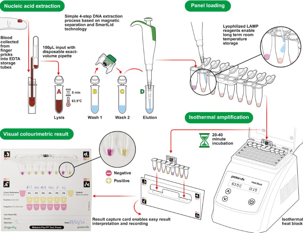
A diagram showing how the Dragonfly technology works (Credit: ProtonDx)
A team of scientists from Imperial College London, the MRC Unit The Gambia, the Clinical Research Unit of Nanoro in Burkina Faso, ProtonDx Ltd, and the NIHR Global Health Research Group have developed and validated a low-cost, point-of-care diagnostic that can rapidly detect low levels of malaria from a finger prick.
The test, called Dragonfly, relies on technology originally created at Imperial and its spinout ProtonDx. The technology allows users to diagnose malaria with high accuracy, without the need for extensive laboratory equipment or infrastructure. Results can be delivered in as little as 45 minutes, and the test is sensitive enough to detect even the lowest levels of malaria parasites in the blood – meaning that people without symptoms of malaria can still be identified.
Malaria is one of the leading causes of preventable deaths worldwide, with around 95% of all deaths occurring in Africa. Asymptomatic infections are a major driver of ongoing transmission, as individuals who carry the disease without showing symptoms do not seek medical treatment. Mosquitos feeding on blood from people without malaria symptoms can still deliver the malaria parasite to other people when they take their next blood meal. The new technology offers hope for combatting this potential spread of infection, by offering a way to identify previously undetectable malaria cases rapidly and on the ground in countries which are most affected by malaria.
The findings, published in Nature Communications, have significant global health implications as this field-deployable molecular diagnostic method offers a sensitive, scalable solution to support test-and-treat strategies for malaria elimination across Africa.
Professor Aubrey Cunnington, from Imperial’s Department of Infectious Disease and Co-Lead of the NIHR Global Health Research Group with Professor Halidou Tinto (from IRSS, Burkina Faso), said: “This is the first time that a diagnostic test for use outside of a laboratory setting has proven sensitive enough to detect low level malaria parasite infections in people who don’t have any symptoms.
“These people are the main source of malaria transmission, and in countries trying to eliminate malaria, there has long been interest in trying to detect these asymptomatically infected people with a screening test performed in their communities, and then giving treatment to those who are positive.
“Until now, no test has been able to detect enough of these infected people to make this a viable proposition, but the Dragonfly test now makes this possible.”
Detecting the undetectable
By collaboratively working as part of the NIHR Global Health Research Group, scientists were able to develop and test this new technology with the help of researchers in the regions affected most by malaria.
Almost 700 blood samples were collected from the community in The Gambia and Burkina Faso to assess the Dragonfly test’s accuracy against gold standard PCR testing and other common methods of testing, including expert microscopy and rapid diagnostic test (e.g., lateral flow immunoassay).
It was found that the Dragonfly tool could detect >95% of all malaria parasite infections, including 95% detection of those where the numbers of parasites were too low to be detected by looking at blood under a microscope.
Although Dragonfly is currently used as a research-used-only device, important progress is being made to understand the potential cost of a final manufactured version – especially when deployed at scale – a critical factor for effective deployment in sub-Saharan Africa. The team is already working closely with the Africa Centres for Disease Control and Prevention to explore opportunities with local manufacturers in the region, ensuring that production and scale-up can be rooted in local capacity. Future studies will also need to assess the robustness of the tool in community settings which are less connected to laboratory facilities.
Dr Jesus Rodriguez-Manzano, last author and technology development lead, from the Department of Infectious Disease, said “This research would not have been possible without the collaborative nature and all the organisations who took part in this study. The technology delivered through this work represents a game changer for malaria control efforts.”
The testing equipment
In the Dragonfly testing process, a capillary blood sample obtained from a simple finger prick is processed in around 10 minutes, without the need for specialised laboratory equipment, to extract high-purity nucleic acids from malaria parasites. The prepared sample is then placed into a detection panel, which is inserted into a portable heater.
After a 30-minute incubation at a constant temperature, results can be read visually using a colour chart: a pink reaction indicates a negative result, while a yellow reaction confirms malaria infection.
The Dragonfly can be manufactured at a fraction of the cost of other platforms, is compact enough to fit into a backpack, and can operate on batteries, an important feature for bringing the tool directly to communities without requiring additional specialised equipment. Testing can be carried out by most people without extensive training, meaning that healthcare providers or scientists do not need to be present for its use.