Day: October 13, 2026

SGLT-2 Inhibitors Lower CVD-Linked Inflammation

Photo by Towfiqu barbhuiya

For the first time in a human trial, SGLT2 inhibitors have been shown to reduce circulating inflammatory cells linked to cardiovascular disease, in addition to their established effects on glycaemia and body weight. The findings were published in Circulation.

“There had been some suggestive studies in animals with these medications indicating they might be modulating the immune system – affecting inflammation – but that hadn’t really been shown in humans,” said Dr Mona Mashayekhi (Division of Diabetes, Endocrinology and Metabolism, Vanderbilt Health). “This study provides the first direct evidence of SGLT2 inhibitors altering human immune cells which may potentially reduce cardiovascular risk.

“This finding is significant because it’s the first time this medication has been shown to have an immune effect at the cellular level with some specificity. If we understand how these medicines are reducing obesity-associated inflammation and heart disease and kidney disease, then we can identify the best patient for this treatment or discover other drug targets in that pathway that could really be beneficial.”

The research team enrolled 16 women with obesity and prediabetes and treated them with empagliflozin. Baseline assessments were performed, followed by three months of treatment. Immune changes were also reviewed in a comparator cohort of women who followed a low-calorie diet for three months. Measurements were taken at two weeks and at three months.

Using genetic sequencing and cellular imaging, investigators found that monocyte–platelet aggregates (MPAs) were significantly decreased. Individuals receiving empagliflozin showed a significant reduction in MPAs within two weeks, with a further decrease by week 12. Only the medication group exhibited reduced MPAs, indicating effects beyond weight loss.

Investigators also documented metabolic reprogramming of circulating monocytes, which shifted from a hyper-inflammatory state toward a more stable phenotype oriented toward normal cellular energy metabolism.

Mashayekhi and colleagues have begun a randomised placebo-controlled trial (planned n = 74) of participants with obesity and metabolic syndrome, currently recruiting at Vanderbilt Health. Immune measures will be assessed over three months as in the pilot; adipose tissue will also be sampled and analysed.

By Jill Clendening

Source: Vanderbilt University Medical Center

What’s the Best Way to Avoid Weight Regain After Stopping GLP-1 Therapy?

Photo by I Yunmai on Unsplash

Glucagon-like peptide-1 (GLP-1) receptor agonists are effective treatments for obesity, but discontinuation is common due to cost, insurance coverage limitations, side effects, patient preference, and other factors – commonly leading to rapid weight regain. Results from a pilot randomised trial, which are published in Obesity Science & Practice, suggest that structured support may attenuate early weight gain after GLP-1 cessation.

In the trial, 39 adults with prior GLP-1-induced weight loss were randomised to usual care, a digital wellness application, or medically tailored meals at the time of GLP-1 cessation, continuing for 4 months.

Surveys showed that diet quality, perceived health, adherence, and satisfaction did not differ significantly by group, though some estimates suggested possible trends favouring the wellness application. Relative to the control group, the other groups experienced less weight gain. Over 4 months, participants in the control group gained an estimated 10.75% body weight, compared with 3.60% in the wellness application group and 3.58% in the medically tailored meals group.

“People invest significant time and effort in improving their health and achieving weight loss,” said corresponding author Kelseanna Hollis-Hansen, PhD, MPH, of Tufts University Food is Medicine Institute. “We need evidence-based strategies to support them if and when they discontinue GLP-1 medication, so they can maintain those gains and continue building long-term health, vitality, and well-being.”

Source: Wiley

Pig Kidney Xenotransplant Successfully Bridges Patient to Human Transplant

Photo by César Badilla Miranda on Unsplash

In January 2026, Tim Andrews, who had received a genetically edited pig kidney in January 2025, became the recipient of a human kidney. A new Mass General Brigham paper, published in The Lancet, details Andrews’ experience and how xenotransplantation could bridge the gap between end-stage kidney failure and a human transplant, allowing a patient to forgo dialysis in the meantime.

Authors report that Andrews, 66 at the time of xenotransplantation, successfully lived with the genetically edited kidney for 271 days – during which he did not need dialysis – before receiving a human kidney from a donor. His case represents the longest dialysis-free survival following porcine kidney xenotransplantation in a living human and the first transition to human kidney transplantation.

A record-setting bridge transplant

“The organ shortage is the greatest crisis we have right now in transplantation,” said lead author Leonardo Riella, MD, PhD, of the Center for Transplantation Sciences and the Division of Nephrology at Mass General Brigham.

“We know that the best treatment option for patients with end-stage kidney disease who have developed renal failure is transplantation, but we simply don’t have enough human organs to transplant in a timely manner. Our vision is that xenotransplantation could help address this gap – initially as a bridge to get patients off dialysis while they wait for a human donor kidney, and potentially, as we establish long-term safety and durability, as a destination therapy in its own right.”

How the pig kidney performed

The world’s first porcine kidney xenotransplant to a living human recipient occurred in 2024 at Massachusetts General Hospital, with the patient surviving 52 days before death from cardiac causes unrelated to the transplant. No previous studies have documented xenograft survival beyond two months, a successful “bridge” to receipt of a human organ, or the mechanisms leading to late xenograft failure in living recipients.

Andrews received the genetically engineered pig kidney under an FDA Expanded Access Investigational New Drug application. He was then monitored for kidney function, rejection, antibody development and potential pig-to-human infection. The kidney functioned immediately. An early episode of T-cell-mediated rejection resolved with treatment, no pig pathogen transmission was detected, and the patient was able to go without dialysis for 271 days.

After approximately six months, immunosuppression was reduced in the setting of infection. The xenotransplant subsequently developed microvascular injury and inflammation that progressed to organ failure. The patient then received a deceased-donor human kidney, with immediate graft function and no evidence of sensitization during follow-up.

Lessons from late graft failure

The findings illustrate both the promise and areas for improvement for xenotransplants as a bridge for patients awaiting human kidneys. Importantly, the microvascular injury to the porcine kidney has implications for the development of immunosuppressive strategies, donor genetic engineering and monitoring in future clinical transplantation studies.

“Mass General Brigham has a strong history of kidney transplantation, beginning with the first live donor kidney transplant in 1954,” said senior author Tatsuo Kawai, MD, PhD, director of the Legorreta Center for Clinical Transplantation Tolerance at Mass General Brigham. “Our findings continue that history by both furthering what we know about xenotransplants and generating knowledge that could be applied to other organ transplantation work.”

“The patients are the true pioneers here,” added Riella. “By stepping forward and being willing to participate in these early-stage studies, they are advancing the science. Without their trust and courage, we would not be able to do what we do.”

Source: Mass General Brigham

Atypical B Cells Found to Switch Roles During Infection

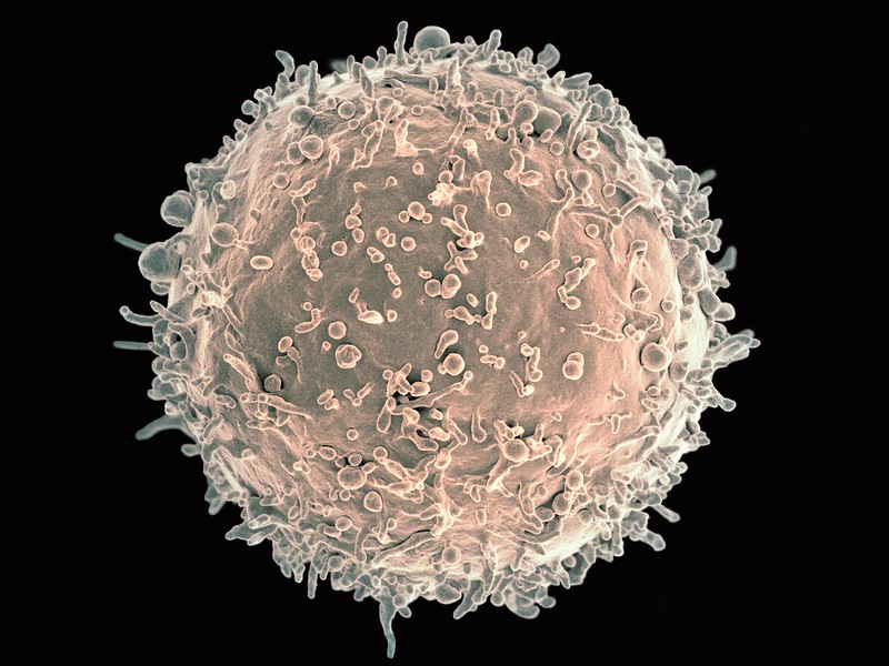
Scanning electron micrograph of a B cell. Credit: NIH

Researchers at Karolinska Institutet have mapped how so-called atypical B cells develop and change function over time. The study, published in The EMBO Journal, shows that the cells do not constitute a uniform group but instead follow several developmental pathways that converge on a common end stage. The findings may help explain why atypical B cells have previously been linked to different roles in the immune system.

B cells are an important part of the immune system and help the body recognise and fight infections. A group of B cells known as atypical B cells has attracted considerable interest among researchers in recent years. They occur in several infections, but also in connection with vaccinations and certain inflammatory diseases. Despite extensive research, it has long remained unclear how these cells arise and what function they serve. 

In the new study, the researchers analysed blood samples from people who had experienced malaria as well as from healthy blood donors. By combining several advanced methods, they were able to track the development of individual B cells and examine how their properties changed over time. 

Atypical B cells develop in several stages

The results show that atypical B cells are not a distinct cell type but rather exist along a continuum with early, intermediate and late developmental stages. The researchers identified three cell-surface markers, CD11c, CD24 and CD27, which made it possible to follow this progression. 

“We show that atypical B cells can be understood as a continuous developmental process rather than as a fixed and clearly defined group of cells,” says Christopher Sundling, researcher at the Department of Medicine, Solna, Karolinska Institutet, and corresponding author of the study. 

The study also shows that the function of the cells changes as they develop. The early stages had a greater capacity to develop into antibody-producing cells and to produce cytokines, signalling molecules that help different parts of the immune system communicate with one another. 

In the later stages, the cells instead acquired characteristics suggesting that they are better able to capture and present foreign substances to other immune cells. The researchers also found that the late-stage cells had a greater capacity to associate with microbial particles. 

May have different origins

Another important observation was that atypical B cells may have several different origins. The researchers found evidence that they arise from different types of memory B cells that follow parallel developmental pathways but eventually reach a similar late stage. 

“Our results show that the function of these cells changes gradually during development. This may help explain why previous studies have described atypical B cells in different ways and assigned them different roles in the immune system,” says Alan-Dine Courey-Ghaouzi, doctoral student and first author of the study. 

The researchers emphasise that the study was carried out in the context of malaria, but that the findings are likely to reflect general principles of how atypical B cells develop in other infections and inflammatory conditions as well. 

Source: Karolinska Institutet

Study may Explain why IBD Patients React to Previously Tolerated Foods

Gut inflammation may alter how bacteria process food.

Irritable bowel syndrome. Credit: Scientific Animations CC4.0

A new study published in Gastroenterology finds that gut inflammation can change how gut bacteria break down food, possibly triggering immune reactions to foods that were previously tolerated. The findings may help explain why many people with IBD react adversely to foods such as dairy or gluten, even in remission.

About two-thirds of patients with IBD report food intolerances. Dairy, wheat and fibre are the most frequently blamed triggers, but the mechanisms have remained unclear.

The gut microbiota may hold the answer. Prior research has shown that patients with IBD often have an altered microbiota. This study identifies a novel mechanism by which that alteration drives food reactions.

“The microbiome acts as a metabolic organ, helping us digest food components, including those that can trigger gut reactions,” said Dr Alberto Caminero. “Our study shows that inflammation impairs this microbial digestive capacity, potentially contributing to adverse food reactions and opening new possibilities for microbiome-based therapies to restore this function in patients with IBD.”

Other key authors from McMaster University Medical Centre included Dr Bruna Barbosa and senior clinical collaborators Dr Neeraj Narula and Dr Premysl Bercik.

Using two mouse models of colitis, researchers found that inflammation made animals more prone to developing food sensitivities. After inflammation, exposure to dairy or gluten triggered an allergic-type immune response in the colon; reintroducing those foods later caused heightened gut sensitivity and worse colitis symptoms.

The cause traced back to the microbiota: inflammation depleted the specific bacteria that normally break down these food triggers. That shift alone was enough to increase sensitisation risk – transferring gut bacteria from inflamed mice into germ-free mice made those mice more prone to sensitisation to the same foods, while restoring the missing bacteria reduced these effects.

The pattern held in humans, too. Among patients with IBD, those who self-reported food intolerances were more likely to have both a reduced ability to break down common trigger foods and lower levels of the bacteria responsible for that job.

The researchers note that microbiota disruption is likely just one piece of a larger picture that also includes genetics, infections and stress – all of which need further research.

Together, the findings point to a possible mechanism behind food sensitivities in IBD: inflammation disrupts the gut bacteria that normally process certain foods, and this disruption – not the foods themselves – helps trigger the immune reactions patients experience.

Source: American Gastroenterological Association