Day: February 10, 2026

Improving Understanding of Female Sexual Anatomy for Better Pelvic Radiotherapy

Female reproductive system. Credit: Scientific Animations CC4.0 BY-SA

Researchers at the Icahn School of Medicine at Mount Sinai, in collaboration with other leading institutions across the country, have published an innovative study that provides radiation oncologists with practical guidance to identify and protect female sexual organs during pelvic cancer treatment.  

Published in the latest issue of Practical Radiation Oncology, this study addresses a long-standing gap in cancer care by bringing key female sexual anatomy into consideration during routine radiotherapy planning and survivorship research. 

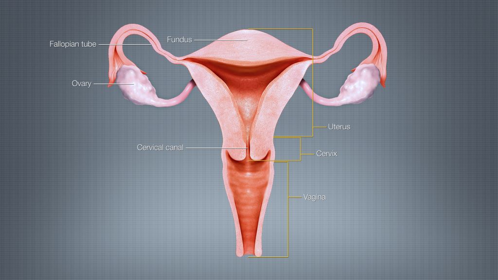
The study, “Getting c-literate: Bulboclitoris functional anatomy and its implications for radiotherapy,” synthesises current scientific knowledge and pairs it with original anatomic dissection, histology, and advanced imaging analysis. The work focuses on the bulboclitoris, a female erectile organ (consisting of the clitoris and the vestibular bulbs) that plays a central role in sexual arousal and orgasm and can be exposed to radiation during treatment for pelvic cancers. 

“Pelvic radiotherapy can be life-saving, but it can also affect sexual function and quality of life,” said Deborah Marshall, MD, MAS, Assistant Professor, Departments of Radiation Oncology and Population Health Science and Policy at the Icahn School of Medicine at Mount Sinai; Division Chief of Women’s Health, Department of Population Health Science and Policy; and senior author of the study. “Compared to male sexual anatomy, female erectile structures have been largely invisible in standard radiation workflows. Our goal was to provide clinicians with a practical anatomy-grounded way to change that.” 

Using detailed anatomic and radiologic correlation, the research team demonstrates how the bulboclitoris and related neurovascular structures can be identified on standard CT and MRI scans and consistently outlined (or “contoured”) for radiotherapy planning. This step-by-step guidance makes it feasible for clinicians to measure radiation dose to these tissues and begin linking exposure to patient-reported outcomes related to arousal and orgasm. 

“This work builds upon our previous knowledge that the clitoris is not just an external structure,” Dr. Marshall said. “It includes an entire internal organ comprised of erectile tissues located just outside the pelvis, and those tissues matter for sexual health and, in particular, for female sexual pleasure. Once clinicians can reliably see and measure them, we can begin to ask better questions, have better conversations with patients, and ultimately deliver better care.” 

Sexual function outcomes after pelvic radiotherapy have historically been understudied in women, limiting counselling, toxicity prevention strategies, and equitable survivorship care. By establishing a shared, standardised approach to identifying the bulboclitoris, the study lays the groundwork for future research to develop dose-volume constraints and mitigation strategies, as other organs at risk are managed in radiation oncology. 

For clinicians, the framework enables routine contouring and dose reporting using CT alone when necessary, with MRI improving soft-tissue visualization when available. In the absence of prospective dose-response data, the authors recommend minimising radiation dose to the bulboclitoris when oncologically appropriate, using an “as low as reasonably achievable” approach. 

For patients, the work supports more informed conversations about potential sexual side effects of pelvic radiotherapy, including changes in arousal, sensation, orgasm, lubrication, or pain. This research also promotes more personalized treatment planning that considers female sexual health and pleasure as a legitimate and important component of cancer survivorship. 

Next steps include prospective research through Mount Sinai’s STAR program, deeper mapping of neurovascular anatomy relevant to sexual function, expanded educational resources for oncology and radiology teams, and improved patient-reported outcome measures that reflect diverse sexual practices and experiences. 

Source: Mount Sinai

Physical Pressure on the Brain Triggers Neurons’ Self-destruction Programming

Gliobastoma (astrocytoma) WHO grade IV – MRI sagittal view, post contrast. 15 year old boy. Credit: Christaras A.

The brain and spinal cord is made up of billions of neurons connected by synapses and managed and modified by glial cells. When neurons die, this communication network is disrupted and since this loss is irreversible, neuron death causes sensory loss, motor impairment and cognitive decline.

An interdisciplinary team of researchers from the University of Notre Dame is investigating the mechanisms of neuron death caused by chronic compression – such as the pressure exerted by a brain tumour – to better understand how to prevent neuron loss.

Published in the Proceedings of the National Academy of Sciences, their study found that chronic compression triggers neuron death by a variety of mechanisms, both directly and indirectly. The research is helping lay the groundwork for identifying therapies to prevent indirect neuron death.

“The impetus for this project was to figure out those underlying mechanisms. In cancer research, most researchers are focused on the tumour itself, but in the meantime, while the tumour is sitting there and growing, it’s damaging the organ that it’s living in,” said Meenal Datta, the Jane Scoelch DeFlorio Collegiate Professor of Aerospace and Mechanical Engineering at Notre Dame and co-lead author of the study. “We fully believe that these growth-induced mechanical forces of the tumor as it expands is part of the reason we see damage in the brain.”

As an engineer who leads the TIME Lab, Datta studies the mechanics of tumors and the microenvironment, specifically for glioblastoma, an incurable brain cancer. She had found in prior work that tumors damage the surrounding brain. But to understand the mechanisms by which tumors kill neurons from compression alone, Datta needed a “hardcore neuroscientist.”

Neurons captured on screen for research experiment.
Imaging of neurons from an experiment with the control group neurons on the left and the neurons impact by chronic compression on the right. (Provided by the Patzke lab.)

That neuroscientist is Christopher Patzke, the John M. and Mary Jo Boler Assistant Professor in the Department of Biological Sciences at Notre Dame and co-lead author of the study. Patzke utilises induced pluripotent stem cells (iPSCs), which are either obtained from external sources or generated directly in his lab. These cells function like embryonic stem cells and can be differentiated or changed in the lab into any cell type in the body, including neurons.

For this study, iPSCs were used to create neural cells and develop a model system of neurons and glial cells that behave as a neuronal network would in the brain. Researchers grew the cells and then applied pressure to the system to mimic the chronic compression of a glioblastoma tumour.

After compressing the cells, graduate students Maksym Zarodniuk and Anna Wenninger, from Datta and Patzke’s labs respectively, compared how many neurons and glial cells died versus lived.

“For the neurons that are still alive, many of them have this programmed self-destruction signaling activated,” Patzke said. “We wanted to understand which molecular pathway was responsible for this; is there a way to save neurons from going down the drain to this cell death mechanism?”

By sequencing and analysing all messenger RNA from the living neuronal and glial cells, the researchers found an increase in HIF-1 molecules, signalling for stress adaptive genes to improve cell survival, which leads to inflammation in the brain. The compression also triggered AP-1 gene expression, a type of neuroinflammatory response.

Both neurological reactions are indicators that neuronal damage and death is underway.

An analysis of data from the Ivy Glioblastoma Atlas Project shows that glioblastoma patients also reflect these compressive stress patterns and gene expression changes as well as synaptic dysfunction in line with the experiment’s results. The researchers confirmed these results by mimicking force via a live compression system applied to preclinical models of brains.

Overall, the findings may help explain why glioblastoma patients experience cognitive impairments, motor deficits and elevated seizure risk. Additionally, the signalling pathways offer opportunities for researchers to explore as drug targets to reduce neuronal death.

“Our approach to this study was disease agnostic, so our research could potentially extend to other brain pathologies that affect mechanical forces in the brain such as traumatic brain injury,” Datta said. “I’m all in on mechanics. Whatever it is that you’re interested in when it comes to cancer, above your question of interest, mechanics is sitting there and many don’t even know they should be considering it.”

The mechanics of compression and its effect on neuron loss is key for future research.

“Understanding why neurons are so vulnerable and die upon compression is critical to prevent excessive sensory loss, motor impairment and cognitive decline,” Patzke said. “This is how we will help patients.”

Source: University of Notre Dame

Obesity Linked to One in 10 Infection Deaths Globally

Image from Rawpixel

Just over one in 10 deaths from a wide range of infectious diseases are associated with obesity worldwide, finds a major new study led by a UCL researcher.

People with obesity face a 70% higher risk of hospitalisation or death from an infection than those of a healthy weight, suggest the findings published in The Lancet.

Obesity is linked to an increase in the risk posed by many different infectious diseases, from flu and COVID to stomach bugs and urinary tract infections, and the researchers found that the higher the BMI, the greater the risk.

The study’s lead author, Professor Mika Kivimaki (UCL Faculty of Brain Sciences), said: “Obesity is well known as a risk factor for metabolic syndrome, diabetes, cardiovascular disease, and many other chronic conditions. Here we have found robust evidence that obesity is also linked to worse outcomes from infectious diseases, as becoming very ill from an infection is markedly more common among people with obesity.”

The researchers studied data from over 540,000 people who participate in large cohort studies in the UK (the UK Biobank dataset) and Finland, to look at the relationship between obesity and severe infectious disease. Participants had their body mass index (BMI) assessed when they entered the studies and were then followed up for an average of 13-14 years.

The researchers found that people with obesity (defined as a BMI of 30 or higher) had a 70% higher risk of hospitalisation or death from any infectious disease in the study period compared to people with a BMI between 18.5 to 24.9 (classified as a healthy weight).

The risk increased steadily as body weight increased. People with a BMI of 40 or higher had three times the severe infection risk compared to people with a healthy weight.

The link between obesity and severe infections was consistent regardless of the measure of obesity used (BMI, waist circumference, or waist-to-height ratio, where data was available) and for a wide range of infection types.

The study included data on 925 bacterial, viral, parasitic, and fungal infectious diseases, and the authors also honed in on 10 common infectious diseases in more detail. For most of these diseases, including flu, Covid-19, pneumonia, gastroenteritis, urinary tract infections, and lower respiratory tract infections, they found that people with obesity were more likely to be hospitalised or die than people with a healthy BMI. However, obesity did not appear to increase the risk of severe HIV or tuberculosis.

The analysis found that the link to severe infections was not explained by obesity-related chronic conditions, as the association was consistent in people with obesity who did not have metabolic syndrome, diabetes, or heart disease, while the association was also not explained by lifestyle factors such as physical activity.

While the study did not investigate the causes of the association, the researchers say that previous studies have suggested that obesity contributes to a general impairment of immune function, including immune dysregulation, chronic systemic inflammation, and metabolic disturbances.

Professor Kivimaki said: “Our findings suggest that obesity weakens the body’s defences against infections, resulting in more serious diseases. People may not get infected more easily, but recovery from infection is clearly harder.”

The researchers found evidence that losing weight can reduce the risk of severe infections as people with obesity who lost weight had a roughly 20% lower risk of severe infections than those who remained obese.

First author Dr Solja Nyberg (University of Helsinki) commented: “As obesity rates are expected to rise globally, so will the number of deaths and hospitalisations from infectious diseases linked to obesity.

“To reduce the risk of severe infections, as well as other health issues linked with obesity, there is an urgent need for policies that help people stay healthy and support weight loss, such as access to affordable healthy food and opportunities for physical activity. Furthermore, if someone has obesity, it is especially important to keep their recommended vaccinations up to date.”

The authors used infectious disease mortality data from the Global Burden of Diseases (GBD) Study to model the impact of obesity on infectious disease deaths for different countries, regions and globally.

The analysis suggested 0.6 million out of 5.4 million (10.8% or one in 10) infectious diseases deaths globally were linked with obesity in 2023.

The researchers estimated that in the UK, one in six (17%) infection-related deaths can be attributable to obesity, and 26% in the US.

Co-author Dr Sara Ahmadi-Abhari (Imperial College London), who conducted the Global Burden of Diseases (GBD) analyses, said: “Estimates of the global impact give a sense of how large the problem may be, but they should be interpreted with caution. Data on infection-related deaths and obesity in the GBD are not always accurate, particularly in low-resource countries.”

Source: University College London

Why Heart Attacks in the Morning Have Worse Outcomes

Human heart. Credit: Scientific Animations CC4.0

It has long been known that heart attacks occurring in the morning are typically more serious than those that happen at night. While daily variations in stress hormone levels and blood pressure affect cardiac health, these are only part of the picture. There is also the diurnal variation in immune response involved: neutrophils, the body’s ‘first responders’, cause more inflammatory damage in the morning, causing havoc even as they neutralise pathogens.

“They’re the first sentinel, but they come fully loaded,” said Douglas Mann, MD, professor at Washington University School of Medicine in St Louis. “They’re shooting at everything and dumping a lot of toxic granules on the environment. They are indiscriminate in terms of their ability to destroy, and they take out healthy cells in the process.”

But exactly why they are more damaging at night has been a mystery. Now, researchers have found the reason behind this diurnal difference in destructiveness, and also how to tweak the ‘internal clocks’ of these white blood cells so that they cause less damage during sterile inflammation while still protecting against pathogens. Their findings are reported in the Journal of Exploratory Medicine, and are summarised in JAMA news.

Finding the pattern

The researchers, from Spain and Yale University, discovered that the timing of heart attacks significantly affects their severity due to a ‘neutrophil clock’ controlled by circadian rhythms. Neutrophils are more active during the day (activated by the Bmal1 protein) and less active at night (inhibited by the CXCR4 receptor).

Analysing more than 2000 patients with ST-segment elevation myocardial infarction, the researchers found that those who had an MI in the morning suffered worse cardiac damage than those who had them at night. Mouse experiments confirmed this pattern and showed that genetically disabling the Bmal1 protein reduced daytime neutrophil activity, protecting against severe cardiac injury.

This suggests a treatment strategy of tricking neutrophils into remaining in their nighttime inactive state, allowing doctors to reduce inflammation and lessen heart attack damage during daytime hours without compromising the immune system’s ability to fight infections.

Reducing cardiac damage without compromising the immune system

Mice engineered to have high levels of CXCR4 were given a drug compound, ATI2341, which bound to CXCR4 receptors. When heart attacks were induced, the mice showed reduced tissue damage. To test the neutrophils’ pathogen-fighting ability, they were also infected with Staphylococcus aureus or Candida albicans, but the mice were able to overcome the infection – the treated mice even tolerated the Candida infection better than the controls.

Mann explained why controlling the neutrophils was a better option. “Prior trials have tried to neutralise neutrophils or reduce neutrophil numbers entirely,” Mann noted. “But when you get rid of neutrophils, you’re also handcuffing the immune system. Before, it was considered an inevitability that neutrophils killing off infection also meant damaging a lot of tissue.”

The crucial question is of course whether this research in mice can translate to humans.

Luigi Adamo, MD, PhD, director of cardiac immunology at Johns Hopkins University who was not involved in the study, said that the study, one of the first use immune circadian rhythms to modulate inflammation, “offers new insight into neutrophils and a new way to look at this cardiac damage that might even apply to other types of sterile inflammation.”

Adamo struck a note of caution: the extremely low success rate in animal-to-human translation in cardioimmunology. “Immune cells are not always the same when you go from mice to humans,” he said.

Treatment implementation is a major obstacle

Even if this neutrophil clock alteration could be applied to humans, it would be difficult to administer since heart attacks strike without warning.

“If everyone took one of these drugs in the morning when they woke up, maybe it would make heart attacks less severe, but ‘preventive’ means you’re giving it chronically, and I don’t know what would happen with long-term stimulation of that receptor and other cell types,” Mann said. “Their data support the acute application, but in the long term, that’s a whole different story.”

As systemic treatment, the off-target effects of ATI2341 would need to be explored. He also struggled to envision a potential therapeutic solution.

“Today, when you have a heart attack, in most places with hospitals and well-developed health care systems, the patient gets an angioplasty,” Mann said. “The only time this drug could be given would be at the time of reperfusion, when you’re blowing up the balloon and opening up the clot.” Typically, ideal reperfusion timing is within two hours – but neutrophils probably do their damage within a matter of 30 minutes, Mann explained. “It’s a race against time, and I’m curious if [the researchers] can demonstrate that.”

Source: JAMA

EthiQal Launches Recognition Programme to Support Practitioners

The newly launched, first of its kind, EthiQal Recognition Programme strives to acknowledge professional conduct that reflects a commitment to the delivery of excellent patient care and the reduction of medicolegal risk.

It is aimed at specialist clinicians in private practice and is based on a point system where defined activities qualify for set points that over time convert to premium refunds.

The Programme underpins EthiQal’s pledge to promoting high-quality healthcare, supporting practitioners in building successful safe practices and managing their medicolegal risk, and aligning individual practitioner’s professional indemnity premiums with their unique insurance risk.

How the Programme works and which activities qualify for point collection are outlined in the Recognition Programme Benefit Guide, with the formal details of the Programme defined in the Terms and Conditions and Benefit Rules documents, which can all be viewed on the EthiQal website: https://ethiqal.co.za/

For more information about EthiQal, click here [https://ethiqal.co.za/contact/] complete the form, and an Advisor will call you back.

Existing EthiQal policyholders can register here:  https://f.insdi.com/ethiqal/ethiqal-reward-and-recognition-program/t?share=dcdb74b1-32a8-4f82-84dc-987066828e32

by completing the online enrolment form. Registration on the Programme is voluntary and requires policyholder enrolment and consent.

For questions about the Programme, contact your Financial Advisor or broker, or email:  recognition@ethiqal.co.za