Day: October 24, 2025

Faster MRI Scans Offer New Hope for Dementia Diagnosis
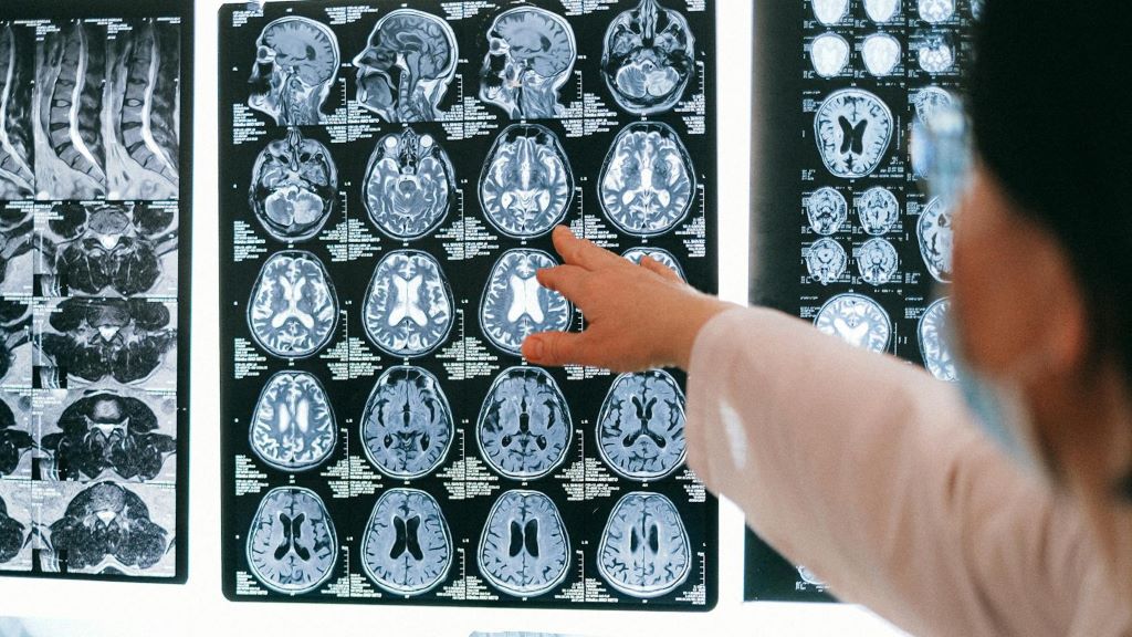
Photo by Anna Shvets

The time to carry out diagnostic MRI scans for dementia can be cut to one third of their standard length, according to a new study led by UCL researchers.

The findings, published in Alzheimer’s & Dementia, have been described as a step towards ending ‘the postcode lottery in dementia diagnosis’. Shorter scans would be easier and more comfortable for patients and also enable more people to be scanned at a lower cost. The team behind the study say this could at least double the number of dementia scans able to be done in one day.

Senior author Professor Nick Fox, Director of the UCL Dementia Research Centre at the UCL Queen Square Institute of Neurology, said: “As more treatments that can slow or change the course of dementia are being developed, it’s important to make sure MRI scans are available to everyone. This is because people living with dementia often need an MRI scan as part of their diagnosis before they can access these treatments.

“To help make this possible, our team carried out the first study looking at how new imaging techniques – called parallel imaging – could speed up MRI scans in clinics. Their goal is to move closer to a future where every person with dementia can get a diagnosis through a scan.”

MRI scans often play a key role in an accurate dementia diagnosis, including ruling out other causes of symptoms and assisting in diagnosing the type of dementia. Emerging disease-modifying treatments such as lecanemab and donanemab also require an MRI scan before starting treatment and for safety monitoring during the course of treatment. Reducing the cost of scanning would contribute to lowering the total cost of delivering for such treatments. 

The ADMIRA study (Accelerated Magnetic Resonance Imaging for Alzheimer’s disease), part funded by Alzheimer’s Society’s Heather Corrie Impact Fund, aimed to understand the reliability of fast MRI scans compared to standard-of-care clinical scans. The neurologists on the study were joined by co-authors from the UCL Hawkes Institute and the UCL Advanced Research Computing Centre in the faculty of Engineering.

The research team scanned 92 people in an outpatient setting where an MRI brain scan was planned as part of their routine clinical assessment. The accelerated scans were carried out and enhanced to increase the quality of the image using new scanning methods. Three neuroradiologists examined these scans, and weren’t aware if they were looking at fast or standard-of-care scans.

Co-author Professor Geoff Parker (UCL Hawkes Institute and UCL Medical Physics and Biomedical Engineering) said: “Our research has taken advantage of recent breakthroughs in scanner technology. Our task was to work out just how fast we could scan while maintaining image quality good enough for diagnosis.”

The team found that the quicker scans reduced time in the scanner by 63% and they were as reliable as the standard-of-care scans for diagnosis and visual ratings.

First author Dr Miguel Rosa-Grilo (UCL Queen Square Institute of Neurology) said: “We were confident that the new scan would prove non-inferior to the standard scan, given the high image quality – but it was remarkable how well it performed.”

Richard Oakley, Associate Director of Research and Innovation at Alzheimer’s Society, said: “Dementia is the UK’s biggest killer, but one in three people living with the condition haven’t had a diagnosis. An early and accurate diagnosis isn’t just a label, it’s the first step to getting vital care, support and treatment.

“While MRIs aren’t the only way to diagnosis dementia, very few people with concerns about their cognitive health are offered one as part of the diagnosis process, mainly because they are expensive and not widely available. These faster MRIs, which take less than half the time of standard scans, could help end this postcode lottery in dementia diagnosis, cut costs and potentially give more people access to them.

“MRI scans can be an uncomfortable and daunting experience for patients, so anything we can do to make it an easier process is really positive.

“So far, this shortened MRI scan has been tested at one specialist centre with one type of MRI scanner, so more research is needed to make sure this works across different types of scanners and a diverse range of people. We’re hugely encouraged by this progress and eager to see how it continues.”

The team will now build on their early results by making sure the approach works across different types of MRI machines, so it can benefit as many hospitals and clinics as possible.

Source: University College London

Number of Steps Matters More for Older Women’s Health than the Frequency

4000 steps once/twice weekly is associated with 26% lower risk of death rising to 40% if done on 3 days of the week – but it’s daily step count rather than number of days that matters, findings suggest 

Photo by Teona Swift on Unsplash

Clocking up at least 4000 daily steps on just 1 or 2 days per week is linked to a lower risk of death and cardiovascular disease among older women, according to research published online in the British Journal of Sports Medicine.

This large prospective study examined not only how many steps older women take but how often they reach their step targets across the week, addressing a key gap in current physical activity guidelines.

Researchers found that achieving at least 4000 steps per day on 1-2 days per week was associated with a significantly lower risk of death and lower risk of cardiovascular disease (CVD), compared with not reaching this level on any day.

A large body of evidence shows that lifelong physical activity is important for improving the healthspan, say the researchers. But it’s not entirely clear how much physical activity people should do as they age to reap appreciable health benefits, particularly in respect of step counts which are yet to make it into physical activity guideline recommendations, they add.

To explore this further, they set out to look at the associations between daily step counts of between 4000 and 7000 and death from all causes and from cardiovascular disease in older women.

They also wanted to find out if the total number of daily steps might drive any observed associations, rather than the frequency of achieving step count thresholds, with a view to informing future guidelines–in particular the US Physical Activity Guidelines, the next edition of which is planned for 2028.

The study followed 13547 women (average age 71) from the US Women’s Health Study who wore accelerometers (activity trackers) for seven consecutive days between 2011–2015 and were tracked for nearly 11 years. The women were free of cardiovascular disease or cancer at the start of the study.

During the monitoring period of nearly 11 years up to the end of 2024, 1765 women (13%) died and 781 (5%) developed cardiovascular disease.

Clocking up at least 4000 steps/day on 1–2 days of the week was associated with a 26% lower risk of death from all causes and a 27% lower risk of a cardiovascular disease death compared with not reaching this threshold on any day of the week.

For those achieving this step count on at least 3 days of the week, the lower risk of death from any cause increased to 40%, but remained at 27% for the risk of cardiovascular death.

But while higher daily step counts of 5000 to 7000 on 3 or more days of the week were associated with a further fall in all-cause mortality risk (32%) they were associated with a levelling out in cardiovascular disease mortality risk (16%).

When the findings were adjusted to take account of average daily steps, previously observed associations weakened, suggesting that [average] steps is the key driver of the protective effect, suggest the researchers.

This is an observational study, and as such, no firm conclusions can be drawn about cause and effect. The researchers also acknowledge that physical activity was assessed only for 1 week, and therefore couldn’t account for variations in behaviour over longer periods, nor did they have information on dietary patterns.

Nevertheless, they suggest: “The present study….suggests that frequency of meeting daily step thresholds is not critical (even 1–2 days/week of ≥4000 steps/day was related to lower mortality and CVD), and that step volume is more important than the frequency of meeting daily step thresholds in the older population.”

And they go on to explain: “An important translational implication of these findings is that since step volume is the important driver of the inverse associations, there is no ‘better’ or ‘best’ pattern to take steps; individuals can undertake [physical activity] in any preferred pattern (eg, ‘slow and steady’ vs ‘bunched patterns’) for lower mortality and CVD risk, at least among older women.”

They conclude: “These findings provide additional evidence for considering including step metrics in the next [physical activity] guidelines, and that ‘bunching’ steps is a viable option for health.”

Source: BMJ Group

Innovative Laser System Measures Scalp and Brain Blood Flow

This optical measurement could offer an affordable and scalable way to diagnose stroke, brain injury and other conditions

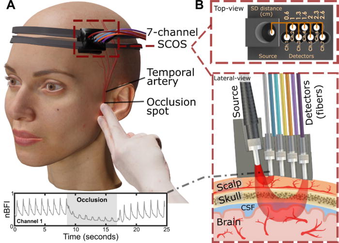
Experimental arrangement of the SCOS system for measuring cerebral blood dynamics during superficial temporal artery (STA) occlusion. (a) 3D visualization of the SCOS device positioned over the temple region and the occlusion site near the ear bone. (b) Top and lateral views of the device, illustrating different detecting channels for sensing the scalp, skull, and brain layers. Credit: Liu et al., APL Bioengineering, 2025

Measuring blood flow in the brain is critical for responding to a range of neurological problems, including stroke, traumatic brain injury (TBI) and vascular dementia. But existing techniques, including magnetic resonance imaging and computed tomography, are expensive and therefore not widely available.

Researchers from the USC Neurorestoration Center and the California Institute of Technology (Caltech) have built a simple, noninvasive alternative. The device takes a technique currently used in animal studies known as speckle contrast optical spectroscopy (SCOS) and adapts it for potential clinical use in humans. It works by capturing images of scattered laser light with an affordable, high-resolution camera.

“It’s really that simple. Tiny blood cells pass through a laser beam, and the way the light scatters allows us to measure blood flow and volume in the brain,” said Charles Liu, MD, PhD, professor of clinical neurological surgery, urology and surgery at the Keck School of Medicine of USC, director of the USC Neurorestoration Center and co-senior author of the new research.

The device has already been tested with humans in small proof of concept studies demonstrating the tool’s utility for assessing stroke risk and detecting brain injury. In the current study, Liu and his team sought to confirm that SCOS is truly measuring blood flow in the brain, rather than in the scalp, which also contains many blood vessels. The question has long plagued researchers who use light-based technology to visualize the brain.

Liu’s team took an innovative approach: By temporarily blocking blood flow to the scalp, they confirmed that SCOS readings were indeed measuring signals from blood vessels in the brain. Readings from 20 participants showed that positioning the detector at least 2.3cm away from the laser source provided the clearest measurement of brain blood flow. The study, funded in part by the National Institutes of Health, the Alfred Mann Foundation and the USC Neurorestoration Center, was just published in the journal APL Bioengineering.

“For the first time in humans, this experimental evidence shows that a laser speckle optical device can probe beyond the scalp layers to access cerebral signals,” said Simon Mahler, PhD, who is now an assistant professor in the Department of Biomedical Engineering at the Stevens Institute of Technology and one of the paper’s coauthors. “This is an important step toward using SCOS to non-invasively measure blood flow in the brain.”

Tracking brain blood flow

For years, researchers measuring brain signals with light-based technology, such as lasers and fiber optics, have used statistical simulations to estimate which signals originate in the brain versus the scalp. The USC Neurorestoration Center team found a direct way to test the difference, thanks to a collaboration between surgeons, engineers and neurologists.

“I perform surgeries to increase blood flow in the brain, and many of these involve temporarily stopping blood flow in the scalp,” said Jonathan Russin, MD, now professor and chief of neurosurgery at the University of Vermont, who continues to collaborate with the USC Neurorestoration Center. “That gave us a simple way to test the technology – by creating a change that affected only the scalp’s circulation while leaving the brain’s blood flow untouched.”

In 20 participants, the researchers temporarily stopped blood flow to the scalp, then collected a series of SCOS readings. By gradually moving the detector further from the head, they captured signals reaching progressively deeper towards the brain. They found that positioning the detector 2.3cm from the head allowed them to measure brain blood flow while minimising interference from the scalp.

The findings confirm the utility of SCOS for non-invasively detecting brain blood flow and provide important guidance for other researchers working with light-based technology, Liu said.

Bringing SCOS to patients

Beyond advancing research, the study helps confirm the clinical potential of SCOS for detecting and responding to stroke, brain injury and dementia. Because all of the team’s research has been done with humans, the tool is poised for rapid translation from the lab to the clinic.

“We look directly at humans in essentially the same way the tool will be applied, so there’s nothing lost in translation,” Liu said. “We are never more than one step away from the problem we’re trying to solve.”

The technique is already being used by some of the team’s collaborators to help diagnose stroke and TBI. Next, the researchers will continue to refine the technology and software, working to improve the resolution of images and the quality of data extracted from readings.

“With the knowledge that we’re now measuring exactly what we intend to measure, we’re also going to expand our testing of this technique with patients in clinical settings,” Liu said.

Source: Keck School of Medicine of USC

Teens More Likely to Use Opioids when Parents Have Prescriptions

Norwegian study of more than 21 000 young people found that those whose parents had persistent opioid prescriptions faced more than double the risk of persistent opioid use

Photo by Danilo Alvesd on Unsplash

If a parent has persistent opioid prescriptions, their adolescent or young adult offspring has more than double the risk of persistent opioid use, according to a new study published October 23rd in the open-access journal PLOS Medicine by Anna Marcuzzi of the Norwegian University of Science and Technology, Norway, and colleagues.

The prescription of strong analgesics such as opioids is not recommended for young people. However, despite potential adverse long-term consequences, opioids are often prescribed for non-malignant pain in this population.

In the new study, researchers analysed data from 21 470 adolescents and young adults aged 13-29 years who participated in the population-based Young-HUNT or HUNT Study in Norway in 2006-2008 or 2017-2019. Each participant was linked with at least one parent who also participated in the HUNT study, and opioid prescription data were obtained from the Norwegian Prescription Database.

24.4% of young people had at least one opioid prescription during the seven-year follow-up period, while 1.3% had persistent opioid prescriptions, defined as prescriptions in at least three out of four quarters of a year. When a mother had persistent opioid prescriptions over a five-year period (two years before and three years after offspring participation), their offspring had 2.60 times the risk of persistent opioid use compared to those whose mothers had no prescriptions. When a father had persistent opioid prescriptions, their offspring had 2.37 times the risk of persistent opioid use. The association was also present but weaker for non-persistent opioid prescription – offspring whose mothers had two or more prescriptions had 1.34 times the risk of receiving any opioid prescription, while those whose fathers had two or more prescriptions had 1.19 times the risk, compared to offspring whose parents had no prescriptions. There was no clear evidence that parental chronic musculoskeletal pain status influenced these associations.

The authors note that because parental opioid prescriptions were measured both before and after offspring HUNT participation, some parental opioid prescriptions could have begun after offspring opioid use. However, they conclude that there is an association between parental and offspring opioid prescriptions.

“The study findings suggest that family-based strategies should be considered when managing pain conditions in adolescents and young adults to avoid potentially unnecessary opioid use,” they say.

The authors add, “Despite restrictive opioid policies, one in four adolescents and young adults received an opioid prescription during the seven years follow-up.”

“Adolescents whose parents had two or more opioid prescriptions had a more than two-fold higher risk of persistent opioid use (ie, multiple prescriptions in a year) than if the parents had no opioid prescriptions.”

Provided by PLOS