New research from the University of Waterloo suggests that men and women should have different kinds of food for breakfast in order to help lose weight.
The study, which employed a mathematical model of men’s and women’s metabolisms, showed that men’s metabolisms respond better on average to a meal laden with high carbohydrates like oats and grains after fasting for several hours, while women are better served by a meal with a higher percentage of fat, such as omelettes and avocados. The findings are out now in Computers in Biology and Medicine.
“Lifestyle is a big factor in our overall health,” said Stéphanie Abo, an Applied Mathematics PhD candidate and the lead author of the study. “We live busy lives, so it’s important to understand how seemingly inconsequential decisions, such as what to have for breakfast, can affect our health and energy levels. Whether attempting to lose weight, maintain weight, or just keep up your energy, understanding your diet’s impact on your metabolism is important.”
The study builds on an existing gap in research on sex differences in how men and women process fat. “We often have less research data on women’s bodies than on men’s bodies,” said Anita Layton, a professor of Applied Mathematics and Canada 150 Research Chair in Mathematical Biology and Medicine.
“By building mathematical models based on the data we do have, we can test lots of hypotheses quickly and tweak experiments in ways that would be impractical with human subjects.”
“Since women have more body fat on average than men, you would think that they would burn less fat for energy, but they don’t,” said Layton. “The results of the model suggest that women store more fat immediately after a meal but also burn more fat during a fast.”
Going forward, the researchers hope to build more complex versions of their metabolism models and extend beyond the consideration of biological sex by incorporating an individual’s weight, age, or stage in the menstrual cycle.
Muscle activation in people suffering from hip osteoarthritis might be a case of ‘mind over matter’, new research from Edith Cowan University (ECU) has shown.
Research undertaken by ECU post-doctoral research fellow Dr Myles Murphy investigated muscle function in people with hip osteoarthritis and found that these patients were unable to activate their muscles as efficiently. The findings are published in Sports Medicine and Health Science.
“Previous research has well established that the degree to which a joint degenerates is not directly related to the amount of pain a person with arthritis will experience. In fact, the stronger your muscles are, the more protected your joint is, and the less pain you will experience.
“Our research has shown that people with hip osteoarthritis were unable to activate their muscles as efficiently, irrespective of strength.”
As part of this research, Dr Murphy and his team studied the brain function of people with hip arthritis, finding that the mind played an enormous part in this equation.
“Basically, people with hip arthritis are unable to activate their muscles properly because the brain is actively putting on the brake to stop them from using the muscle. We don’t know why that is, yet. But the brain seems to really be hampering the progress of rehabilitation and the muscles to protect the joint,” Dr Murphy said.
“We suspect that it is a short-term, protective response gone wrong. Unlike a rolled ankle or a hurt knee, chronic pain like osteoarthritis tends to hang around for a long time. Instead of being a protective response in the short term, the brain’s protective response becomes a really problematic and maladaptive response in the long term.”
Hip osteoarthritis is more prevalent in people over the age of 45, and women are much more like to develop the condition. People who have reported previous joint damage, from a sports injury or accident, are more likely to present with hip osteoarthritis, as are those with joint abnormalities, such as developmental dysplasia of the hip.
People living with hip arthritis often presents with different walking patterns than those without and could struggle with everyday activities like getting out of a chair, or vehicle.
“The impact on their daily lives is the biggest burden of osteoarthritis. The condition also results in substantial time-loss from work, and is associated with a high economic cost,” Dr Murphy said.
“The level of disability for normal activity within our study cohort was about 25%, compared to the 0% reported in our healthy control group.”
Dr Murphy is currently investigating novel ways in which to overcome this automatic muscle inhibition to effectively rehabilitate patients.
In the meantime, those living with hip osteoarthritis have been urged to continue strength training and to work with a qualified physiotherapist or exercise physiologist.
“You will need to work quite hard to build the strength in those muscles, but it can be done. There is no quick fix. Staying strong is something that people with hip osteoarthritis will need to actively keep working on,” he said.
World Mental Health Day, celebrated internationally on 10 October, is not just another commemorative day, but in fact, a time to truly reflect on the need to break the stigma associated with seeking mental health support.
Today, many may acknowledge that mental health issues are common and can adversely affect a significant portion of the population. “In fact, according to the fourth annual Mental State of the World Report 2023, published in March 2024 by Sapien Labs, Brazil, South Africa and the United Kingdom all show the greatest proportion of respondents who are distressed or struggling with their mental health, which indicates that there is still a dire need for more open conversations about mental health in families, workplaces and communities,” says Madelein O’Connell,Executive: Marketing, Sales and Corporate Relations at Bestmed Medical Scheme.
“We already know that our mental wellbeing can affect emotions, physical health, relationships and overall quality of life,” adds O’Connell. “Beyond this, neglecting your mental health can also lead to burnout, anxiety, and an array of serious health conditions. However, prioritising your mental health can be daunting, with so many not knowing where to start. It’s important to remember that it starts with, and is built on, small, consistent steps, which can make a significant difference.”
From a medical scheme perspective, there is often a range of mental health services covered as supplementary benefits by the medical scheme, such as access to psychologists, psychiatrists, counsellors, and support for conditions like anxiety, depression, and trauma. It’s important for members to understand what their medical scheme provides.
“At Bestmed, we offer a free Tempo wellness programme for our members, which can be accessed via the Bestmed App or online Member portal. As part of the Tempo wellness programme, members have access to free Tempo Wellness Webinars, hosted by mental healthcare experts, who discuss and give advice on various relevant topics,” says O’Connell.
“We really want to support the integration of mental, nutritional, and physical health in overall wellbeing for our members, as we recognise that mental health covers a wide array of aspects. In fact, the World Health Organisation (WHO), defines mental health as ‘a state of mental well-being that enables people to cope with the stresses of life, realise their abilities, learn well and work well, and contribute to their community’, so giving people the support and skills to navigate their life is vital, particularly those more vulnerable to mental health challenges, such as adolescents, the elderly, or individuals dealing with long-term physical illness.”
Madelein O’Connell concludes, “of course, we also recognise that limited mental health service availability, a shortage of mental health professionals and financial constraints can impact a person’s ability to find the right support they need, when they need it. However, there are also some incredible mental health support organisations, locally, that can assist. LifeLine and The South African Depression and Anxiety Group (SADAG), for example, are confidential, free, and offer a starting point for anyone in need.”
Positive expectations facilitate reward processing and negative expectations prime pain processing
Photo by Ryan Quintal on Unsplash
The expectations humans have of a pleasurable sensation asymmetrically shape neuronal responses and subjective experiences to hot sauce, according to a study published October 8th, in the open-access journal PLOS Biologyby Yi Luo from East China Normal University, Kenneth Kishida from Wake Forest School of Medicine, US, and colleagues.
Expectations shape our perception, profoundly influencing how we interpret the world. Positive expectations about sensory stimuli can alleviate distress and reduce pain through what’s known as the placebo effect, while negative expectations may heighten anxiety and exacerbate pain. In the new study, Luo, Kishida, and colleagues investigated the impact of the hedonic aspect of expectations on subjective experiences.
Specifically, the researchers measured neurobehavioral responses to the taste of hot sauce among individuals with a wide range of taste preferences. In total, 47 participants completed the tasks while undergoing functional magnetic resonance imaging scanning. The researchers identified participants who liked versus those who strongly disliked spicy flavors and provided contextual cues about the spiciness of the sauce to be tasted. That way, they were able to dissociate the effects of positive and negative expectations from sensory stimuli (i.e., visual and taste stimuli), which were the same across all participants.
The results showed that positive expectations lead to modulations in the intensity of subjective experience. These modulations were accompanied by increased activity in brain regions previously linked to pleasure, information integration, and the placebo effect, including the anterior insula, dorsolateral prefrontal cortex, and dorsal anterior cingulate cortex. By contrast, negative expectations decreased hedonic experience and increased neural activity in the Neurological Pain Signature network.
Taken together, these findings demonstrate that hedonic aspects of one’s expectations asymmetrically shape how the brain processes sensory input and associated behavioral reports of one’s subjective experiences of intensity, pleasure, and pain. The results suggest a dissociable impact of hedonic information. While positive expectations facilitate higher-level information integration and reward processing, negative expectations prime lower-level processes related to pain and emotions. According to the authors, this study demonstrates the powerful role of hedonic expectations in shaping subjective reality and suggests potential avenues for consumer and therapeutic interventions targeting expectation-driven neural processes.
The authors add, “Our study highlights how hedonic expectations shape subjective experiences and neural responses, offering new insights into the mechanisms behind pain perception.”
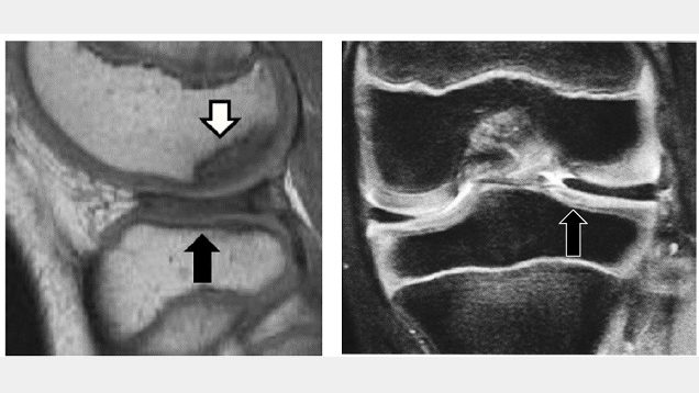
Discoid lateral meniscus and osteochondritis dissecans in adolescent patients. The black arrow represents DLM and the white arrow represents osteochondritis dissecans. Credit: Osaka Metropolitan University
Growing pains are common in maturing children, but sometimes this growth can be irregular and cause injury. Discoid lateral meniscus (DLM), a misshapen knee cartilage, is one such occurrence that can degenerate into osteochondritis dissecans, a joint disorder where the bone and joint begin to separate from the rest of the bones. It has been reported that osteochondritis dissecans of the femoral condyle occurs in approximately 14.5% of cases of DLM, but there has been little analysis of its treatment to date.
Dr Ken Iida and Specially Appointed Professor Yusuke Hashimoto’s team at Osaka Metropolitan University’s Graduate School of Medicine analysed the incidence of post-surgery osteochondritis dissecans. This analysis consisted of two groups, a pre-osteochondritis group with DLM and osteochondritis dissecans of the outer femoral epicondyle, and a non-osteochondritis dissecans DLM group. They studied 95 cases of DLM patients under the age of 15 who underwent surgery between 2003 and 2017 and had five years of post-surgery records. There were 15 cases in the pre-osteochondritis dissecans group and 80 non-osteochondritis dissecans cases.
Their analysis found that the surgical results for osteochondritis dissecans were good in pre-osteochondritis cases, but 28.5% had a recurrence of the joint disorder. In the non-osteochondritis dissecans group, 8.8% were diagnosed with the disorder after surgery. Additionally, age was found to be a risk factor for relapse or post-surgical osteochondritis dissecans, and surgery on patients ages 9 and under was also involved in the occurrence of osteochondritis dissecans.
“Patients with DLM accompanied by osteochondritis dissecans of the femoral condyle often have difficulty in deciding on a treatment method,” Dr Iida explained. “Based on the results of this study, we believe for patients ages 9 years or younger, it is necessary to consider conservative treatment methods rather than immediate surgery.”
Deep brain stimulation (DBS) may provide immediate improvement in arm and hand strength and function weakened by traumatic brain injury or stroke, according to research from the University of Pittsburgh School of Medicine.
Encouraging results from extensive tests in monkeys and humans open a path for a new clinical application of an already widely used brain stimulation technology and offer insights into neural mechanisms underlying movement deficits caused by brain injury. The results are published in Nature Communications.
“Arm and hand paralysis significantly impacts the quality of life of millions of people worldwide,” said senior and corresponding author Elvira Pirondini, Ph.D., assistant professor of physical medicine and rehabilitation at Pitt. “Currently, we don’t have effective solutions for patients who suffered a stroke or traumatic brain injury but there is a growing interest in the use of neurotechnologies that stimulate the brain to improve upper-limb motor functions.”
Brain lesions caused by serious brain trauma or stroke can disrupt neural connections between the motor cortex, a key brain region essential for controlling voluntary movement, and the muscles. Weakening of these connections prevents effective activation of muscles and results in movement deficits, including partial or complete arm and hand paralysis.
To boost the activation of existing, but weakened, connections, researchers proposed to use deep brain stimulation (DBS), a surgical procedure that involves placing tiny electrodes in specific areas of the brain to deliver electrical impulses that regulate abnormal brain activity. Over the past several decades, DBS has revolutionised the treatment of neurological conditions such as Parkinson’s disease by providing a way to control symptoms that were once difficult to manage with medication alone.
“DBS has been life-changing for many patients. Now, thanks to ongoing advancements in the safety and precision of these devices, DBS is being explored as a promising option for helping stroke survivors recover their motor functions,” said senior author Jorge González-Martínez, MD, PhD, neurosurgery professor at Pitt. “It offers new hope to millions of people worldwide.”
Taking cues from another successful Pitt project that used electrical stimulation of the spinal cord to restore arm function in individuals affected by stroke, scientists hypothesised that stimulating the motor thalamus – a key relay hub of movement control – using DBS could help restore movements that are essential for tasks of daily living, such as object grasping. However, because the theory has not been tested before, they first had to test it in monkeys, which are the only animals that have the same organization of the connections between the motor cortex and the muscles as humans.
To understand the mechanism of how DBS of the motor thalamus helps improve voluntary arm movement and to finesse the specific location of the implant and the optimal stimulation frequency, researchers implanted the FDA-approved stimulation device into monkeys that had brain lesions affecting how effectively they could use their hands.
As soon as the stimulation was turned on, it significantly improved activation of muscles and grip force. Importantly, no involuntary movement was observed.
To verify that the procedure could benefit humans, the same stimulation parameters were used in a patient who was set to undergo DBS implantation into the motor thalamus to help with arm tremors caused by brain injury from a serious motor vehicle accident that resulted in severe paralysis in both arms.
As soon as the stimulation was turned on again, the range and strength of arm motion was immediately improved: The participant was able to lift a moderately heavy weight and reach, grasp and lift a drinking cup more efficiently and smoothly than without the stimulation.
To help bring this technology to more patients in the clinic, researchers are now working to test the long-term effects of DBS and determine whether chronic stimulation could further improve arm and hand function in individuals affected by traumatic brain injury or stroke.
By Reo Botes, Managing Executive at Essential Employee Benefits
Cross-subsidising medical aid contributions is a long-standing practice in South Africa and is and one of the benefits companies can use to make themselves stand out as employers of choice. This approach allows employers to support their employees in managing healthcare costs, which can be particularly burdensome in a country where healthcare expenses continue to rise. However, despite this subsidy, medical aid remains unaffordable for many individuals, especially those in lower income brackets and even for middle-income earners. The reality is that even if half of the cost is subsidised by their employer, many employees find it challenging to allocate a significant portion of their income toward medical aid contributions.
The challenge of affordability
The affordability challenge is exacerbated by the annual increases in medical aid contributions, which frequently exceed the rate of salary increases. For instance, a medical aid plan that costs R2,000 a month will still require the employee to pay R1,000 a month should the employer subsidise at least 50%. For someone earning a modest salary, such as entry-level employees, this R1,000 can represent a substantial chunk of their monthly income, making it an untenable option, this means that the employee then loses out on this benefit if the only employee benefit option is medical aid.
Furthermore, medical inflation has continued to soar, leading to dramatic increases in the cost of even entry-level medical aid plans. These plans, which were once within reach for some, have become prohibitively expensive. The rising cost of living, coupled with stagnant wages, has forced many individuals to reconsider their insurance cover. With limited options available when employers subsidise the costs, employees often find themselves in a difficult position, needing to balance health needs with financial realities.
The need for alternative solutions
Given these challenges, it has become increasingly important for businesses to explore additional more affordable healthcare options in the mix. While this may introduce some administrative tasks, the decision ultimately boils down to whether the cost of the employee not being able to perform their tasks optimally outweighs the costs of a Health Insurance solution. The key is to find the balance between keeping people healthy and productive, which necessitates a shift towards enhancing accessible health products.
There is an obvious and direct correlation between employee health and productivity, and so the primary objective of employee healthcare benefits should always be to maximise employee health. Including alternative subsidised healthcare options, particularly for lower-income earners and those looking to step down their cover, allows employers to provide greater choice and flexibility. This not only benefits employees but can positively impact the company’s bottom line.
Health insurance products offer a cost-effective solution that enhances access to healthcare at a fraction of the cost of medical aid. While this type of insurance is not as comprehensive as medical aid, it is significantly more affordable. When subsidised by employers, health insurance can cost employees just a few hundred Rands a month, making it a feasible option for many.
Depending on the provider and product suite, health insurance can supply access to primary or day-to-day healthcare services, including optometry and dentistry, as well as cover for in-hospital procedures in a private hospital. This accessibility empowers employees to seek the treatment they need without the additional stress associated with financial strain and affordability, the outcome being a healthier, happier, and ultimately more productive and profitable workforce.
The importance of employee health
Healthy employees are more engaged and productive, which ultimately benefits the employer. Ensuring that employees have access to preventive care and timely treatment, allows companies to reduce absenteeism and increase job satisfaction This creates a mutually beneficial situation where employees feel their health needs are supported, and employers benefit from a stable, healthy and productive employee base.
Moreover, as the landscape of healthcare continues to evolve, businesses must remain agile and responsive to the changing needs of their employees. This includes recognising the importance of mental health and wellness programmes as part of a comprehensive employee benefits package. By prioritising employee health, companies can foster a positive workplace culture that attracts and retains top talent.
The role of employers in promoting wellness
Employers play a crucial role in promoting employee wellness beyond just providing healthcare benefits. By fostering a work environment that encourages healthy habits, employers can positively impact the overall well-being of their workforce. This can include initiatives such as:
Providing healthy snack options and encouraging regular breaks
Organising fitness challenges or subsidising gym memberships
Offering mental health support and resources
Promoting work-life balance and flexible work arrangements
Educating employees on the importance of preventive care and regular check-ups
When employees feel supported in their health and wellness goals, they are more likely to be engaged, motivated, and productive in their work. This, in turn, contributes to the overall success and competitiveness of the organisation.
The impact on employee retention and recruitment
Offering comprehensive and affordable healthcare benefits can significantly impact employee retention and recruitment. In today’s competitive job market, potential employees often prioritise companies that demonstrate a commitment to their well-being. By providing a robust healthcare benefits package that includes subsidised medical aid and health insurance options, employers can position themselves as an employer of choice.
Moreover, retaining talented employees becomes easier when they feel valued and supported by their employer. By investing in their employees’ health, companies can foster a sense of loyalty and commitment, reducing costly staff turnover rates, which ensures continuity in their workforce.
The ongoing challenges surrounding medical aid affordability in South Africa highlight the need for innovative solutions that prioritise employee health and well-being. By expanding the range of healthcare options available to employees, businesses can enhance access to necessary medical services while also addressing the financial burdens that many individuals face.
As the healthcare landscape continues to change, it is crucial for employers to stay informed and proactive in their approach to employee benefits. By investing in the health of their workforce, companies not only contribute to the well-being of their employees but also position themselves as desirable employers in a competitive job market. Ultimately, the goal should be to create a healthier, more productive workforce that can thrive in the face of ongoing economic challenges.
Incorporating health insurance into employee benefit packages is a cost-effective strategy to achieve this objective. While it’s not necessary to complicate matters with an array of product options, offering more affordable choices aligned to the employee segment is crucial. Partnering with an independent advisor or engaging with different product suppliers can assist businesses in understanding the broader spectrum of available products and selecting a basket that will offer the best balance between benefit and affordability for all parties concerned.
Princeton-led team of researchers and gamers has mapped every neuron and synapse in the brain of an adult fruit fly.
Video still from Amy Sterling / FlyWire / Princeton
For many heartbreaking diseases of the brain, such as dementia, Parkinson’s, Alzheimer’s, doctors can only treat the symptoms. Medical science does not have a cure. Why? Because it’s difficult to cure what we don’t understand, and the human brain, with its billions of neurons connected by a hundred trillion synapses, is almost hopelessly complex.
“FlyWire,” a Princeton-led team of scientists and citizen scientists, has now made a massive step toward understanding the human brain by building a neuron-by-neuron and synapse-by-synapse roadmap – scientifically speaking, a “connectome” – through the brain of an adult fruit fly (Drosophila melanogaster). The FlyWire Consortium comprises members from more than 146 labs at 122 institutions, with major contributions from teams at the University of Cambridge and the University of Vermont.
“Any brain that we truly understand tells us something about all brains,” said Sebastian Seung, Princeton’s Evnin Professor in Neuroscience and a professor of computer science. “With the fly wiring diagram, we have the potential for an unprecedented, detailed and deep understanding.”
Previous researchers have mapped the brain of a C. elegans worm, with its 302 neurons, and the brain of a larval fruit fly, which had 3000 neurons, but the adult fruit fly is several orders of magnitude more complex, with almost 140 000 neurons and tens of millions of synapses connecting them.
“This is a major achievement,” said Mala Murthy, director of the Princeton Neuroscience Institute and, with Seung, a leader of the research team. “There is no other full brain connectome for an adult animal of this complexity.”
A pathway toward tailored treatments
“How the brain functions depends critically on which neurons connect to which other neurons and the strength of their connections,” said Murthy, Princeton’s Karol and Marnie Marcin ’96 Professor of Neuroscience. “To have a full wiring diagram of the fly brain – as a Drosophila neurobiologist, this is something I’ve dreamed of since I started my lab in 2010.”
Mala has pursued that vision, a full connectome of the fruit fly brain, since beginning her collaboration with Seung in 2018. “As neuroscientists, we have a habit of simplifying, of saying, ‘Hey, can I find that one neuron or cell type and figure out its function, how it contributes to animal decisions and behaviours? If I can simplify the problem down to that one neuron, then I can figure out what it does,’” said Murthy. “But when you take a step back, you realise ‘that one neuron’ is in a complex network of almost 140 000 neurons collectively coordinating this behaviour.”
Neuroscientists like to point out that the human brain is the body’s most complex organ, and possibly the most complex neural network anywhere. “In many respects, it is more powerful than any human-made computer, yet for the most part we still do not understand its underlying logic,” said John Ngai, director of the U.S. National Institutes of Health’s BRAIN Initiative, which provided partial funding for the connectome project. “Without a detailed understanding of how neurons connect with one another, we won’t have a basic understanding of what goes right in a healthy brain or what goes wrong in disease.
“The collaborations across diverse areas of expertise in this type of team science consortium have brought the Drosophila brain map to light at an unprecedented pace, paving the way for detailed maps of the human brain and the tailored treatments that could follow,” Ngai said.
How gamers and AI helped make it happen
Sven Dorkenwald, the lead author on the flagship Nature paper, spearheaded the FlyWire consortium that mapped the fly brain.
“What we built is, in many ways, an atlas,” said Dorkenwald, a 2023 Ph.D. graduate of Princeton, now at the University of Washington and the Allen Institute. “Just like you wouldn’t want to drive to a new place without Google Maps, you don’t want to explore the brain without a map. What we have done is build an atlas of the brain, and added annotations for all the businesses, the buildings, the street names. With this, researchers are now equipped to thoughtfully navigate the brain, as we try to understand it.”
The map was built from 21 million images taken of the fruit fly brain by a team of scientists in Davi Bock’s lab, then at the Howard Hughes Medical Institute’s Janelia Research Campus. Using an AI model built by the Seung lab, the lumps and blobs in those images were turned into a labeled, three-dimensional map by the FlyWire Consortium — an unlikely collaboration among gamers, professional tracers, and neuroscientists who are collectively listed as last author on the flagship paper.
FlyWire took inspiration from the earlier EyeWire project, a crowdsourced gamer project that mapped neurons in a mouse retina. When EyeWire was launched, about 10 years ago, artificial intelligence hadn’t advanced to a point where it could accurately trace out each neuron, so gamers painstakingly assembled millions of tiny puzzles to solve the 3D structure of each mouse neuron, revealing each point of connection between them.
In the intervening decade, AI models trained on their work improved their ability to trace out neurons and synapses. Now humans serve as proofreaders, checking the AI-generated products and assembling the countless pieces into one massive whole, as well as annotators, adding cell type labels to each neuron. A team led by Gregory Jefferis at the MRC Laboratory of Molecular Biology and the University of Cambridge, and Bock, now at the University of Vermont, led the effort to add hierarchical annotations to all neurons in the connectome; their work appears in a companion paper in the special issue of Nature and completes the description of the FlyWire resource.
“FlyWire expanded on EyeWire not only in the AI improvements but also in the type of contributions that members could make,” said Amy Sterling, executive director of EyeWire and the crowdsourcing manager of FlyWire. “Members of EyeWire only mapped cells. Members of the FlyWire Consortium were able to both map neurons and contribute labels, both of which they did by the tens of thousands. Labs that originally competed ended up collaborating. Community members built apps and plug-ins to enhance the usage of FlyWire. Great beauty is revealed both in a map of a brain and in the idea that hundreds of human brains worked together make it all possible.”
This collaborative work was made possible through advances in computational infrastructure running in the cloud that the Seung and Murthy labs developed in collaboration with the Allen Institute for Brain Science. Since 2019, the researchers and gamers of FlyWire have collectively contributed 33 person-years to proofreading and annotating the results of the AI model. Without AI, Seung said, the project would have taken almost 50 thousand person-years.
“This dataset is a remarkable story of the power of open team science,” said Forrest Collman, associate director at the Allen Institute for Brain Science. “A dataset was produced and released by the Bock lab, picked up by researchers at Princeton, combined with open-source software to distribute the data to people spread across the world for proofreading, and then collaboratively analysed by the Drosophila neuroscience community.”
Mapping the forest and the trees
The collaboration of online gamers, tracers, neuroscientists and cutting-edge artificial intelligence resulted in a map of every one of the fruit fly’s 139 255 neurons and 50 million of their synaptic connections. The word connectome highlights that it is those connections, ie synapses, between neurons where the brain’s most vital work takes place.
Most neurons look a bit like a tree, with a trunk, branches, roots and twigs. Just as a tree affects its neighbours, its roots connecting to surrounding organisms and its branches battling for sunlight, neurons connect with each other via synapses. But a whole brain is even more connected than a forest, because neurons can reach each other across comparatively massive distances.
“It would be like a tree in New York interacting with a tree in Los Angeles,” said Dorkenwald. “Some of these neurons span the entire brain, from one eye to the other eye. There’s a diversity of sizes, from tiny neurons to others 100 times as big.”
And just like a map that traces out every tiny alley as well as every superhighway, the fly connectome shows connections within the fruit fly brain at every scale.
Once the connections were fully mapped, the team wanted to make it useful to the thousands of scientists conducting research in the field. To address this need, PNI research scientist Arie Matsliah, second author on the flagship paper, together with Sterling and a team of PNI developers, developed the FlyWire Codex. Matsliah calls it “Google for the connectome.” With the Codex, anyone with internet access can navigate every neuron and synaptic pathway in the brain map, without having to download massive amounts of data or knowing any advanced data analysis techniques. It has been already used by 10 000 people worldwide, with thousands of new searches processed daily.
The humble fruit fly
“It might surprise people that flies have brains, but they do,” said Seung. “And their brains have neurons, and while their neurons don’t look exactly the same as ours, they do look more or less like trees, like human neurons. It’s amazing. Our last common ancestor might have been half a billion years ago, and yet fruit flies have recognisable neurons and the same neurotransmitters we have: glutamate, acetylcholine, dopamine.”
Most of us don’t think about fruit flies unless they’re circling over our bananas. With a whole body that’s basically a speck, their brains are almost incomprehensibly tiny – about 750 microns across, 350 microns tall, and 250 microns deep. That’s significantly smaller than a poppy seed.
But this tiny insect has many behaviors that our larger, more complex bodies share, from complicated actions like communicating with romantic partners to simpler ones like moving rapidly, navigating, foraging for food, avoiding predators, responding to light and dark – indeed, fruit flies were responsible for the discovery of circadian rhythms, which spawned a whole field of brain and behavioural science.
Six different Nobel Prizes have honoured 10 researchers studying D. melanogaster, including the 1995 Nobel Prize in Physiology or Medicine for Princeton’s Eric Wieschaus, who discovered the genes controlling embryonic development in fruit flies, and which were later found to be important in cancer research as well.
“Fruit flies are a wonderful model organism, because it’s quite small, but at the same time it has very complex behaviors,” Dorkenwald said. “As humans, we can relate to how male fruit flies sing to females, how they court them and follow them, and the competition between them.” Fruit fly behaviour and physiology have been studied extensively for more than a century, so researchers have a wealth of pre-existing knowledge to tie to this new connectome of neurons and synapses.
‘A massive, interdisciplinary effort’
“This extraordinary accomplishment is the result of a massive, interdisciplinary team effort,” said Murthy. “We brought together Drosophila neuroscientists, with crowdsourced gamers and BRAIN Initiative funds and the ingenuity of our people here at Princeton.” The University’s endowment supported the effort via the Bezos Center for Neural Circuit Dynamics and the McDonnell Center for Systems Neuroscience.
At the University and across the nation and the world, the network of collaborators was vast. “At Princeton alone, we’ve had many postdocs and students working together with software engineers and full-time proofreaders,” Murthy said.
“Worldwide, there are more than 280 members in the FlyWire community building the connectome, most from the neuroscience and Drosophila science communities,” she continued. “And why are they motivated to do this for us? Because they can use the data for their own science.”
Instead of keeping their data confidential, the Princeton researchers opened their in-progress neural map to the scientific community from the beginning. “It took us about two years to correct all the errors,” said Murthy. “We released the data openly from Day One to the entire fly community and said, ‘Do whatever science you want, but help us proofread and annotate it as you go.’”
“Now that we have this brain map, we can close the loop on which neurons relate to which behaviours ,” Dorkenwald said. “It’s wonderful, because new experiments will prompt new hypotheses, and we can relate things to the whole connectome, and we can iterate. I think the hard work is ahead. This is a beginning, not the end of the work.”
A study led by Johns Hopkins Medicine researchers concludes that commonly used ways of positioning the patient’s arm during blood pressure (BP) screenings can substantially overestimate test results and may lead to a misdiagnosis of hypertension.
In a report on the study, published in JAMA Internal Medicine, investigators examined the effects of three different arm positions: an arm supported on a desk, arm supported on a lap, and an unsupported arm hanging at the patient’s side. Researchers found that lap support overestimated systolic pressure by nearly 4mmHg, and an unsupported arm hanging at the side overestimated systolic pressure by nearly 7mmHg.
The findings confirm that arm position makes a “huge difference” when it comes to an accurate blood pressure measurement, says Tammy Brady, MD, PhD, senior author of the study. And they underscore the importance of adhering to clinical guidelines calling for firm support on a desk or other surface when measuring blood pressure, the investigators add.
The latest clinical practice guidelines from the American Heart Association emphasise several key steps for an accurate measurement – including appropriate cuff size, back support, feet flat on the floor with legs uncrossed, and an appropriate arm position, in which the middle of an adjustable BP cuff is positioned at mid-heart level on an arm supported on a desk or table.
Despite these recommendations, the researchers say BP is too often measured with patients seated on an exam table without any, or inadequate, arm support. In some cases, a clinician holds the arm, or the patient holds an arm in their lap. In the new Johns Hopkins study, the researchers recruited 133 adult participants (78% Black, 52% female) between Aug. 9, 2022, and June 1, 2023. Study participants, who ranged from age 18 to 80, were sorted at random into one of six possible groups that differed by order of the three seated arm positions. Measurements were taken during a single visit between 9 a.m. and 6 p.m. Before BP measures were taken, all participants first emptied their bladders and then walked for two minutes to mimic a typical clinical scenario in which people walk into a clinic or office before screening takes place. They then underwent a five-minute, seated rest period with their backs and feet supported. Each person, wearing an upper arm BP cuff selected and sized based on their upper arm size, had three sets of triplicate measurements taken with a digital blood pressure device 30 seconds apart.
Upon completion of each set of three measurements, the cuff was removed, participants walked for two minutes and rested for five minutes. In the same visit, they then underwent a fourth set of triplicate measurements with their arm supported on a desk, a set used to account for well-known variations in BP readings. All of the measurements were conducted in a quiet and private space, and participants were asked not to talk to researchers or use their phones during the screening.
Researchers found that BP measurements obtained with arm positions frequently used in clinical practice – an arm on the lap or unsupported at the side – were markedly higher than those obtained when the arm was supported on a desk, the standard, recommended arm position. Supporting the arm on the lap overestimated systolic and diastolic BP by 3.9mmHg and 4.0mmHg, respectively. An unsupported arm at the side overestimated systolic by 6.5mmHg and diastolic by 4.4mmHg.
“If you are consistently measuring blood pressure with an unsupported arm, and that gives you an overestimated BP of 6.5mmHg, that’s a potential difference between a systolic BP of 123 and 130, or 133 and 140 – which is considered stage 2 hypertension,” says study author Sherry Liu, MHS, an epidemiology research coordinator at Johns Hopkins Bloomberg School of Public Health.
Investigators caution that their study results may only apply during screenings with automated BP devices, and may not apply to readings done with other BP devices.
However, Brady says, the findings suggest that clinicians need to pay better attention to best practice guidelines, and that patients “must advocate for themselves in the clinical setting and when measuring their BP at home.”
Life expectancy increased dramatically over the 19th and 20th centuries, thanks to improvements such as healthier diets and medical advances. But after nearly doubling over the course of the 20th century, the rate of increase has slowed considerably in the last three decades, according to a new study led by the University of Illinois Chicago.
Despite frequent breakthroughs in medicine and public health, life expectancy at birth in the world’s longest-living populations has increased only an average of six and a half years since 1990, the analysis found. That rate of improvement falls far short of some scientists’ expectations that life expectancy would increase at an accelerated pace in this century and that most people born today will live past 100 years.
The Nature Aging paper offers new evidence that humans are approaching a biologically based limit to life. The biggest boosts to longevity have already occurred through successful efforts to combat disease, said lead author S. Jay Olshansky of the UIC School of Public Health. That leaves the damaging effects of aging as the main obstacle to further extension.
“Most people alive today at older ages are living on time that was manufactured by medicine,” said Olshansky, a professor of epidemiology and biostatistics. “But these medical Band-Aids are producing fewer years of life even though they’re occurring at an accelerated pace, implying that the period of rapid increases in life expectancy is now documented to be over.”
That also means extending life expectancy even more by reducing disease could be harmful, if those additional years aren’t healthy years, Olshansky added. “We should now shift our focus to efforts that slow aging and extend healthspan,” he said. Healthspan is a relatively new metric that measures the number of years a person is healthy, not just alive.
Life expectancy increased rapidly through the 19th century and first half of the 20th century. In 1990, some scientists predicted those rapid gains would continue, leading to “radical life extension.” But a new analysis proposes that we may be nearing the limit of human longevity. (Strategic Marketing and Communications / UIC)
The analysis, conducted with researchers from the University of Hawaii, Harvard and UCLA, is the latest chapter in a three-decade debate over the potential limits of human longevity.
In 1990, Olshansky published a paper in Science that argued humans were approaching a ceiling for life expectancy of around 85 years of age and that the most significant gains had already been made. Others predicted that advances in medicine and public health would accelerate 20th-century trends upward into the 21st century.
Thirty-four years later, the evidence reported in the 2024 Nature Aging study supports the idea that life expectancy gains will continue to slow as more people become exposed to the detrimental and immutable effects of aging. The study looked at data from the eight longest-living countries and Hong Kong, as well as the United States – one of only a handful of countries that has seen a decrease in life expectancy in the period studied.
“Our result overturns the conventional wisdom that the natural longevity endowment for our species is somewhere on the horizon ahead of us – a life expectancy beyond where we are today,” Olshansky said. “Instead, it’s behind us – somewhere in the 30- to 60-year range. We’ve now proven that modern medicine is yielding incrementally smaller improvements in longevity even though medical advances are occurring at breakneck speed.”
While more people may reach 100 years and beyond in this century, those cases will remain outliers that won’t move average life expectancy significantly higher, Olshansky said.
That conclusion pushes back against products and industries, such as insurance and wealth-management businesses, which increasingly make calculations based on assumptions that most people will live to be 100.
“This is profoundly bad advice because only a small percentage of the population will live that long in this century,” Olshansky said.
But the finding doesn’t rule out that medicine and science can produce further benefits, he said. There may be more immediate potential in improving quality of life at older ages instead of extending life, the authors argue. More investment should be made in geroscience – the biology of aging, which may hold the seeds of the next wave of health and life extension.
“This is a glass ceiling, not a brick wall,” Olshansky said. “There’s plenty of room for improvement: for reducing risk factors, working to eliminate disparities and encouraging people to adopt healthier lifestyles – all of which can enable people to live longer and healthier. We can push through this glass health and longevity ceiling with geroscience and efforts to slow the effects of aging.”