Year: 2023

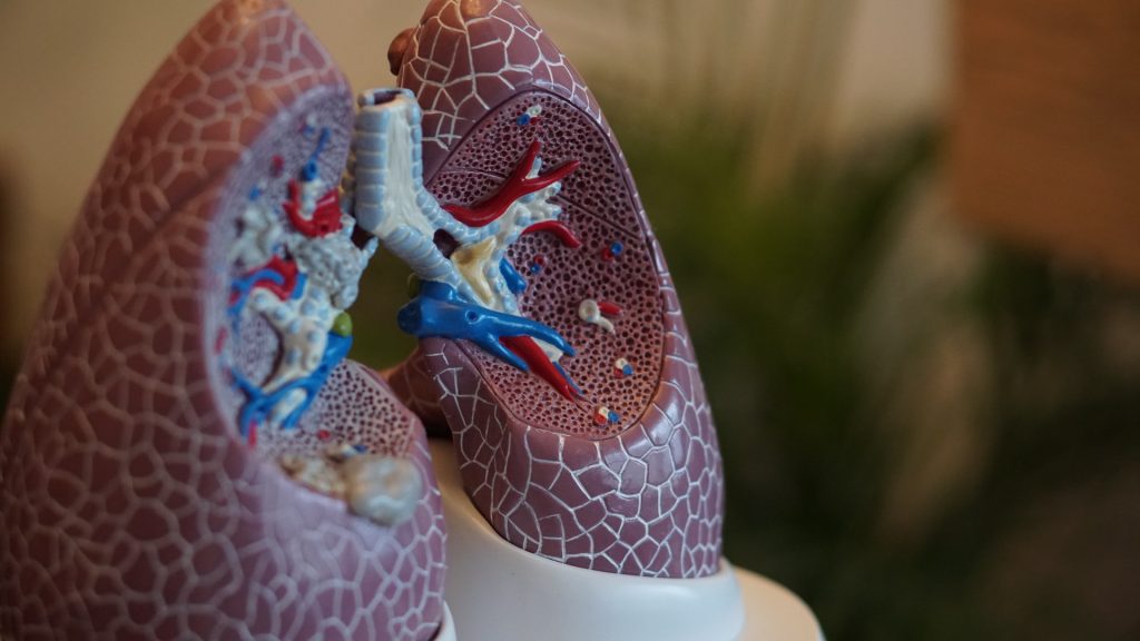
Don’t Teach us Resilience – Fix the Problem, Doctors and Nurses Say

Source: Pixabay CC0

A unique collaborative study on hospital clinician wellbeing by teams at 60 of the best US hospitals, was published in JAMA Health Forum. The study found that physicians and nurses, even at hospitals known to be good places to work, experienced adverse outcomes during the pandemic and want hospital management to make significant improvements in their work environments and in patient safety.

The solutions to high hospital clinician burnout and turnover, they say, are not resilience training for clinicians to better cope with adverse working conditions but organisational improvements that provide safe workloads and better work-life balance.

Researchers sought information in 2021 from 21 050 physicians and registered nurses practicing in 60 Magnet recognised hospitals in 22 states. Forty-seven percent of nurses and 32% of physicians experienced high burnout. Twenty-three percent of physicians and 40% of nurses said they would leave their jobs if possible. Less than 10% of physicians and nurses reported experiencing joy in their work.

Many clinicians are downright hostile to programmes – like resilience training – that are designed to adapt them to poor work conditions; clinicians want the working conditions improved.

Linda H Aiken, PhD, study lead author

Not having enough nurses to care for patients, having little control over workloads, lack of confidence in management to resolve problems in patient care, and concerns about patient safety were all associated with higher burnout, job dissatisfaction, and intent to leave among both nurses and physicians.

Lead author Linda H Aiken, PhD, at the University of Pennsylvania said, “Physicians and nurses largely agree about what hospital management could do to address their burnout, job dissatisfaction, and plans to leave their current jobs; they want improved staffing, modern working conditions in which they can spend more time in direct patient care, greater control over their workloads and work schedules, and a higher priority on patient safety.”

Eighty-seven percent of nurses and 45% of physicians said improving nurse staffing was very important to their own mental health and wellbeing. Other high priorities for clinicians were health breaks without interruption and reduced time spent on documentation. Aiken added, “Many clinicians are downright hostile to programmes – like resilience training – that are designed to adapt them to poor work conditions; clinicians want the working conditions improved.”

Clinicians are concerned about quality and safety of care. Half of physicians and nurses lack confidence that their patients can safely manage their care after discharge highlighting the need for improvement in discharge planning. Patient safety remains a concern with 26% of nurses and 12% of physicians giving their own hospitals an unfavorable patient safety grade. Thirty-nine percent of nurses and 33% of physicians feel mistakes are held against them contrary to recommendations of the National Academy of Medicine to search for and correct system deficiencies that cause most medical errors.

The study was carried out by Penn Nursing’s Center for Health Outcomes and Policy Research in collaboration with the US Clinician Wellbeing Study Consortium composed of 60 Magnet Hospitals. The study took place in 2021 during the COVID pandemic, a time when all US hospitals were severely challenged. Previous research shows that clinicians in hospitals with better work environments prior to the pandemic had better outcomes during the pandemic. The Consortium committed to this study to learn from their experiences during the pandemic how to sustain and further improve their favourable work environments to better withstand external threats and to rebound rapidly.

Source: University of Pennsylvania School of Nursing

B Cells Are Elevated in PCOS – But Are not The Cause

Phot by Sora Shimazaki on Pexels

While previous research have shown that while more B cells are present in women in polycystic ovary syndrome (PCOS), a new study by researchers at Karolinska Institutet has ruled out B cells as the cause of this common syndrome. The study is published in eLife.

Affecting 10–15% of women of fertile age, PCOS is linked to irregular ovulation and menstruation, pregnancy complications, and insulin resistance, all of which are worsened by being overweight. PCOS increases with rising body mass index, and in women with severe obesity, it is around 25%. While the root cause of the condition is unknown, a driver is a surplus of androgens.  

A recent study has shown that women with PCOS have a higher number of B cells in their blood and proposed that these contribute to the development of PCOS through the production of autoimmune antibodies.

“We’ve now examined how B cells affect the development of PCOS with the goal to find new ways of treating the conditions,” says the study’s last author Elisabet Stener-Victorin, research group leader for reproductive endocrinology and metabolism at the Department of Physiology and Pharmacology at Karolinska Institutet.

First, blood from women with PCOS was examined and showed abnormal variations in the frequency of different populations of B cells compared to healthy women. Among these were the so called double negative B cells, a heterogeneous cluster, where some have been described as poised to develop autoimmune functions.

To study whether B cells may cause PCOS, the researchers transferred antibodies from women with PCOS to mice to see if they developed the syndrome. While this proved not to be the case, they did put on weight.

Mice unaffected by B cells

The next step was to transfer B cells from PCOS-like mice (induced by continuous exposure to androgens) to mice lacking B cells to test the hypothesis that B cells drive the development of the disease. However, the recipient mice were unaffected by this transfer.

To see if B cells play an essential role in the development of PCOS, mice lacking B cells were exposed to androgens. These mice were not protected as expected, but developed the same characteristics as normal mice acquire when exposed to androgens.

Finally, androgen exposed PCOS-like mice displayed altered B cell frequencies, as women with PCOS, and simultaneous treatment with a drug that blocks androgen receptors, prevents these alterations of B cells in both blood and tissues, such as ovaries and endometrium.

The researchers conclude that androgens are necessary for the condition to form, but not B cells, the role of which remains unclear.

“B cells are clearly affected in the syndrome, which could contribute to a higher susceptibility to some comorbidities, but they don’t cause PCOS,” says Sara Torstensson, PhD student at Institutet and shared first author.

“We are now studying how other immune cells are affected and how this influences reproductive and metabolic function in PCOS,” says Angelo Ascani, guest PhD student at Graz University, Austria and also first author.

Source: Karolinska Institutet

Steaks are OK? Global Study Challenges Current Advice on High-fat Diets

Photo by Jose Ignacio Pompe on Unsplash

In a study conducted across 80 countries, researchers found that unprocessed red meat and whole grains can be included or left out of a healthy diet. Published in the European Heart Journal, the findings showed that diets emphasising fruit, vegetables, dairy (mainly whole-fat), nuts, legumes and fish were linked with a lower risk of cardiovascular disease (CVD) and premature death in all world regions. The addition of unprocessed red meat or whole grains had little impact on outcomes.

“Low-fat foods have taken centre stage with the public, food industry and policymakers, with nutrition labels focused on reducing fat and saturated fat,” said study author Dr Andrew Mente of the Population Health Research Institute, McMaster University, Hamilton, Canada. “Our findings suggest that the priority should be increasing protective foods such as nuts (often avoided as too energy dense), fish and dairy, rather than restricting dairy (especially whole-fat) to very low amounts. Our results show that up to two servings a day of dairy, mainly whole-fat, can be included in a healthy diet. This is in keeping with modern nutrition science showing that dairy, particularly whole-fat, may protect against high blood pressure and metabolic syndrome.”

The study examined the relationships between a new diet score and health outcomes in a global population. A healthy diet score was created based on six foods that have each been linked with longevity. The PURE diet included 2-3 servings of fruit per day, 2-3 servings of vegetables per day, 3-4 servings of legumes per week, 7 servings of nuts per week, 2-3 servings of fish per week, and 14 servings of dairy products (mainly whole fat but not including butter or whipped cream) per week. A score of 1 (healthy) was assigned for intake above the median in the group and a score of 0 (unhealthy) for intake at or below the median, for a total of 0 to 6. Dr Mente explained: “Participants in the top 50% of the population – an achievable level – on each of the six food components attained the maximum diet score of six.”

Associations of the score with mortality, myocardial infarction, stroke and total CVD (including fatal CVD and non-fatal myocardial infarction, stroke and heart failure) were tested in the PURE study which included 147 642 people from the general population in 21 countries. The analyses were adjusted for factors that could influence the relationships such as age, sex, waist-to-hip ratio, education level, income, urban or rural location, physical activity, smoking status, diabetes, use of statins or high blood pressure medications, and total energy intake.

The average diet score was 2.95. During a median follow-up of 9.3 years, there were 15 707 deaths and 40 764 cardiovascular events. Compared with the least healthy diet (score of 1 or less), the healthiest diet (score of 5 or more) was linked with a 30% lower risk of death, 18% lower likelihood of CVD, 14% lower risk of myocardial infarction and 19% lower risk of stroke. Associations between the healthy diet score and outcomes were confirmed in five independent studies including a total of 96 955 patients with CVD in 70 countries.

Dr Mente said: “This was by far the most diverse study of nutrition and health outcomes in the world and the only one with sufficient representation from high-, middle- and low-income countries. The connection between the PURE diet and health outcomes was found in generally healthy people, patients with CVD, patients with diabetes, and across economies.”

“The associations were strongest in areas with the poorest quality diet, including South Asia, China and Africa, where calorie intake was low and dominated by refined carbohydrates. This suggests that a large proportion of deaths and CVD in adults around the world may be due to undernutrition, that is, low intakes of energy and protective foods, rather than overnutrition. This challenges current beliefs,” said Professor Salim Yusuf, senior author and principal investigator of PURE.

In an accompanying editorial, Dr Dariush Mozaffarian of the Friedman School of Nutrition Science and Policy, Tufts University, USA, stated: “The new results in PURE, in combination with prior reports, call for a re-evaluation of unrelenting guidelines to avoid whole-fat dairy products. Investigations such as the one by Mente and colleagues remind us of the continuing and devastating rise in diet-related chronic diseases globally, and of the power of protective foods to help address these burdens. It is time for national nutrition guidelines, private sector innovations, government tax policy and agricultural incentives, food procurement policies, labelling and other regulatory priorities, and food-based healthcare interventions to catch up to the science. Millions of lives depend on it.”

Source: European Society of Cardiology

Peptides May Solve the Sticky Problem of Bacterial Biofilms

This image shows an intricate colony of Pseudomonas aeruginosa. The bacteria have self-organised into a sticky, mat-like colony called a biofilm, which allows them to cooperate with each other, adapt to changes in their environment, and ensure their survival.
Credit: Scott Chimileski and Roberto Kolter, Harvard Medical School, Boston

Researchers have developed peptides that can help combat bacteria growing in biofilms, which occur in up to 80% of human infections. Their results, published in Nature Chemical Biology, may offer a way to tackle antimicrobial-resistant infections. 

Treating infections becomes significantly more challenging when biofilms are present, as they not only reduce the effectiveness of antibiotics but also give rise to several medical complications. These complications include infections following joint replacements, prosthetic devices, as well as contamination in catheters and other medical equipment. The lack of specific treatments makes the management and treatment of biofilms exceptionally difficult.

The team of researchers, led by Dr Clarissa Melo Czekster and Dr Christopher Harding from the School of Biology at St Andrews, in collaboration with researchers at University of Dundee, developed antimicrobial peptides that can target the harmful bacteria growing in biofilms.

The team determined how a key enzyme (PaAP) in biofilms work and developed a revolutionary new strategy to inhibit the protein. Their inhibitor is potent and targets cells from the human pathogen Pseudomonas aeruginosa in biofilms. P. aeruginosa, a WHO pathogen of concern, causes chronic infections in patients with cystic fibrosis and other conditions, which means a biofilm inhibitor is urgently needed.

Dr Czekster and the team are currently working in collaboration with the University of St Andrews Technology Transfer Centre and industry partner Locate Bio, a biomedicine spinout of the University of Nottingham, to commercialise the technology. The Locate Bio team are trialling the peptides to see how they work with the company’s Programmed Drug Release technology to develop new orthobiologic solutions and products. The Technology Transfer Centre has filed a UK priority patent application.

Dr Czekster said: “Our research reveals how designed inhibitors can target a key enzyme in bacterial virulence, offering molecular insights applicable to aminopeptidases in diverse organisms.

“This remarkable new research presents an innovative strategy to target bacterial biofilms and pave the way for better treatment of bacterial infection.”

Source: University of St. Andrews

A Sensor Printed onto Clothing that Monitors Sweat Electrolytes

Photo by Ketut Subiyanto on Pexels

Researchers from Japan have developed a novel wearable chemical sensor capable of measuring the concentration of chloride ions in sweat. The technology, described in the journal ACS Sensors, uses a ‘heat-transfer printing’ technique, the proposed sensor can be applied to the outer surface of common textiles to prevent skin irritation and allergies, and could also be useful in the early detection of heat stroke and dehydration.

Advances in miniaturisation have led to science-fiction like technologies such as wearable sensors which are usually placed directly on the skin. They can monitor important bodily parameters, including heart rate, blood pressure, and muscle activity and are often incorporated into devices such as smart watches.

Some wearable sensors can also detect chemicals in bodily fluids. For instance, sweat biosensors can measure the concentration of ions in sweat, providing information on their levels in blood. However, designing such chemical sensors is more complex than physical sensors. Direct contact between a wearable chemical sensor and skin can cause irritation and allergies. In contrast, if the sensor is fabricated directly on a wearable textile, its accuracy decreases due to surface irregularities.

In a recent study, a research team, led by Associate Professor Isao Shitanda of the Tokyo University of Science (TUS) in Japan, has developed an innovative sweat biosensor that addresses the aforementioned problems. Their technique involves ‘heat-transfer printing’ to fix a thin, flexible chloride ion sensor onto a textile substrate.

“The proposed sensor can be transferred to fibre substrates, and thus can be incorporated into textiles such as T-shirts, wristbands, and insoles,” explains Dr Shitanda. “Further, health indicators such as chloride ion concentration in sweat can be measured by simply wearing them.”

The heat-transfer printing approach offers several advantages. For one thing, the sensor is transferred outside of the piece of clothing, which prevents skin irritation. In addition, the wicking effect of the textile helps spread the sweat evenly between the electrodes of the sensor, creating a stable electrical contact. Moreover, printing the sensor on a flat surface and then transferring it prevents the formation of blurred edges that commonly occur when printing directly onto a textile.

The researchers carefully selected non-allergenic materials and electrochemical mechanisms of the sensor. After developing the sensor, they conducted various experiments using artificial sweat to verify its accuracy in measuring chloride ion concentration. The change in the electromotive force of the sensor was −59.5 mTV/log CCl−. Additionally, it displayed a Nernst response and a linear relationship with the concentration range of chloride ions in human sweat. Moreover, no other ions or substances typically present in sweat were found to interfere with the measurements.

Lastly, the team tested the sensor on a volunteer who exercised on a static bicycle for 30 minutes, by measuring their perspiration rate, chloride ion levels in blood, and saliva osmolality every five minutes to compare with the data previously gathered by the sensor. The proposed wearable sensor could reliably measure the concentration of chloride ions in sweat.

The sensor can also transmit data wirelessly, making real-time health monitoring easier. “Since chloride is the most abundant electrolyte in human sweat, measuring its concentration provides an excellent indicator of the body’s electrolyte balance and a useful tool for the diagnosis and prevention of heat stroke,” remarks Dr Shitanda.

This research thus demonstrates the potential of using wearable ion sensors for the real-time monitoring of sweat biomarkers, facilitating personalised healthcare development and athlete training management.

Source: Tokyo University of Science

Long-term Improvement in Cystic Fibrosis Symptoms Using Triple Therapy

Photo by Robina Weermeijer on Unsplash

Triple combination therapy can achieve positive, lasting effects in patients with cystic fibrosis (CF), according to the results of a study published in the European Respiratory Journal. Researchers from Charité – Universitätsmedizin Berlin and the Max Delbrück Center found that, in many patients, the therapy reduced mucus stickiness and lung inflammation, improving lung function and quality of life.

Two years ago, a research group headed by Charité and lead researcher Prof Marcus Mall showed that combination therapy involving three drugs – elexacaftor, tezacaftor, and ivacaftor – is effective in a large portion of patients with cystic fibrosis, a hereditary disease, meaning that the treatment noticeably improves both lung function and quality of life. Now, the team has investigated whether this form of treatment is also helpful in the long term, meaning over a period of 12 months or more. To examine this, the researchers focused on the sputum.

“In patients with cystic fibrosis, the mucus in the airways is very sticky because it doesn’t contain enough water and the mucins, the molecules that form mucus, adhere too much due to their chemical properties. This results in thick, sticky mucus, which clogs the airways, making it harder for patients to breathe and leading to chronic bacterial infection and inflammation of the lungs,” explains Mall, Director of the Department of Pediatric Respiratory Medicine, Immunology and Critical Care Medicine and the Christiane Herzog Cystic Fibrosis Center at Charité.

In the current study, the researchers showed that a combination of elexacaftor, tezacaftor, and ivacaftor results in less viscous respiratory secretions and decreasing inflammation and bacterial infection in the lungs of cystic fibrosis patients. “What’s more, the effects lasted over the entire one-year study period. This is really important because previous medications caused a rebound in the bacterial load in the airways,” explains Dr Simon Gräber, who also works in the Department of Pediatric Respiratory Medicine, Immunology and Critical Care Medicine at Charité and was one of the co-leaders of the study. 79 adolescents and adults with cystic fibrosis and chronic lung disease participated in the trial.

A major step in treating cystic fibrosis, further research important

“This is a major step forward in treating cystic fibrosis,” Mall says. “At the same time, it would be premature to say that patients have been normalised, let alone cured. Chronic lung changes arising over many years of living with the disease cannot be reversed, unfortunately.” This means patients with advanced lung disease will still need to rely on established treatments involving inhaling mucus-thinning medications, taking antibiotics, and physical therapy.

“We plan to forge ahead with our research on how to make treatments that address cystic fibrosis via the molecular defects that cause the disease — like the triple medication combination studied here — even more effective. This includes starting treatment in early childhood with the goal of preventing chronic lung changes wherever possible,” Mall notes. “Aside from that, this therapy is not available to about ten percent of our patients right now due to their genetic conditions,” Gräber adds. “That’s why we are also hard at work on research involving new molecular treatments so we can treat all people with cystic fibrosis effectively.”

The researchers are also working to advance their understanding of mucus defects in cystic fibrosis and develop new mucolytics, drugs that thin and loosen the mucus. This research could also benefit patients with common chronic inflammatory lung diseases such as asthma and COPD.

Cystic fibrosis

Cystic fibrosis is one of the most common fatal hereditary diseases worldwide. As many as 8000 children, teens, and adults are living with the disease in Germany today. An imbalance in salt and water transport across mucosal surfaces of the body causes people with cystic fibrosis to produce thick, sticky secretions that harm organs such as the lungs, intestine and pancreas. This leads to progressive loss of lung function and shortness of breath, which still significantly lowers life expectancy despite advances in treatment. Some 150 to 200 children are born with this rare disease in Germany each year.

About the triple combination therapy

A combination of three drugs – elexacaftor, tezacaftor, and ivacaftor – became available in Europe in August 2020. The therapy noticeably improves lung function and quality of life in patients with the most common genetic defect involved in CF, F508del. This means the treatment is an option for nearly 90% of those living with cystic fibrosis. The combination therapy was approved for children starting at the age of six years in early 2022.

Source: Charité – Universitätsmedizin Berlin

Thinning of the Retina is an Early Marker of MS

Retina and nerve cells. Credit: NIH

For the first time, a study has shown that diagnosis of multiple sclerosis (MS) can be significantly improved by additionally measuring the thickness of retinal layers in the eye in a currently existing procedure. Use of the procedure helps to detect the condition at an earlier stage and predict its progression more accurately, which can help to improve patient outcomes. The findings have been published in the journal Neurology.

As part of their investigation, the research team headed by Gabriel Bsteh and Thomas Berger collaborated with ophthalmology colleagues examine 267 MS patients over five years. Their research built on study results published in 2022, which showed that MS relapse-related damage to the retina reflects the degree of damage caused to the patient’s brain. The previous study also demonstrated that a 5µm reduction in the thickness of the retinal layer following optic neuritis indicated a doubling of the risk of permanent disability after the next relapse. Thanks to the latest research with the large cohort of MS patients, the research team has confirmed that the thickness of the retinal layer can be used as a precise biomarker to assist early diagnosis.

Diagnostic procedure already available

The researchers used a procedure known as optical coherence tomography (OCT) to measure the thickness of the retinal layer. An imaging method that uses infrared light, OCT allows for the generation of high-resolution, three-dimensional images of extremely thin layers of tissue measuring just a few µm. OCT is also a tool for diagnosing and evaluating the progression of eye diseases such as glaucoma. “So we already have this procedure at our disposal,” commented Gabriel Bsteh, first author of the study. He added: “If we use optical coherence tomography alongside the current criteria to diagnose MS, we obtain significantly more accurate results at a much earlier stage. This means we can initiate treatment measures sooner, which considerably improves the long-term prognosis for patients.”

The retina: a window to the brain

Multiple sclerosis is an autoimmune, chronic inflammatory disease that causes inflammation and loss of nerve cells throughout the nervous system. For the most part, patients are unable to feel the consequences of this damage to begin with, so the condition often goes undiagnosed until a late stage, meaning that valuable time is lost during which effective treatment could have been administered. Given that early detection and prognosis of the disease’s progression play a decisive role in MS cases, medical researchers have been trying to find improved detection methods for some time now to help avert serious consequences such as impaired mobility and blindness as far as possible. “We have identified a new biomarker for MS diagnosis, namely the retinal layer thickness, which can be likened to a window to the brain,” said Gabriel Bsteh, summing up the study’s key finding. In the next phases of research, the focus will turn to the importance of retinal layer thickness in measuring responses to MS treatment.

Source: Medical University of Vienna

Obesity Genetic Risk Could be Curbed by Practising Restraint

Photo by Jonathan Borba

Obesity risk genes make people feel hungrier and lose control over their eating, but practisng dietary restraint could counteract this, according to new research from University of Exeter. Published in the International Journal of Epidemiology, the study found that those with higher genetic risk of obesity can reduce the effects that are transmitted via hunger and uncontrolled eating by up to half through dietary restraint.

Lead author psychology PhD student, Shahina Begum said: “At a time when high calorie foods are aggressively marketed to us, it’s more important than ever to understand how genes influence BMI. We already know that these genes impact traits and behaviours such as hunger and emotional eating, but what makes this study different is that we tested the influence of two types of dietary restraint – rigid and flexible – on the effect of these behaviours. What we discovered for the first time was that increasing both types of restraint could potentially improve BMI in people genetically at risk; meaning that restraint-based interventions could be useful to target the problem.”

Genes linked to obesity increase BMI, with up to a quarter of this effect explained by increases in hunger and uncontrolled (including emotional) eating. There are over 900 genes that have so far been identified by researchers as being associated with BMI and several studies suggest these risk genes influence feelings of hunger and loss of control towards food.

This study examined 3780 adults aged between 22 and 92 years old from two UK cohorts: the Genetics of Appetite Study, and Avon Longitudinal Study of Parents and Children. Their weight and height were measured, and they provided a DNA sample via their blood to calculate an overall score for their genetic risk of obesity. They then completed questionnaires to measure 13 different eating behaviours, including disinhibition (a tendency to engage in binge or emotional eating) and over-eating due to hunger.

As expected, researchers found that a higher genetic risk score was associated with a higher BMI, partly due to increased disinhibition and hunger. However, results also found that those who had high levels of dietary restraint reduced those effects by almost half for disinhibition and a third for hunger, suggesting that restraint may counteract some of the effects of genetic risk.

There are different types of dietary restraint, including flexible strategies to rigid strategies, like calorie counting. The study tested the influence of both types of restraint for the first time and found both could potentially improve BMI in people genetically at risk.

Interventions to facilitate dietary restraint could include changing the food environment (by reducing the calorie content or portion size of food) or supporting individuals. To this end, members of the research team have developed a Food Trainer app (https://www.exeter.ac.uk/research/foodt/) to help achieve that. The app works as a game that trains people to repeatedly stop to high calorie food and research suggests this training may be particularly beneficial for those with a higher BMI.

Source: University of Exeter

Cardiac Arrest Survivors have Better Outcomes if Cerebrovascular Regulation Kicks in

Source: CC0

A study of out-of-hospital cardiac arrest patients has shown that they have better neurological outcomes if a protective cerebrovascular regulation system reasserts itself. The research, published in the Journal of Cerebral Blood Flow and Metabolism, shows that this information can be used to assign more intensive rehabilitation, and also can be used to develop new interventions to improve cerebral perfusion.

Despite advances in treatment for out-of-hospital cardiopulmonary arrest and efforts to improve outcomes, many patients still suffer neurological sequelae (hypoxic-ischaemic brain injury, HIBI) even after return of spontaneous circulation. It is known that if brain function is maintained normally, there is a mechanism, cerebrovascular autoregulation (CVAR), that tries to maintain cerebral blood flow at a constant level even with changes in systemic blood pressure, but until now, it was unclear whether such a reaction occurs in the brain after resuscitation. Cerebral regional oxygen saturation (crSO2), a measure of oxygen supply and demand balance in the brain, is affected by blood pressure, and we focused on a method to evaluate the presence or absence of CVAR using this correlation. The researchers used this correlation to evaluate the presence or absence of CVAR in the post-resuscitated brain and assessed its relationship to life expectancy.

In this study, the research group analysed 100 patients with out-of-hospital cardiac arrest who were transported to the trauma and acute critical care centre of the Osaka University Graduate School of Medicine. CVAR was determined by calculating the moving Pearson correlation coefficient and by continuously monitoring crSO2 and mean blood pressure for 96 hours after return of spontaneous circulation. Assuming undetected CVAR time as a bad exposure for the organism (time-dependent covariate), the researchers evaluated the association of life prognosis using Cox proportional hazards model. CVAR was detected in all 24 patients with good neuroprognosis (Cerebral Performance Scale5: CPC 1-2) out of 100 analysed subjects and in 65 (88%) of 76 patients with poor neuroprognosis (CPC 3-5). The analysis using the Cox proportional hazards model showed that the survival rate decreased significantly as the undetected time of CVAR increased.

The results of this study have two major implications. First, the ability to identify subgroups with high mortality from early post-resuscitation clinical data can help identify populations that should receive enhanced therapeutic intervention. In addition, it may help to avoid early withdrawal of treatment from those who may recover. Secondly, we believe that intensive therapeutic management that maintains proper cerebral perfusion suggests improved life outcomes, and that developing a systemic management approach based on cerebral perfusion may be a breakthrough in reducing post-resuscitation neurological sequelae.

Source: EurekAlert!

Schizophrenia Might Stem From Genetic Mutations In Utero

Photo by Alex Green on Pexels

As an adult-onset psychiatric disorder, schizophrenia is thought to be triggered by some combination of environmental factors and genetics, although the exact cause remains unclear. In a study published in the journal Cell Genomics, researchers find a correlation between schizophrenia and somatic copy-number variants, a type of mutation that occurs early in development but after genetic material is inherited. This study is one of the first to rigorously describe the relationship between somatic genetic mutations and schizophrenia risk.

“We originally thought of genetics as the study of inheritance. But now we know that genetic mechanisms go way beyond that,” says senior author Chris Walsh, an investigator at the Howard Hughes Medical Institute and chief of genetics and genomics at Boston Children’s Hospital. “We’re looking at mutations that are not inherited from the parents.”

The researchers analysed genotype-marker data from over 20,000 blood samples of people with or without schizophrenia from the Psychiatric Genomics Consortium. They ultimately identified two genes, NRXN1 and ABCB11, that correlated with schizophrenia cases when disrupted in uteroNRXN1, a gene that helps transmit signals throughout the brain, has been associated with schizophrenia before. However, this is the first study to associate somatic, not inherited, NRXN1 mutations with schizophrenia.

Unlike inherited mutations, which are present in all the cells of the body, somatic mutations are only present in a fraction of cells based on when and where a mutation occurred. If a mutation occurs early in development, it is expected that the variant is present throughout the body in a mosaic pattern. On the basis of this principle, researchers can identify somatic mutations that occurred early in development and are present not only in the brain but also in a fraction of cells in the blood.

“If a mutation occurs after fertilisation when there are only two cells, the mutation will be present in half of the cells of the body,” says Walsh. “If it occurs in one of the first four cells, it will be present in about a quarter of the cells of the body, and so on.”

The second gene the researchers identified, ABCB11, is most known to encode a liver protein. “That one came out of nowhere for us,” says Eduardo Maury, a student in Harvard-MIT’s MD-PhD program. “There have been some studies associating mutations in this gene with treatment-resistant schizophrenia, but it hasn’t been strongly implicated in schizophrenia per se.”

When the team investigated further, they found that ABCB11 is also expressed in very specific subsets of neurons that carry dopamine from the brainstem to the cerebral cortex. Most schizophrenia drugs are thought to act on these cells to decrease an individual’s dopamine levels, so this might explain why the gene is associated with treatment resistance.

Next, the team is working towards identifying other acquired mutations that might be associated with schizophrenia. Given that the study analysed blood samples, it will be important to look at more brain-specific mutations that might have been too subtle or recent in a patient’s life for this analysis to detect. In addition, somatic deletions or duplications might be an under-investigated risk factor associated with other disorders.

“With this study, we show that it is possible to find somatic variants in a psychiatric disorder that develops in adulthood,” says Maury. “This opens up questions about what other disorders might be regulated by these kinds of mutations.”

Source: Cell Press via ScienceDaily