Year: 2023

MRI Scans Reveal How Horror Movies Terrify Us

Photo by Daniel Jensen on Unsplash

Finnish researchers at the University of Turku mapped the brain activity of (un)lucky participants who watched two of the highest rated horror movies of the last 100 years.

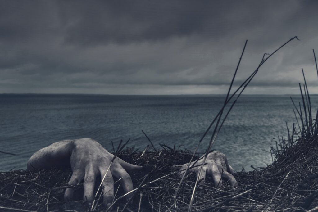
Humans are fascinated by things that scare them, such as death-defying stunts and true crime documentaries, provided these sources of fear at a safe distance. Horror movies are no different, providing a relentless villain, such as Jason in Friday the 13th or a supernatural threat.

For their study into cinematic terror, published in the journal NeuroImage, the researchers first established the 100 best and scariest horror movies of the past century, and how they made people feel.

Unseen threats are the scariest

Firstly, 72% of people report watching at last one horror movie every 6 months, and the reasons for doing so, besides the feelings of fear and anxiety, was primarily that of excitement. Watching horror movies was also an excuse to socialise, with many people preferring to watch horror movies with others than on their own.

People found horror that was psychological in nature and based on real events the scariest, and were far more scared by things that were unseen or implied rather than what they could actually see.

“This latter distinction reflects two types of fear that people experience. The creeping foreboding dread that occurs when one feels that something isn’t quite right, and the instinctive response we have to the sudden appearance of a monster that make us jump out of our skin,” says principal investigator, Professor Lauri Nummenmaa from Turku PET Centre.

MRI reveals different types of fear

Researchers wanted to know how the brain copes with fear in response to this complicated and ever changing environment. The group had people watch two horror movies (The Conjuring 2, 2016, and Insidious, 2010; both directed by James Wan) whilst measuring neural activity in a magnetic resonance imaging scanner.

During those times when anxiety is slowly increasing, regions of the brain involved in visual and auditory perception become more active, as the need to attend for cues of threat in the environment become more important. After a sudden shock, brain activity is more evident in regions involved in emotion processing, threat evaluation, and decision making, enabling a rapid response.

However, these regions are in continuous talk-back with sensory regions throughout the movie, as if the sensory regions were preparing response networks as a scary event was becoming increasingly likely.

“Therefore, our brains are continuously anticipating and preparing us for action in response to threat, and horror movies exploit this expertly to enhance our excitement,” explains Researcher Matthew Hudson.

Source: University of Turku

Researchers Find an Obesity-related Trigger for Diabetes

Obesity
Image source: Pixabay CC0

A new study may help explain how excess weight can contribute to diabetes, which may lead to targeted treatment and prevention. The findings suggest that many people with elevated insulin levels, an early marker of diabetes risk, also have defects in an enzyme important to the processing of a key fatty acid from the diet. The research was published in the journal Cell Metabolism.

“Between 30 million and 40 million people in the United States have Type 2 diabetes, and another 90 million to 100 million have risk factors that make them likely to develop Type 2 diabetes in the future,” said senior investigator Clay F. Semenkovich, MD, at the Washington University School of Medicine in St. Louis. “Many at risk for diabetes have elevated levels of insulin, a hallmark of insulin resistance and a signal that means trouble may be brewing. If we could intervene before they actually develop diabetes, we might be able to prevent significant health problems – such as heart disease, chronic kidney disease, nerve damage, vision loss and other problems – in a great number of people.”

When there is excessive body fat, beta cells in the pancreas ae signalled to secrete more insulin. When insulin levels become elevated and remain high, the body can become resistant to insulin, and eventually the beta cells that secrete insulin can fail, leading to diabetes.

Studying human tissue samples, Washington University researchers found that the overproduction of insulin involves a process called palmitoylation. This is the process by which cells attach the fatty acid palmitate to proteins.

Thousands of human proteins can be attached to palmitate, but the researchers found that when this fatty acid isn’t removed from proteins in beta cells, diabetes is the end result. Examining tissue samples from people who were thin or overweight, and with and without diabetes, the researchers found that the people with diabetes were deficient in an enzyme that removes palmitate from beta cells.

“They hyper-secrete insulin because this process goes awry, and they can’t appropriately regulate the release of insulin from beta cells,” Semenkovich explained. “Regulating insulin release is controlled in part by this palmitoylation process.”

The research team also genetically engineered a mouse that was deficient in the APT1 enzyme, which is responsible for palmitate removal from proteins. The engineered mice went on to develop diabetes.

Because impaired APT1 function contributed to diabetes risk, the researchers worked with the university’s Center for Drug Discovery to screen and identify compounds that can increase the activity of the APT1 enzyme.

“We’ve found several candidate drugs, and we’re pursuing those,” Semenkovich said. “We think that by increasing APT1 activity, we might reverse this process and potentially prevent people at risk from progressing to diabetes.”

Although he said the new findings identifying APT1 as a target are an important step, Semenkovich explained that APT1 is only one treatment target among many.

“There are several ways that Type 2 diabetes may develop,” he said. “This enzyme is not the answer, but it’s an answer, and it appears we have some promising tools that might keep some people with prediabetes from developing diabetes.”

Source: Washington University School of Medicine

Why do Older Fathers Pass on More Mutations?

Photo by Muhammad Daudy on Unsplash

It is not known exactly why older fathers pass on more mutations than younger ones do, even though the male reproductive system is a hotpot for evolution. The mechanisms that might underlie these well-documented trends have long remained a mystery. Now, a new study in the journal Nature Ecology & Evolution describes why older male fruit flies are more likely to pass mutations onto their offspring, which may hold clues for inherited-disease risk in humans.

Researchers in Li Zhao’s lab at Rockefeller University studied mutations that occur during the production of sperm from germline cells, known as spermatogenesis. They found that mutations are common in the testes of both young and old fruit flies, but more abundant in older flies from the outset. Moreover, many of these mutations seem to be removed in younger fruit flies during spermatogenesis by the body’s genomic repair mechanisms – but they fail to be fixed in the testes of older flies.

“We were trying to test whether the older germline is less efficient at mutation repair, or whether the older germline just starts out more mutated,” says first author Evan Witt. “Our results indicate that it’s actually both. At every stage of spermatogenesis, there are more mutations per RNA molecule in older flies than in younger flies.”

Genetic self-repair

Genomes have a few repair mechanisms. When it comes to testes, they have to work overtime; testes have the highest rate of gene expression of any organ. Moreover, genes that are highly expressed in spermatogenesis tend to have fewer mutations than those that are not. This sounds counterintuitive, but it makes sense: One theory to explain why the testes express so many genes holds that it might be a sort of genomic surveillance mechanism – a way to reveal, and then weed out, problematic mutations.

But when it comes to older sperm, the researchers found, the weed-whacker apparently sputters out. Previous research suggests that a faulty transcription-coupled repair mechanism, which only fixes transcribed genes, could be to blame.

Inherited or new mutations?

To get these results, scientists in the Laboratory of Evolutionary Genetics and Genomics did single-cell sequencing on the RNA from the testes of about 300 fruit flies, roughly half of them young (48 hours old) and half old (25 days old), advancing a line of inquiry they began in 2019. In order to understand whether the mutations they detected were somatic, or inherited from the flies’ parents, or de novo they then sequenced the genome of each fly. They were able to document that each mutation was a true original. “We can directly say this mutation was not present in the DNA of that same fly in its somatic cells,” says Witt. “We know that it’s a de novo mutation.”

This unconventional approach – inferring genomic mutations from single-cell RNA sequencing and then comparing them to the genomic data – allowed the researchers to match mutations to the cell type in which they occurred. “It’s a good way to compare mutational load between cell types, because you can follow them throughout spermatogenesis,” Witt says.

Applicability to humans

The next step is to expand the analysis to more age groups of flies and test whether or not this transcription repair mechanism can occur – and if it does, identify the pathways responsible, Witt says. “What genes,” he wonders, “are really driving the difference between old and young flies in terms of mutation repair?”

Because fruit flies have a high reproductive rate, investigating their mutation patterns can offer new insights into the effect of new mutations in human health and evolution, says Zhao.

Witt adds, “It’s largely unknown whether a more mutated male germline is more or less fertile than a less mutated one. There’s not been very much research on it except for at a population level. And if people inherit more mutations from ageing fathers, that increases the odds of de novo genetic disorders or certain types of cancers.”

Source:

Brain Structures Predict Risk of Awareness under Anaesthesia

Photo by Anna Shvets on Pexels

Awareness during anaesthesia is an extremely rare but horrific risk for patients. Now, for the first time, neuroscientists have identified brain structures which could predict an individual’s predisposition this phenomenon. The findings, just published in the journal Human Brain Mapping, could help identify patients who need larger anaesthetic doses.

Although anaesthesia has been used in clinical medicine for over 150 years, scientists do not fully understand why its effect on people is so varied. One in four patients presumed to be unconscious during general anaesthesia may in fact have subjective experiences, such as dreaming. Estimated to occur in 1:1000 to 1:20 000 cases, some patients may have awareness under general anaesthetic. These experiences may range from hearing sounds to the pain of surgery combined with the sensation of suffocation and paralysis in the setting of neuromuscular blockade.

The researchers from Trinity College Dublin found that one in three participants were unaffected by moderate propofol sedation in their response times, thus thwarting a key aim of anaesthesia – the suppression of behavioural responsiveness.

The research also showed, for the first time, that the participants who were resistant to anaesthesia had fundamental differences in the function and structures of the fronto-parietal regions of the brain to those who remained fully unconscious. Crucially, these brain differences could be predicted prior to sedation.

Lorina Naci, Associate Professor of Psychology, Trinity who lead the research said:

“The detection of a person’s responsiveness to anaesthesia prior to sedation has important implications for patient safety and wellbeing. Our results highlight new markers for improving the monitoring of awareness during clinical anaesthesia. Although rare, accidental awareness during an operation can be very traumatic and lead to negative long-term health outcomes, such as post-traumatic stress disorder, as well as clinical depression or phobias.”

“Our results suggest that individuals with larger grey matter volume in the frontal regions and stronger functional connectivity within fronto-parietal brain networks, may require higher doses of propofol to become nonresponsive compared to individuals with weaker connectivity and smaller grey matter volume in these regions.”

The research, conducted in Ireland and Canada, investigated 17 healthy individuals who were sedated with propofol, the most common clinical anaesthetic agent. The participants’ response time to detect a simple sound was measured when they were awake and as they became sedated. Brain activity of 25 participants as they listened to a simple story in both states was also measured.

Source: Trinity College Dublin

Even Placebos Given Openly can Reduce Feelings of Guilt

Photo by Caleb Woods on Unsplash

While guilt is usually an appropriate emotional reaction, usually in response to doing something negative or hurtful, sometimes it can be unwarranted and persistent. Researchers at the University of Basel have shown that placebos can help assuage feelings of guilt, even when the placebo is administered openly, ie the participants are aware of the treatment being a placebo.

Guilt is considered an important moral emotion, as long as it is adaptive – in other words, appropriate and in proportion to the situation. “It can improve interpersonal relationships and is therefore valuable for social cohesion,” says Dilan Sezer, researcher at the Division of Clinical Psychology and Psychotherapy at the University of Basel. Previous research had demonstrated that placebos – even given openly – can still be effective in provoking a beneficial response.

In order to arouse feelings of guilt, healthy participants were recruited and asked to write about a time when they had disregarded important rules of conduct, or treated someone close to them unfairly, hurt or even harmed them. The idea was that the study participants should still feel bad about the chosen situation.

Participants were then randomised to three conditions: Participants in one group received placebo pills with being deceptively told that this was a real medication while participants in another group were told that they are given a placebo. Both groups were told that what they had been given will be effective against feelings of guilt. The control group received no treatment at all. The results, published in Scientific Reports, showed that feelings of guilt were significantly reduced in both placebo groups compared with those without medication.

This was also the case when the subjects knew they had been given a placebo.  “Our study therefore supports the intriguing finding that placebos work even when they are administered openly, and that explanation of the treatment is key to its effectiveness,” states the study’s lead author, Dilan Sezer.

Where feelings of guilt are irrational and continue for longer periods of time, they are considered maladaptive – in other words, disproportionate. These emotions can affect people’s health and are also, among other things, a common symptom of depression.

Scientific studies have shown that placebo effects can be powerful in treating depression. But the finding that open-label placebos can also be useful for such strong emotions as guilt is new. It stands to reason, says Dilan Sezer, that we should try to harness these effects to help those affected. “The administering of open-label placebos, in particular, is a promising approach, as it preserves patient autonomy by allowing patients to be fully aware of how the intervention works.” The results of the study are an initial promising step in the direction of symptom-specific and more ethical treatments for psychological complaints using open-label placebos, Sezer continues.

Further research will need to be done into whether it is possible to treat maladaptive guilt with placebos. And it is still not known whether similar effects are also possible with other feeling states. For Dilan Sezer, one thing is certain: “Using open-label placebos would be an inexpensive and straightforward treatment option for many psychological and physical complaints.”

Source: University of Basel

Heparin Combination Extends Half-life to 24 Hours

Source: CC0

The anticoagulant drug heparin is widely given to patients with blood clotting disorders or after surgery to prevent complications. But it remains difficult to dose correctly, potentially leading to overdosing or underdosing. A team of Penn State researchers combined heparin with a protein fragment, peptide, to slow down the release of the drug and convey the medication directly to the site of a clot. They published their findings in the journal Small.

“We wanted to develop a material that can gradually deliver heparin over time rather than the current iteration that gets cleared from the body in a couple of hours,” said corresponding author Scott Medina, Penn State associate professor of biomedical engineering. “We also wanted to deliver the drug through the skin instead of through an IV.”

When mixed, positively charged peptides and negatively charged heparin bind to create a nanogranular paste that can be injected under the skin, forming a cache of material that is then diffused in the circulatory system and travels to blood clots when they appear. The turbulent flow of fluid near a blood clot triggers the two materials to separate, allowing heparin to begin its anticoagulating action.

“The peptide is ideal for pairing with heparin because it essentially blocks heparin’s action until it is needed in the body,” said Atip Lawanprasert, doctoral student in biomedical engineering and first author on the paper. “The peptide also has some anticoagulating properties on its own: It binds to platelets in the blood, enabling action at the clotting site.”

Without an added bonding agent, heparin applies its anti-clotting properties indiscriminately, not just at blood clot sites, and clears quickly, its half-life only 60 to 90 minutes. Using preclinical animal trials, researchers determined that the addition of peptide allows for a dramatic increase of heparin’s half-life, to up to nearly 24 hours.

“The peptide increases heparin’s effects by more than ten times longer than what is currently being used,” Medina said. “The increased half-life allows for sustained treatments for patients, less medication waste and more accurate dosing. Eventually, this could allow the medication to be injected under the skin just once a day, rather than an all-day IV drip.”

Next, researchers plan to replicate the study in a clinical setting, as well as study the effect of the medication’s toxicity with multi-day administration.

Source: Penn State

Mucosal Vaccines Could be the Next Step Against Respiratory Viruses

Image by Arek Socha from Pixabay

Vaccines that provide long-lasting protection against influenza, coronaviruses and respiratory syncytial virus (RSV) have proved exceptionally difficult to develop. In a new review article in Cell Host & Microbe, NIH researchers explore the challenges and outline approaches to improved vaccines and describe a promising path forward: mucosal vaccines.

Unlike the respiratory viruses that cause measles, mumps and rubella — for which vaccination or recovery from illness provides decades-long protection against future infection – flu, RSV, SARS-CoV-2 and “common cold” coronaviruses share several characteristics that enable them to cause repeated re-infections. These include very short incubation periods, rapid host-to-host transmission and replication in the nasal mucosa rather than throughout the body. This last feature – non-systemic replication – means these viruses do not stimulate the full force of the adaptive immune response, which typically takes a week or more to mount.

A next generation of improved vaccines for mucosa-replicating viruses will require advances in understanding on several fronts, the authors say. For instance, more must be learned about interactions between flu viruses, coronaviruses and RSV and the components of the immune response that operate largely or exclusively in the upper respiratory system. Over time, these interactions have evolved and led to “immune tolerance,” wherein the human host tolerates transient, limited infections by viruses that are generally non-lethal to avoid the destructive consequences of an all-out immune system attack.

The authors note that mucosal immunisation appears to be an optimal route of vaccination for the viruses of interest, when feasible. However, to develop useful mucosal vaccines, significant knowledge gaps must be filled including finding ideal vaccine formulations; determining dosage size, frequency and timing; and developing techniques for overcoming immune tolerance.

Source: NIH/National Institute of Allergy and Infectious Diseases

Artificial Pancreas Successfully Trialled for Type 2 Diabetes

Diabetes - person measures blood glucose
Photo by Photomix Company from Pexels

Cambridge scientists have successfully trialled an artificial pancreas for use by patients living with type 2 diabetes. They report in Nature Medicine that the device doubled the amount of time patients were in the target range for glucose compared to standard treatment and halved the time spent experiencing high glucose levels.

The artificial pancreas developed by University of Cambridge researchers combines an off-the-shelf glucose monitor and insulin pump with an app developed by the team, known as CamAPS HX. This app is run by an algorithm that predicts how much insulin is required to maintain glucose levels in the target range.

The researchers have previously shown that an artificial pancreas run by a similar algorithm is effective for patients living with type 1 diabetes, from adults through to very young children. They have also successfully trialled the device in patients with type 2 diabetes who require kidney dialysis.

Today, in Nature Medicine, the team report the first trial of the device in a wider population living with type 2 diabetes (not requiring kidney dialysis). Unlike the artificial pancreas used for type 1 diabetes, this new version is a fully closed loop system, whereas patients with type 1 diabetes need to tell their artificial pancreas that they are about to eat to allow adjustment of insulin, for example, with this version they can leave the device to function entirely automatically.

The researchers recruited 26 patients who were randomised to one of two groups – the first group would trial the artificial pancreas for eight weeks and then switch to the standard therapy of multiple daily insulin injections; the second group would take this control therapy first and then switch to the artificial pancreas after eight weeks.

The team used several measures to assess how effectively the artificial pancreas worked. The first was the proportion of time that patients spent with their glucose levels within a target range of between 3.9 and 10.0mmol/L. On average, patients using the artificial pancreas spent two-thirds (66%) of their time within the target range, compared to control (32%).

A second measure was the proportion of time spent with glucose levels above 10.0mmol/L. Over time, high glucose levels raise the risk of potentially serious complications. Patients taking the control therapy spent two-thirds (67%) of their time with high glucose levels — this was halved to 33% when using the artificial pancreas.

Average glucose levels fell from 12.6mmol/L when taking the control therapy to 9.2mmol/L while using the artificial pancreas.

The app also reduced levels of a molecule known as glycated haemoglobin, or HbA1c. Glycated haemoglobin develops when haemoglobin, a protein within red blood cells that carries oxygen throughout the body, joins with glucose in the blood, becoming ‘glycated’. By measuring HbA1c, clinicians are able to get an overall picture of what a person’s average blood sugar levels have been over a period of weeks or months. For people with diabetes, the higher the HbA1c, the greater the risk of developing diabetes-related complications. After the control therapy, average HbA1c levels were 8.7%, while after using the artificial pancreas they were 7.3%.

No patients experienced dangerously-low blood sugar levels (hypoglycaemia) during the study. One patient was admitted to hospital while using the artificial pancreas, due to an abscess at the site of the pump cannula.

Dr Charlotte Boughton from the Wellcome-MRC Institute of Metabolic Science at the University of Cambridge, who co-led the study, said: “Many people with type 2 diabetes struggle to manage their blood sugar levels using the currently available treatments, such as insulin injections. The artificial pancreas can provide a safe and effective approach to help them, and the technology is simple to use and can be implemented safely at home.”

Dr Aideen Daly, also from the Wellcome-MRC Institute of Metabolic Science, said: “One of the barriers to widespread use of insulin therapy has been concern over the risk of severe ‘hypos’ — dangerously low blood sugar levels. But we found that no patients on our trial experienced these and patients spent very little time with blood sugar levels lower than the target levels.”

Feedback from participants suggested that participants were happy to have their glucose levels controlled automatically by the system, and nine out of ten (89%) reported spending less time managing their diabetes overall. Users highlighted the elimination of the need for injections or fingerprick testing, and increased confidence in managing blood glucose as key benefits. Downsides included increased anxiety about the risk of hypoglycaemia, which the researchers say may reflect increased awareness and monitoring of glucose levels, and practical annoyances with wearing of devices.

The team now plan to carry out a much larger multicentre study to build on their findings and have submitted the device for regulatory approval with a view to making it commercially available for outpatients with type 2 diabetes.

Source: University of Cambridge

Complex Surface Features on B Cells may Hold Immune Secrets

Image credit: Albert Ludwigs University of Freiburg

Using new microscopic methods in combination with machine learning-based image analysis, researchers from Albert Ludwigs University of Freiburg have discovered new structures on the surface of living B cells that affect the distribution and possibly the function of their antigen receptors. The researchers’ study has been published in The EMBO Journal.

B cells recognise pathogens through specialised receptors on their surface. For the first time, scientists were able to observe how these receptors are distributed on the surface of living and moving cells. They found that the B cell surface is shaped into a characteristic landscape of interconnected ridges and protrusions. On this landscape, the IgM-class B cell antigen receptors (IgM-BCR) accumulate in specific areas. The precision of the receptors’ localisation and their clustering into larger units likely constitute a mechanism that controls receptor signalling and facilitates antigen sensing and thereby the activation of B cells.

In most immunological textbooks, lymphocytes are depicted as round, ball-like cells whose smooth surface carries randomly distributed receptors. The notion of a smooth unstructured B cell surface has already been challenged by electron micrographs of fixed and frozen lymphocytes, revealing thin membrane protrusions called microvilli on the cells’ surface. These tentacle-like structures help immune cells to search for molecular markers of pathogens, so-called antigens. B lymphocytes recognise such antigens through different classes of their B cell antigen receptors (BCR). These antigen receptors are complex molecular machines that, when activated, interact with other molecules to initiate a signalling cascade, leading to the differentiation of B cells into plasma cells and the production of protective antibodies.

The research group of Prof. Dr. Michael Reth collaborated with other imaging specialists to analyse how the IgM-BCR is distributed across the 3D surface of living B cells. For this, they used a technique called lattice light sheet microscopy, LLSM for short. “This method can capture volumetric images of living cells at a very high speed,” explains Dr. Deniz Saltukoglu from Freiburg University, the first author of the study. “In other types of high-resolution microscopy, cells need to be attached to a flat surface, which completely alters the B cells’ outer structures. LLSM allowed us to observe the cells in an environment that mimics biological tissues, meaning that the structures and movements that we saw were largely undisturbed,” she says.

The researchers then developed custom image analysis tools to quantify and objectively characterise the microscopic data. “We needed to segment the images and isolate morphological features,” says Saltukoglu. “So far this had only been done with two-dimensional data, so we had to develop new computational tools for volumetric, time course data.” For this, the researchers drew inspiration from algorithms that are used to map geographical data for archaeological surveys. With this approach, they found that the B cell surface carries a network of elevated ridges, with microvilli growing from the intersections of the network. Within this “cellular landscape”, the IgM-BCRs form clusters that concentrate along the ridges, in close proximity to the bases of the microvilli. The position of these clusters was linked with the dynamic movement of the ridges on the cells’ surface.

“We think that the 3-D location of the antigen receptors controls their activity,” says Reth. “Localisation at the microvilli base may prevent their unwanted activation. Once B cells receive a danger signal, they extent their microvilli and we assume that the IgM-BCR clusters then get recruited to the tip where they are localized in an optimal position for antigen sensing.”

This hypothesis is in line with other findings from Reth’s group, which suggest that the IgM-BCRs are regulated via lateral interactions with regulatory coreceptors. This means that the position and distribution of antigen receptors likely represent additional control mechanisms that affect signalling and activation of cells of the immune system.

Source: Albert Ludwigs University of Freiburg

Patients Themselves may be the Source of New Strains of HAIs

E. Coli bacteria. Image by CDC

Despite stringent infection-control efforts around the world, hospital-acquired infections (HAIs)keep on popping up from new strains of bacteria. In Science Translational Medicine, researchers report evidence pointing to an unexpected source of such bacteria: the hospitalised patients themselves.

From experiments with mice, researchers at Washington University School of Medicine in St. Louis discovered that urinary tract infections (UTIs) can arise after sterile tubes, called catheters, are inserted into the urinary tract, even when no bacteria are detectable in the bladder beforehand. Such tubes are commonly used in hospitals to empty the bladders of people undergoing surgery. In the mice, inserting the tubes activated dormant Acinetobacter baumannii bacteria hidden in bladder cells, triggering them to emerge, multiply and cause UTIs, the researchers said.

The findings suggest that screening patients for hidden reservoirs of dangerous bacteria could supplement infection-control efforts and help prevent deadly HAIs.

“You could sterilise the whole hospital, and you would still have new strains of A. baumannii popping up,” said co-senior author Mario Feldman, PhD, a professor of molecular microbiology. “Cleaning is just not enough, and nobody really knows why. This study shows that patients may be unwittingly carrying the bacteria into the hospital themselves, and that has implications for infection control. If someone has a planned surgery and is going to be catheterised, we could try to determine whether the patient is carrying the bacteria and cure that person of it before the surgery. Ideally, that would reduce the chances of developing one of these life-threatening infections.”

The notoriously multidrug-resistant A. baumannii is a major threat to patients, causing many cases of UTIs in people with urinary catheters, pneumonia in people on ventilators, and bloodstream infections in people with central-line catheters into their veins.

The researchers set out to investigate why so many A. baumannii UTIs develop after people receive catheters.

Most UTIs among otherwise healthy people are caused by the bacterium Escherichia coli. Research has shown that E. coli can hide out in bladder cells for months after a UTI seems to have been cured, and then re-emerge to cause another infection.

The researchers investigated whether A. baumannii can hide inside cells like E. coli can. They studied mice with UTIs caused by A. baumannii. They used mice with weakened immune systems because, like people, healthy mice can fight off A. baumannii.

Once the infections had resolved and no bacteria were detected in the mice’s urine for two months, the researchers inserted catheters into the mice’s urinary tracts with a sterile technique. Within 24 hours, about half of the mice developed UTIs caused by the same strain of A. baumannii as the initial infection.

“The bacteria must have been there all along, hiding inside bladder cells until the catheter was introduced,” said co-senior author Scott J. Hultgren, PhD, a professor and expert on UTIs. “Catheterisation induces inflammation, and inflammation causes the reservoir to activate, and the infection blooms.”

Since A. baumannii rarely causes symptoms in otherwise healthy people, many people who carry the bacteria may never know they’re infected, the researchers said. According to the researchers’ literature search, 2% of healthy people carry A. baumannii in their urine.

“I wouldn’t put much weight on the precise percentage, but I think we can say with certainty that some percentage of the population is walking around with A. baumannii,” Feldman said. “As long as they’re basically healthy, it doesn’t cause any problems, but once they’re hospitalised, it’s a different matter. This changes how we think about infection control. We can start considering how to check if patients already have Acinetobacter before they receive certain types of treatment; how we can get rid of it; and if other bacteria that cause deadly outbreaks in hospitals, such as Klebsiella, hide in the body in the same way. That’s what we’re working on figuring out now.”

Source: Washington University School of Medicine