Tag: white matter

Childhood Experiences Shape White Matter with Cognitive Effects Seen Years Later

Photo by Caleb Woods on Unsplash

Mass General Brigham investigators have linked difficult early life experiences with reduced quality and quantity of the white matter communication highways throughout the adolescent brain. This reduced connectivity is also associated with lower performance on cognitive tasks. However, certain social resiliency factors like neighbourhood cohesion and positive parenting may have a protective effect. Results are published in Proceedings of the National Academy of Sciences (PNAS).

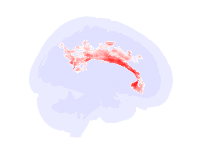
White matter are the communication highways that allow the brain networks to carry out the necessary functions for cognition and behaviour. They develop over the course of childhood, and childhood experiences may drive individual differences in how white matter matures. Lead author Sofia Carozza, PhD, and senior author Amar Dhand, MD, PhD, of the Department of Neurology at Brigham and Women’s Hospital, a founding member of the Mass General Brigham healthcare system, wanted to understand what role this process plays in cognition once children reach adolescence.

“The aspects of white matter that show a relationship with our early life environment are much more pervasive throughout the brain than we’d thought. Instead of being just one or two tracts that are important for cognition, the whole brain is related to the adversities that someone might experience early in life,” said Carozza.

The team studied data from 9082 children (about half of them girls, with an average age of 9.5) collected in the Adolescent Brain Cognitive Development (ABCD) study. This study, funded by the National Institutes of Health and conducted at 21 centres across the U.S., gathered information on brain activity and structure, cognitive abilities, environment, mood and mental health. The researchers looked at several categories of early environmental factors, including prenatal risk factors, interpersonal adversity, household economic deprivation, neighbourhood adversity, and social resiliency factors.

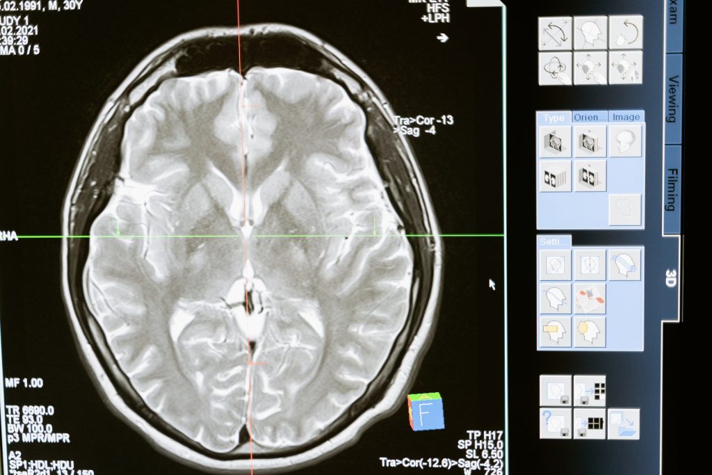
Carozza and Dhand used diffusion imaging scanning of the brain to measure fractional anisotropy (FA)—a way of estimating the integrity of the white matter connections—and streamline count, an estimate of their strength. They then used a computational model to compare how these features of white matter were related to both childhood environmental factors and current cognitive abilities such as language skills and mental arithmetic.

Their analysis revealed widespread differences in white matter connections throughout the brain depending on the children’s early-life environments. In particular, the researchers found lower quality of white matter connections in parts of the brain tied to mental arithmetic and receptive language. These white matter differences accounted for some of the relationship between adverse life experiences in early childhood and lower cognitive performance in adolescence.

“We are all embedded in an environment, and features of that environment such as our relationships, home life, neighbourhood, or material circumstances can shape how our brains and bodies grow, which in turn affects what we can do with them,” said Carozza. “We should work to make sure that more people can have those stable, healthy home lives that the brain expects, especially in childhood.”

The researchers note that their study is based on observational data, which means they cannot draw strong causal conclusions. Brain imaging was also only available at a single timepoint, offering a snapshot but not allowing researchers to track changes over time. Prospective studies—following children over time and collecting brain imaging information at multiple time points—would be needed to more definitively connect adversity and cognitive performance.

Source: Mass General Brigham

Sex Differences in Brain Structure Present at Birth

Photo by Chayene Rafaela on Unsplash

Sex differences in brain structure are present from birth, research from the Autism Research Centre at the University of Cambridge has shown.

While male brains tended to be greater in volume than female brains, when adjusted for total brain volume, female infants on average had significantly more grey matter, while male infants on average had significantly more white matter in their brains.

Grey matter is made up of neuron cell bodies and dendrites and is responsible for processing and interpreting information, such as sensation, perception, learning, speech, and cognition.  White matter is made up of axons, which are long nerve fibres that connect neurons together from different parts of the brain. 

Yumnah Khan, a PhD student at the Autism Research Centre, who led the study, said: “Our study settles an age-old question of whether male and female brains differ at birth. We know there are differences in the brains of older children and adults, but our findings show that they are already present in the earliest days of life.

“Because these sex differences are evident so soon after birth, they might in part reflect biological sex differences during prenatal brain development, which then interact with environmental experiences over time to shape further sex differences in the brain.”

One problem that has plagued past research in this area is sample size. The Cambridge team tackled this by analysing data from the Developing Human Connectome Project, where infants receive an MRI brain scan soon after birth. Having over 500 newborn babies in the study means that, statistically, the sample is ideal for detecting sex differences if they are present.

A second problem is whether any observed sex differences could be due to other factors, such as differences in body size.  The Cambridge team found that, on average, male infants had significantly larger brain volumes than did females, and this was true even after sex differences in birth weight were taken into account.

After taking this difference in total brain volume into account, at a regional level, females on average showed larger volumes in grey matter areas related to memory and emotional regulation, while males on average had larger volumes in grey matter areas involved in sensory processing and motor control.

The findings of the study, the largest to date to investigate this question, are published in the journal Biology of Sex Differences.

Dr Alex Tsompanidis who supervised the study, said: “This is the largest such study to date, and we took additional factors into account, such as birth weight, to ensure that these differences are specific to the brain and not due to general size differences between the sexes.

“To understand why males and females show differences in their relative grey and white matter volume, we are now studying the conditions of the prenatal environment, using population birth records, as well as in vitro cellular models of the developing brain. This will help us compare the progression of male and female pregnancies and determine if specific biological factors, such as hormones or the placenta, contribute to the differences we see in the brain.”

The researchers stress that the differences between males and females are average differences.

Dr Carrie Allison, Deputy Director of the Autism Research Centre, said: “The differences we see do not apply to all males or all females, but are only seen when you compare groups of males and females together. There is a lot a variation within, and a lot of overlap between, each group.”  

Professor Simon Baron-Cohen, Director of the Autism Research Centre, added: “These differences do not imply the brains of males and females are better or worse. It’s just one example of neurodiversity. This research may be helpful in understanding other kinds of neurodiversity, such as the brain in children who are later diagnosed as autistic, since this is diagnosed more often in males.”

The research was funded by Cambridge University Development and Research, Trinity College, Cambridge, the Cambridge Trust, and the Simons Foundation Autism Research Initiative.

Reference
Khan, Y.T., Tsompanidis, A., Radecki, M.A. et al. Sex differences in human brain structure at birth. Biol Sex Differ; 17 Oct 2024; DOI: 10.1186/s13293-024-00657-5

Republished under a Creative Commons Attribution-NonCommercial-ShareAlike 4.0 International License.

Source: University of Cambridge

White Matter may Aid Recovery from Spinal Cord Injuries

View of the spinal cord. Credit: Scientific Animations CC4.0

Injuries, infection and inflammatory diseases that damage the spinal cord can lead to intractable pain and disability but some degree of recovery may be possible. The question is, how best to stimulate the regrowth and healing of damaged nerves.

At the Vanderbilt University Institute of Imaging Science (VUIIS), scientists are focusing on a previously understudied part of the brain and spinal cord – white matter, which is made up of axons that relay signals. Their discoveries could lead to treatments that restore nerve activity through the targeted delivery of electromagnetic stimuli or drugs.

In a recent paper published in the Proceedings of the National Academy of SciencesAnirban Sengupta, PhD, John Gore, PhD, and their colleagues report the detection of signals from white matter in the spinal cord in response to a stimulus that are as robust as grey matter signals.

“In the spinal cord, the white matter signal is quite large and detectable, unlike in the brain, where it has less amplitude than the grey matter (signal),” said Sengupta, research instructor in Radiology and Radiological Sciences at Vanderbilt University Medical Center.

“This may be due to the larger volume of white matter in the spinal cord compared to the brain,” he added. Alternatively, the signal could represent “an intrinsic demand” in metabolism within the white matter, reflecting its critical role in supporting grey matter.

For several years, Gore, who directs the VUIIS, and his colleagues have used functional magnetic resonance imaging (fMRI) to detect blood oxygenation-level dependent (BOLD) signals, a key marker of nervous system activity, in white matter.

Last year, they reported that when participants undergoing fMRI perform a task, like wiggling their fingers, BOLD signals increase in white matter throughout the brain.

The current study monitored changes in BOLD signals in the white matter of the spinal cord at rest and in response to a vibrotactile stimulus applied to the fingers in an animal model. In response to stimulation, white matter activity was higher in “tracts” of ascending fibres that carry the signal from the spine to the brain.

This result is consistent with white matter’s known neurobiological function, the researchers noted. White matter contains non-neuronal glial cells that do not produce electrical impulses, but which regulate blood flow and neurotransmitters, the signaling molecules that transmit signals between nerve cells.

Much remains to be learned about the function of white matter in the spinal cord. But the findings from this research may help in improved understanding of diseases that affect white matter in the spinal cord, including multiple sclerosis, Sengupta said.

“We will be able to see how activity in the white matter changes in different stages of the disease,” he said. Researchers also may be able to monitor the effectiveness of therapeutic interventions, including neuromodulation, in promoting recovery following spinal cord injury.

Source: Vanderbilt University Medical Center

Significant White Matter Changes in Autism Revealed by MRI

Significant alterations in the brain’s white matter in adolescents with autism spectrum disorder (ASD). Credit: RSNA and researcher, Clara Weber

Using specialised MRI, researchers found significant changes in the microstructure of the brain’s white matter, especially in the corpus callosum in adolescents and young adults with autism spectrum disorder (ASD) compared to controls. This research will be presented next week at the annual meeting of the Radiological Society of North America (RSNA).

“One in 68 children in the U.S. is affected by ASD, but high variety in symptom manifestation and severity make it hard to recognise the condition early and monitor treatment response,” explained Clara Weber, postgraduate research fellow at Yale University School of Medicine. “We aim to find neuroimaging biomarkers that can potentially facilitate diagnosis and therapy planning.”

Researchers reviewed diffusion tensor imaging (DTI) brain scans from a large dataset of patients between the age of six months and 50 years. DTI is an MRI technique that measures connectivity in the brain by detecting how water moves along its white matter tracts. Water molecules diffuse differently through the brain, depending on the integrity, architecture and presence of barriers in tissue.

“If you think of gray matter as the computer, white matter is like the cables,” Weber said. “DTI helps us assess how connected and intact those cables are.”

For the study, clinical and DTI data from 583 patients from four existing studies of distinct patient populations were analysed: infants (median age 7 months), toddlers (median age 32 months), adolescents, and young adults.

“One of the strengths of our study is that we looked at a wide range of age groups, not just school-aged children,” Weber said.

To assess the influences of age and ASD diagnosis on white matter microstructure, the research team created fractional anisotropy, mean diffusivity and radial diffusivity maps using data from the four studies.

Fractional anisotropy is the extent water diffusion is restricted to just one direction. A value of zero means that diffusion is unrestricted in all directions, while one means that diffusion is unidirectional. Mean diffusivity is the overall mobility of water molecules, indicating how densely cells are packed together. Radial diffusivity is the extent water diffuses perpendicular to a white matter tract.

“When white matter integrity is disrupted, we see more water diffusing perpendicularly, which translates to a higher radial diffusivity,” Weber said.

The key finding of the analysis was reduced fractional anisotropy within the anterior/middle tracts of the corpus callosum in adolescent and young adult ASD patients compared to individuals in the control group. The corpus callosum is a thick bundle of nerve fibers that connects and allows the two sides of the brain to communicate. Corresponding increases in ASD-related mean diffusivity and radial diffusivity were found in young adults.

“In adolescents, we saw a significant influence of autism,” Weber said. “In adults, the effect was even more pronounced. Our results support the idea of impaired brain connectivity in autism, especially in tracts that connect both hemispheres.”

Compared to controls, no reduction in fractional anisotropy was seen in the same tracts in toddlers and infants with ASD.

The researchers hope the findings can help improve early diagnosis of ASD and provide potential objective biomarkers to monitor treatment response.

“We need to find more objective biomarkers for the disorder that can be applied in clinical practice,” Weber said.

Source: EurekAlert!

White Matter Changes Uncovered in Repeated Brain Injury

Photo by MART PRODUCTION from Pexels

A new study has uncovered insights into white matter changes that occur during chronic traumatic encephalopathy (CTE), a progressive brain disease associated with repetitive head impacts. This discovery may help in identifying new targets for therapies.

CTE been diagnosed after death in the brains of American football players and other contact sport athletes as well as members of the armed services. The disease has been identified as causing impulsivity, explosivity, depression, memory impairment and executive dysfunction.

Though much prior research focused on repetitive head trauma leading to the development of abnormal tau, this study focused on white matter changes, particularly the oligodendrocytes which myelinate nerve sheaths. The results have been published online [PDF] in the journal Acta Neuropathologica.

“Research to date has focused on the deposition of abnormal tau in the gray matter in CTE. This study shows that the white matter undergoes important alterations as well.  There is loss of oligodendrocytes and alteration of oligodendrocyte subtypes in CTE that might provide new targets for prevention and therapies,” explained corresponding author Ann McKee, MD, chief of neuropathology at VA Boston Healthcare, director of the BU CTE Center.

Dr McKee and her team isolated cellular nuclei from the postmortem dorsolateral frontal white matter in eight cases of CTE and eight matched controls. They conducted single-nucleus RNA-seq (snRNA-seq) with these nuclei, revealing transcriptomic, cell-type-specific differences between the CTE and control cases. In doing so, they discovered that the white matter in CTE had fewer oligodendrocytes and the oligodendroglial subtypes were altered compared to control tissue.

Since previous studies have largely focused on the CTE-specific tau lesion located in the cortex in the brain, these findings are particularly informative as they explain a number of features of the disease. “In comparison, the cellular death process occurring in white matter oligodendrocytes in CTE appears to be separate from the accumulation of hyperphosphorylated tau,” she said. “We know that the behavioural and mood changes that occur in CTE are not explained by tau deposition. This study suggests that white matter alterations are also important features of the disease, and future studies will determine whether these white matter changes play a role in the production of behavioral or mood symptoms in CTE, such as explosivity, violence, impulsivity, and depression.”

Source: Boston University School of Medicine

Journal information: Chancellor, K. B., et al. (2021) Altered oligodendroglia and astroglia in chronic traumatic Encephalopathy. Acta Neuropathologica. doi.org/10.1007/s00401-021-02322-2.