

Tag: radiotherapy

FLASH Radiation Treatment for Tumours a Step Closer
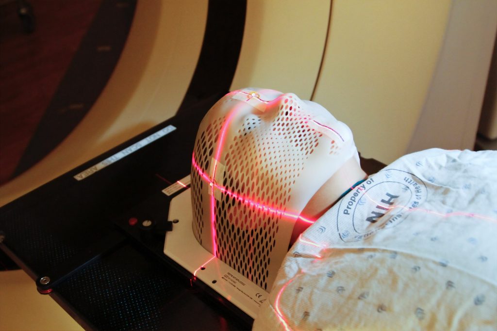
Heavy ion bombardment in FLASH radiation treatment could be the future of radiotherapy, with encouraging findings from a German lab.

The GSI Helmholtzzentrum für Schwerionenforschung and the future accelerator centre FAIR succeeded in performing a carbon ion FLASH experiment for the first time in their Phase 0 experiment. 

The scientists involved were able to achieve the very high dose rates required to irradiate tumours. The success was a collective effort of the GSI Biophysics Department and the accelerator crew on the GSI/FAIR campus in close collaboration with the German Cancer Research Center DKFZ and the Heidelberg Ion Therapy (HIT) center.

FLASH irradiation involves utra-short and ultra-high radiation, delivering the treatment dose in fractions of a second. Traditional radiation therapy, as well as proton or ion therapy, deliver smaller doses of radiation to a patient over an extended time period, whereas FLASH radiotherapy is thought to require only a few short irradiations, all lasting less than 100 milliseconds.

In the field of electron radiation, recent in vivo investigations have shown that the FLASH method’s ultra-high dose rate is less harmful to healthy tissue, but just as efficient as conventional dose-rate radiation to inhibit tumour growth. Such an effect has not yet been demonstrated for proton and for ion beam irradiation, which is the basis of the tumour therapy with carbon ions developed at GSI. There is still a lot of research to be done here. The results of the current experiment at GSI are now being evaluated and will contribute to new knowledge.

There have also been technical barriers to FLASH radiation. Until now, FLASH technique has only been applicable using electron and proton accelerators. While the required dose rates for electrons and protons can be achieved with a cyclotron (circular accelerator), this is more difficult with the synchrotrons required in heavy ion therapy, such as the SIS18 at GSI.

That is why the current FAIR Phase 0 experiment is a very crucial step: Thanks to the improvements at the existing GSI accelerator facility as part of the preparations for FAIR, the necessary dose rate in millisecond range can now also be achieved for carbon ions. However,  much development work remains to be done before this procedure is mature enough to be routinely used on patients in the field of electron radiation.

Professor Marco Durante, Head of the GSI Biophysics Research Department, was very pleased with this important milestone in  the development of FLASH irradiation:

“It is a forward-looking method that could significantly increase the therapeutic window in radiotherapy. I am very happy that the researchers and the accelerator team were able to demonstrate the possibility to create conditions with carbon beams that are necessary for FLASH therapy of tumors. If we can combine the great effect and precision of heavy ion therapy with FLASH irradiation while maintaining efficacy and causing little damage to healthy tissue, this could pave the way of a future radiation therapy several years from now.”

Professor Paolo Giubellino, The Scientific Managing Director of GSI and FAIR, expressed his delight at the results: “The combination of expertise in biophysics and medicine as well as engineering excellence allows the first world-class experiments FLASH irradiation with ion beams to be performed. This could result in important complements to existing radiation therapies. Applications in tumour therapy are one of the research areas that can benefit from the recent increased intensities of GSI accelerators. However, modern radiobiology will substantially benefit from beams with even higher intensities, such as we will have at the FAIR facility currently under construction. FLASH is a first example of these future directions of work”.

Source: GSI Helmholtzzentrum für Schwerionenforschung GmbH

New Radiotherapy Treatment for Metastatic Cancers is Safe

MRI machine. Image by Michal Jarmoluk from Pixabay

A phase I trial showed that stereotactic body radiotherapy (SBRT) proved safe for treating cancer patients with multiple metastases. However, some late adverse effects underscored the need for long-term follow-up.

SBRT is a new radiotherapy treatment method that delivers high doses of ionising radiation to tumours with great precision, minimising damage to other parts of the body while killing tumour cells.

Increasingly used by clinicians, SBRT can improve survival in cancer patients with multiple metastases, explained first author Steve Chmura, MD, PhD, of University of Chicago Medicine, and colleagues, but there is little evidence as to its safety in this application.

“Existing data include mostly treatment of 1 or 2 metastases separated widely from each other and use of differing radiation doses, toxicity reporting, image guidance, and normal tissue constraints,” they wrote. “Given the critical need, NRG Oncology NRG-BR001 trial sought to determine the safety of delivering curative-intent SBRT to patients with 3 to 4 metastases or 2 metastases within close proximity to each other.”

Patients in the study had metastatic breast, prostate, or non-small-cell lung carcinoma (NSCLC). Each metastasis was assigned to a metastatic location based on the potential for toxicity.

Dose-limiting toxicity (DLT; side effects severe enough to discontinue treatment) was the primary study outcome, defined as specific AEs of grades 3 to 5 related to SBRT within 180 days of treatment. Dose levels were considered to be safe if DLTs were seen in no more than one of six patients per location with metastases.

Of the evaluable 35 patients, 12 (34.3%) had breast cancer, 10 NSCLC (28.6%), and 13 (37.1%) prostate cancer, with a median of three metastases per patient.

DLT analysis WAS based on six evaluable patients in all of the metastatic locations save the liver (five evaluable patients). The authors reported there were no protocol-specified DLTs in any of the seven metastatic locations within 180 days of the initiation of treatment.

There were 50 grade 3 or 4 AEs reported in 18 patients, and eight were deemed to be linked to the treatment. Of those eight, six (including bone pain, pulmonary fibrosis, bronchial fistula, bronchial stenosis, spinal fracture, and humeral fracture) were reported in six patients over 180 days from the start of the treatment. No treatment-related deaths occurred, according to the authors.

The authors suggested that, with the number of late AEs reported in this trial, patients should be monitored closely for late toxic effects.

“Given the potential for ablative radiotherapy to improve outcomes of patients with oligometastatic cancer, the finding that SBRT is safe when delivered to 3 to 4 metastases or 2 metastases in close proximity to one another is important, and serves as the foundation for ongoing randomized trials,” wrote the authors. They noted that these include studies such as the phase II/III NCI-sponsored NRG-BR002 trial.

Source: MedPage Today

Journal information: Chmura S, et al “Evaluation of safety of stereotactic body radiotherapy for the treatment of patients with multiple metastases” JAMA Oncol 2021; DOI: 10.1001/jamaoncol.2021.0687.

Tumour Weakened against Radiation by Tweaking a Certain Protein

The vulnerability of specific tissues to ionising radiation has been linked to the time-varying levels of a tumour-suppressing protein, opening new avenues for cancer combination therapy.

The ability for cells to survive radiation damage has been known to be connected to p53, but tissues with vastly different levels of p53 have been shown to have great differences in sensitivity. In the face of this apparent paradox, researchers from Blavatnik Institute at Harvard Medical School, Massachusetts General Hospital, and the Novartis Institutes for BioMedical Research investigated the behaviour of p53 in irradiated tissues.

“Dynamics matter. How things change over time matters,” said co-corresponding author Galit Lahav, the Novartis Professor of Systems Biology at HMS. “Our ability to understand biology is limited when we only look at snapshots. By seeing how things evolve temporally, we gain much richer information that can be critical for dissecting diseases and creating new therapies.”

Ionising radiation randomly damages a cell’s molecular machinery, causing it to initiate cell death if it is too serious. The arbitrator of cellular suicide is p53, which is also involved in tumour suppression. 

The findings opened new avenues for combination cancer therapies, as they discovered that administering a drug that blocks p53 levels from oscillating resulted in tumours in mice being more susceptible to radiation. 
Administering to the mice an experimental anti-cancer drug that inhibits MDM2, a protein which degrades p53, they forced p53 to stay elevated. The large intestine, which is normally radiation resistant, showed increased vulnerability.

Testing out this enhanced vulnerability on human tumours transplanted into mice, a significant tumour shrinkage was seen following radiation and then MDM2 inhibitor administration.

Co-corresponding author Galit Lahav, Novartis Professor of Systems Biology, Harvard Medical School explained: “By irradiating first, we force the cancer cells to activate p53, and by adding MDM2 inhibitor on top of that, we can keep p53 active longer. This combination has a much stronger effect than either alone.”

The findings showed the importance of understanding the role of p53 in cancer, the dynamic nature of which is not being looked at in studies testing MDM2. More research into the biological pathways of p53 is called for. 
“For a lab studying p53, cancer is always a major motivation. Our goal is to acquire knowledge to help develop better and more efficient therapies,” Lahav said. “Understanding how p53 behaves over time in different conditions is a critical piece of the puzzle.”

Source: News-Medical.Net

Journal information: Stewart-Ornstein, J., et al. (2021) p53 dynamics vary between tissues and are linked with radiation sensitivity. Nature Communications.doi.org/10.1038/s41467-021-21145-z