Tag: mitochondria

Topping up Mitochondrial Content to Fight Kidney Cancer

Anatomic model of a kidney
Photo by Robina Weermeijer on Unsplash

Researchers at Karolinska Institutet in Sweden have linked resistance to treatment for VHL syndrome-induced kidney cancer to low mitochondrial content in the cell. When the researchers increased the mitochondrial content with an inhibitor, the cancer cells responded to the treatment. Their findings, which are published in Nature Metabolism, may lead to more targeted cancer drugs.

Mitochondria are the most oxygen-demanding component of the cell, but it was not known how mitochondria adapt in a low-oxygen environment and how they are linked to cancer therapy resistance.

“We’ve shown for the first time how the formation of new mitochondria is regulated in cells that lack oxygen and how this process is altered in cancer cells with VHL mutations,” explained Associate Professor Susanne Schlisio, group leader at the Karolinska Institutet.

A gene called von Hippel-Lindau (VHL) prevents healthy cells from turning cancerous. The 2019 Nobel Prize in Physiology or Medicine was awarded to the discovery that VHL was part of the cell’s oxygen detection system. Normally, VHL breaks down another protein called HIF – but when VHL is mutated, HIF accumulates and causes a disease called VHL syndrome in which the cells react as if they were lacking oxygen. This syndrome greatly increases the risk of tumours, both benign and malignant. VHL syndrome-induced kidney cancer has a poor prognosis, with a five-year survival rate of just 12%.

Researchers analysed the protein content of cancer cells from patients with different variants of VHL syndrome, to see how they differed from another group of individuals with a special VHL mutation called Chuvash, a mutation involved in hypoxia-sensing disorders without any tumour development. Those with the Chuvash VHL-mutation had normal mitochondria in their cells, while those with VHL syndrome mutation had few.

To increase the amount of mitochondrial content in VHL related kidney cancer cells, the researchers treated these tumours with an inhibitor of a mitochondrial protease called “LONP1.” This resulted in the cells becoming susceptible to the cancer drug sorafenib, which they had previously resisted. In mouse studies, this combination treatment led to reduced tumour growth.

The study’s first author Shuijie Li, postdoctoral researcher in the Schlisio’s group, suggested that the findings could be applied to more than just VHF syndromic kidney cancers.

“We hope that this new knowledge will pave the way for more specific LONP1 protease inhibitors to treat VHL-related clear cell kidney cancer,” Dr Li said. “Our finding can be linked to all VHL syndromic cancers, such as the neuroendocrine tumours pheochromocytoma and paraganglioma, and not just kidney cancer.”

Source: Karolinska Institutet

Micronutrients Could Replenish Mitochondria in Cardiac Cells

There is convincing evidence that micronutrients, such as iron, selenium, zinc, copper, and coenzyme Q10, can impact the function of cardiac cells’ energy-producing mitochondria to contribute to heart failure according to a review published in the Journal of Internal Medicine.

Research has established a relationship between poor cardiac performance and metabolic perturbations, including deficits in substrate uptake and utilisation, reduction in mitochondrial oxidative phosphorylation and excessive reactive oxygen species production. Together, these disturbances result in depletion of cardiac adenosine triphosphate (ATP) and loss of cardiac energy. Delivering more energy substrates such as fatty acids to the mitochondria will be worthless if the mitochondria can’t turn them into fuel. 

Micronutrients are required to efficiently convert macronutrients to ATP. However, studies have shown that up to 50% of patients with heart failure have deficiencies in one or more micronutrients. “Micronutrient deficiency has a high impact on mitochondrial energy production and should be considered an additional factor in the heart failure equation,” the authors argued. Their findings suggest that micronutrient supplementation could represent an effective treatment for heart failure.

“Micronutrient deficiency has a high impact on mitochondrial energy production and should be considered an additional factor in the heart failure equation, moving our view of the failing heart away from ‘an engine out of fuel’ to ‘a defective engine on a path to self-destruction’,” said co–lead author Nils Bomer, PhD, of the University Medical Center Groningen, in The Netherlands.

An accompanying editorial suggests a large trial to see if there is indeed a clinical benefit.

Source: Wiley

Red Blood Cell Abnormalities May Trigger Lupus

Credit: CC0

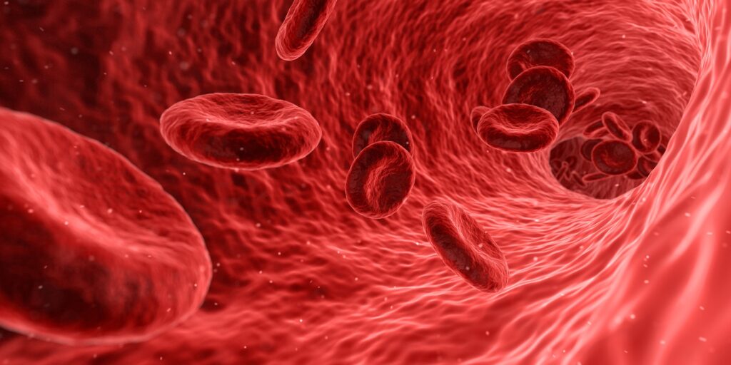
A new study revealed that lupus may be triggered by a defective process in the development of red blood cells (RBCs) which leaves mitochondria remnants. The study was published in Cell.

The researchers found that in a number of lupus patients, maturing red blood cells fail to get rid of their mitochondria, which are normally excluded from red blood cells. This abnormal retention of mitochondria can trigger the cascade of immune hyperactivity characteristic of this disease.

“Our findings support that red blood cells can play a really important role in driving inflammation in a subgroup of lupus patients. So this adds a new piece to the lupus puzzle, and could now open the door to new possibilities for therapeutic interventions,” said the study’s senior author, Dr Virginia Pascual, the Drukier Director of the Gale and Ira Drukier Institute for Children’s Health and the Ronay Menschel Professor of Pediatrics at Weill Cornell Medicine

Lupus is a chronic disorder with no cure that features intermittent and sometimes debilitating attacks by the immune system on the body’s own healthy tissues, including skin, joints, hair follicles, heart and kidneys. A common underlying factor in lupus is the abnormally elevated production of immune-activating proteins called type I interferons. Treatments aim to suppress immune activity, including interferon-driven inflammation.

Previous research found defective mitochondria in the immune cells of lupus patients. In the current study, the researchers focussed on red blood cells, which should lack mitochondria. Many lupus patients had red blood cells with detectable levels of mitochondria, and more common in patients with worse symptoms. By contrast, healthy controls had no mitochondria-containing red blood cells.

Lead author of the study, Dr. Simone Caielli, assistant professor of immunology research at the Drukier Institute and the Department of Pediatrics at Weill Cornell Medicine, then studied how human red blood cells normally get rid of mitochondria as they mature, as prior studies had mainly examined this in mice, and why this process could be defective in lupus patients.

Subsequent experiments showed these abnormal red blood cells cause inflammation. Normally, when red blood cells age or display signs of damage they are removed by macrophages, with binding antibodies helping removal. When the macrophages ingest them, the mitochondrial DNA in the red blood cells triggers a powerful inflammatory pathway called the cGAS/STING pathway, in turn driving type I interferon production. These findings show that “those lupus patients with mitochondria-containing red blood cells and evidence of circulating anti-RBC antibodies had higher interferon signatures compared to those who didn’t,” Dr Caielli said.

The researchers are now investigating how the mitochondria is retained in these cells. Identifying lupus patients with these cells could help predict when they are likely to undergo lupus flares and to develop therapies.

Source: Weill Cornell Medicine

Intense Training Results in Temporary Mitochondrial Impairment

Photo by Quino Al on Unsplash

Elite athletes have temporary mitochondrial impairment following intense workouts, according to new research, which suggests they may need to be mindful about overtraining. 

Mitochondria are organelles that are responsible for most of the useful energy derived from the breakdown of carbohydrates and fatty acids, which is converted to ATP by the process of oxidative phosphorylation. Mitochondrial capacity is a term used to describe the body’s ability to generate energy, and is one factor associated with increased athletic performance during endurance exercise. Previous research found that untrained recreational athletes had a decrease in mitochondrial capacity after sprinting exercises.

In this study, the researchers worked with a small group of male elite athletes, many of whom were national title holders or had international recognition for their performance in cycling and triathlon. The athletes participated in a four-week training programme in their primary sport, which consisted of two to four days of low-to-moderate–intensity endurance workouts, followed by three days of more intense training. These intense workouts included high-intensity interval training in the morning, followed by a seven-hour break and then a moderate-intensity cycling session in the afternoon. Each volunteer did between 12 and 20 hours of activity per week. The athletes, though used to heavy training, were not accustomed to this specific workout schedule.

The researchers were surprised to observe that the highly trained participants’ mitochondrial capacity was impaired after the month-long training period. “We thought that elite athletes should be more resistant against [these] kind of alterations,” said Filip Larsen, PhD, of the Swedish School of Sport and Health Sciences and corresponding author of the study.

However, elite athletes may be able to prevent temporary mitochondrial impairment by listening to their bodies, the researchers suggested. By paying attention to changes such as “mood disturbances, reductions in maximal heart rate [during exercise] and muscles that feel heavy and unresponsive” top athletes may be able to pull back and avoid overtraining situations that could contribute to reduced mitochondrial content and function, Larsen explained. “Exercise is good for you, but too much unaccustomed training might have mitochondrial consequences.”

The study also found that reduced mitochondrial capacity did not affect exercise performance, suggesting that oxygen delivery from the heart to the muscles plays a more important role than mitochondrial function in performance. Expression of three proteins with strong antioxidant properties were also found to be increased in the muscles after intense training.

Source: American Physiological Society

Journal information: Daniele A. Cardinale et al, Short term intensified training temporarily impairs mitochondrial respiratory capacity in elite endurance athletes, Journal of Applied Physiology (2021). DOI: 10.1152/japplphysiol.00829.2020

Mitochondria Dump DNA into Cells, Triggering Inflammation

Researchers have discovered that when building blocks for DNA in cells are in short supply, mitochondria— the powerhouses of cells — release their own DNA, triggering an inflammatory response. Targeting this process can now open up new avenues of treatment into ageing-related diseases.

Mitochondria, the producers of energy for cells, , have their own genetic material: mitochondrial DNA. In certain situations, however, mitochondria are known to release their DNA into the interior of the cell, provoking a reaction from the cell’s own immune system. Some cardiac and neurodegenerative diseases as well as the ageing process are associated with the mitochondrial genome.

To find out when mitochondria release their DNA, the researchers have focused on the mitochondrial protein YME1L. “In cells lacking YME1L, we observed the release of mitochondrial DNA into the cell interior and a related immune response in the cells,” explained Thomas MacVicar, one of the study’s two first authors.  
“If the cells lack YME1L, there is a deficiency of DNA building blocks inside the cell,” he continued. “This deficiency triggers the release of mitochondrial DNA, which in turn causes an inflammatory response in the cell: the cell stimulates similar inflammatory reactions as it does during a bacterial or viral infection. If we add DNA building blocks to the cells from the outside, that also stops the inflammation.”

This newly discovered link between cellular inflammatory response and the metabolism of DNA building blocks could have far-reaching consequences, MacVicar explained. “Some viral inhibitors stop the production of certain DNA building blocks, thereby triggering an inflammatory response. The release of mitochondrial DNA could be a crucial factor in this, contributing to the effect of these inhibitors,” he said. 
Mitochondrial DNA is associated with a number of ageing-associated inflammatory diseases, including cardiac and neurodegenerative diseases, as well as obesity and cancer. The authors hope that new therapeutic opportunities in such diseases can be created by modulating the metabolism of DNA building blocks.

Source: Medical Xpress

Journal information: Hans-Georg Sprenger et al, Cellular pyrimidine imbalance triggers mitochondrial DNA–dependent innate immunity, Nature Metabolism (2021). DOI: 10.1038/s42255-021-00385-9