Tag: brain waves

Brain Training Game Offers New Hope for Drug-free Pain Management

An animated jellyfish floats through water in the PainWaive game. Image: PainWaive

The first trial of an interactive game that trains people to alter their brain waves has shown promise as a treatment for nerve pain – offering hope for a new generation of drug-free treatments.

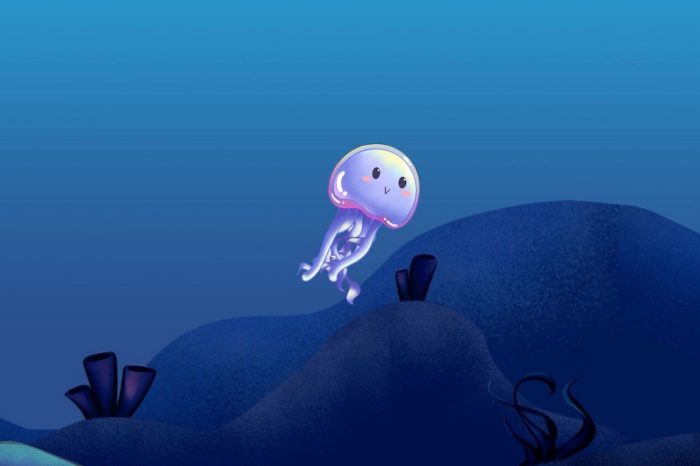
You’re staring at a jellyfish drifting through inky black water on a screen. As your mind calms, the water turns turquoise. Nothing else seems to change, but the headset you’re wearing has picked up a subtle shift in your brainwaves and the game responds by altering the imagery. Now, for the first time, you can see your brain activity change. And by seeing it, you can practice making it happen again.

The game is part of PainWaive, a drug-free treatment for nerve pain developed by UNSW. Combining a game-like app and a brain-monitoring headset, PainWaive teaches users how to regulate the abnormal brain activity linked to chronic nerve pain, offering a potential in-home, non-invasive alternative to opioids.

The first trial of the treatment, led by Professor Sylvia Gustin and Dr Negin Hesam-Shariati from UNSW’s NeuroRecovery Research Hub, has delivered promising results, published in the Journal of Pain.

The study compared hundreds of measures across participants’ pain and related issues like pain interference before, during and after four weeks of interactive game play. Their brain activity was tracked via EEG (electroencephalogram) headsets, with the app responding in real time to shifts in brainwave patterns.

Three out of the four participants showed significant reductions in pain, particularly nearing the end of the treatment. Overall, the pain relief achieved by the three was comparable to or greater than that offered by opioids.

“Restrictions in the study’s size, design and duration limit our ability to generalise the findings or rule out placebo effects,” Dr Hesam-Shariati says. 

“But the results we’ve seen are exciting and give us confidence to move to the next stage and our larger trial.”

The PainWaive project builds on UNSW Professor Sylvia Gustin’s seminal research into changes in the brain’s thalamus – a central relay hub in the brain – associated with nerve (neuropathic) pain.

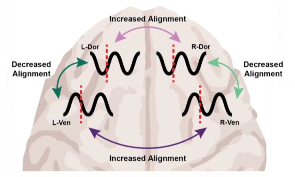
“The brainwaves of people with neuropathic pain show a distinct pattern: more slow theta waves, fewer alpha waves, and more fast, high beta waves,” Prof. Gustin says. 

“We believe these changes interfere with how the thalamus talks to other parts of the brain, especially the sensory motor cortex, which registers pain.

“I wondered, can we develop a treatment that directly targets and normalises these abnormal waves?”

The challenge was taken up by an interdisciplinary team at UNSW Science and Neuroscience Research Australia (NeuRA), led by Prof. Gustin and Dr Hesam-Shariati, and resulted in PainWaive.

The four participants in its first clinical trial received a kit with a headset and a tablet preloaded with the game app, which includes directions for its use. They were also given tips for different mental strategies, like relaxing or focusing on happy memories, to help bring their brain activity into a more “normal” state.

The user data, meanwhile, was uploaded to the research team for remote monitoring.

“After just a couple of Zoom sessions, participants were able to run the treatment entirely on their own,” says Dr Hesam-Shariati.

“Participants felt empowered to manage their pain in their own environment. That’s a huge part of what makes this special.”

Initially, Dr Hesam-Shariati says, the team planned to use existing commercial EEG systems, but they were either too expensive or didn’t meet the quality needed to deliver the project. Instead, they developed their own.

“Everything except the open-source EEG board was built in-house,” says Dr Hesam-Shariati. “And soon, even that will be replaced by a custom-designed board.”

Thanks to 3D printing, Prof. Gustin says, the team has cut the cost of each headset to around $300 – a fraction of the $1000 to $20 000 price tags of existing systems.

The headset uses a saline-based wet electrode system to improve signal quality and targets the sensorimotor cortex.

“We’ve worked closely with patients to ensure the headset is lightweight, comfortable, and user-friendly,” says Prof. Gustin.

“Owning the technology offers us the potential to one day offer PainWaive as a truly affordable, accessible solution for at-home pain management, especially for those with limited access to traditional treatments.”

The team is currently focussed on a randomised controlled trial of the PainWaive technology, aiming to recruit 224 people with nerve pain following spinal cord injury. 

It’s part of more than a dozen active collaborations between UNSW Science and the Centre for Pain IMPACT at NeuRA, all building on Prof. Gustin’s foundational research into the brain. 

Among these is a clinical trial of an eHealth therapy, called Pain and Emotion Therapy, that was shown to reduce chronic pain by retraining the brain to process emotions more effectively.

Another major project, Project Avatar, is inspired by Prof Gustin’s discovery that half of people with complete spinal cord injuries still have touch signals reaching the brain – though the brain can’t identify them. 

The trial uses immersive virtual reality and real-world touch stimulation to help the brain relearn how to feel.

“Many of our team are clinician-scientists, and we’re focused on developing practical treatments that can be integrated into the healthcare system,” says Prof Gustin.

“It’s incredibly inspiring to see results that help unlock the brain’s potential to heal itself and bring back hope to people living with pain.” 

The researchers are now calling for participants to register their interest in two upcoming trials of the neuromodulation technology: The Spinal Pain Trial, investigating its potential to reduce chronic spinal pain, and the StoPain Trial, exploring its use in treating chronic neuropathic pain in people with a spinal cord injury.

Source: University of New South Wales

Under Different Anaesthetics, Same Result: Unconsciousness by Shifting Brainwave Phase

MIT study finds that an easily measurable brain wave shift may be a universal marker of unconsciousness under anaesthesia

Photo by Anna Shvets on Pexels

At the level of molecules and cells, ketamine and dexmedetomidine work very differently, but in the operating room, they do the same exact thing: anaesthetise the patient. By demonstrating how these distinct drugs achieve the same result, a new study in animals by neuroscientists at The Picower Institute for Learning and Memory at MIT identifies a potential signature of unconsciousness that is readily measurable to improve anaesthesiology care.

What the two drugs have in common, the researchers discovered, is the way they push around brain waves, which are produced by the collective electrical activity of neurons. When brain waves are in phase, meaning the peaks and valleys of the waves are aligned, local groups of neurons in the brain’s cortex can share information to produce conscious cognitive functions such as attention, perception and reasoning, said Picower Professor Earl K. Miller, senior author of the new study in Cell Reports. When brain waves fall out of phase, local communications, and therefore functions, fall apart, producing unconsciousness.

The finding, led by graduate student Alexandra Bardon, not only adds to scientists’ understanding of the dividing line between consciousness and unconsciousness, Miller said, but also could provide a common new measure for anesthesiologists who use a variety of different anesthetics to maintain patients on the proper side of that line during surgery.

“If you look at the way phase is shifted in our recordings, you can barely tell which drug it was,” said Miller, a faculty member in The Picower Institute and MIT’s Department of Brain and Cognitive Sciences. “That’s valuable for medical practice.  Plus if unconsciousness has a universal signature, it could also reveal the mechanisms that generate consciousness.”

A figure from the paper summarises the main findings. Under either ketamine or dexmedetomidine general anaesthesia, brain waves become shifted out of phase within a hemisphere and more into phase across hemispheres.

If more anesthetic drugs are also shown to affect phase in the same way, then anaesthesiologists might be able to use brain wave phase alignment as a reliable marker of unconsciousness as they titrate doses of anesthetic drugs, Miller said, regardless of which particular mix of drugs they are using. That insight could aid efforts to build closed-loop systems that can aid anaesthesiologists by constantly adjusting drug dose based on brain wave measurements of the patient’s unconsciousness.

Miller has been collaborating with study co-author Emery N. Brown, an anaesthesiologist and Professor of Computational Neuroscience and Medical Engineering, on building such a system. In a recent clinical trial with colleagues in Japan, Brown demonstrated that monitoring brain wave power signals using EEG enabled an anaesthesiologist to use much less sevoflurane during surgery with young children. The reduced doses proved safe and were associated with many improved clinical outcomes, including a reduced incidence of post-operative delirium.

Phase findings

Neuroscientists studying anaesthesia have rarely paid attention to phase, but in the new study, Bardon, Brown and Miller’s team made a point of it as they anaesthetised two animals.

After the animals lost consciousness, the measurements indicated a substantial increase in “phase locking,” especially at low frequencies. Phase locking means that the relative differences in phase remained more stable. But what caught the researchers’ attention were the differences that became locked in: Within each hemisphere, regardless of which anesthetic they used, brain wave phase became misaligned between the dorsolateral and ventrolateral regions of the prefrontal cortex.

Surprisingly, brain wave phase across hemispheres became more aligned, not less. But Miller notes that case is still a big shift from the conscious state, in which brain hemispheres are typically not aligned well, so the finding is a further indication that major changes of phase alignment, albeit in different ways at different distances, are a correlate of unconsciousness compared to wakefulness.

“The increase in interhemispheric alignment of activity by anesthetics seems to reverse the pattern observed in the awake, cognitively engaged brain,” the Bardon and Miller team wrote in Cell Reports.

Determined by distance

Distance proved to be a major factor in determining the change in phase alignment. Even across the 2.5 millimetres of a single electrode array, low-frequency waves moved 20-30 degrees out of alignment. Across the 20 or so millimetres between arrays in the upper (dorsolateral) and lower (ventrolateral) regions within a hemisphere, that would mean a roughly 180-degree shift in phase alignment, which is a complete offset of the waves.

The dependence on distance is consistent with the idea of waves traveling across the cortex, Miller said. Indeed in a 2022 study, Miller and Brown’s labs showed that the anaesthetic propofol induced a powerful low-frequency traveling wave that swept straight across the cortex, overwhelming higher-frequency straight and rotating waves.

The new results raise many opportunities for follow-up studies, Miller said. Does propofol also produce this signature of changed phase alignment? What role do travelling waves play in the phenomenon? And given that sleep is also characterised by increased power in slow wave frequencies, but is definitely not the same state as anaesthesia-induced unconsciousness, could phase alignment explain the difference?

Source: Picower Institute

Brain Fluid Dynamics is Key to the Mysteries of Migraine

Credit: University of Rochester Medical Center

New research describes how a spreading wave of disruption and the flow of fluid in the brain triggers headaches, detailing the connection between the neurological symptoms associated with aura and the migraine that follows. The study, which appears in Science, also identifies new proteins that could be responsible for headaches and may serve as foundation for new migraine drugs.

“In this study, we describe the interaction between the central and peripheral nervous system brought about by increased concentrations of proteins released in the brain during an episode of spreading depolarization, a phenomenon responsible for the aura associated with migraines,” said lead author Maiken Nedergaard, MD, DMSc, co-director of the University of Rochester Center for Translational Neuromedicine. “These findings provide us with a host of new targets to suppress sensory nerve activation to prevent and treat migraines and strengthen existing therapies.”

It is estimated that one out of 10 people experience migraines and in about a quarter of these cases the headache is preceded by an aura, a sensory disturbance that can includes light flashes, blind spots, double vision, and tingling sensations or limb numbness. These symptoms typically appear five to 60 minutes prior to the headache.

The cause of the aura is a phenomenon called cortical spreading depression, a temporary depolarization of neurons and other cells caused by diffusion of glutamate and potassium that radiates like a wave across the brain, reducing oxygen levels and impairing blood flow. Most frequently, the depolarization event is located in the visual processing centre of the brain cortex, hence the visual symptoms that first herald a coming headache.

While migraines auras arise in the brain, the organ itself cannot sense pain. These signals must instead be transmitted from the central nervous system to the peripheral nervous system. The process of communication between the brain and peripheral sensory nerves in migraines has largely remained a mystery.

Fluid dynamics models shed light on migraine pain origins

Nedergaard and her colleagues at the University of Rochester and the University of Copenhagen are pioneers in understanding the flow of fluids in the brain. In 2012, her lab was the first to describe the glymphatic system, which uses cerebrospinal fluid (CSF) to wash away toxic proteins in the brain. In partnership with experts in fluid dynamics, the team has built detailed models of how the CSF moves in the brain and its role in transporting proteins, neurotransmitters, and other chemicals.

The most widely accepted theory is that nerve endings resting on the outer surface of the membranes that enclose the brain are responsible for the headaches that follow an aura. The new study, which was conducted in mice, describes a different route and identifies proteins, many of which are potential new drug targets, that may be responsible for activating the nerves and causing pain.

As the depolarization wave spreads, neurons release a host of inflammatory and other proteins into CSF. In a series of experiments in mice, the researchers showed how CSF transports these proteins to the trigeminal ganglion, a large bundle of nerves that rests at the base of the skull and supplies sensory information to the head and face.

It was assumed that the trigeminal ganglion, like the rest of the peripheral nervous system, rested outside the blood-brain-barrier, which tightly controls what molecules enter and leave the brain. However, the researchers identified a previously unknown gap in the barrier that allowed CSF to flow directly into the trigeminal ganglion, exposing sensory nerves to the cocktail of proteins released by the brain.

Migraine-associated proteins double during brain wave activity

Analysing the molecules, the researchers identified twelve proteins called ligands that bind with receptors on sensory nerves found in the trigeminal ganglion, potentially causing these cells to activate. The concentrations of several of these proteins found in CSF more than doubled following a cortical spreading depression. One of the proteins, calcitonin gene-related peptide (CGRP), is already the target of a new class of drugs to treat and prevent migraines called CGRP inhibitors. Other identified proteins are known to play a role in other pain conditions, such as neuropathic pain, and are likely important in migraine headaches as well.

“We have identified a new signaling pathway and several molecules that activate sensory nerves in the peripheral nervous system. Among the identified molecules are those already associated with migraines, but we didn’t know exactly how and where the migraine inducing action occurred,” said Martin Kaag Rasmussen, PhD, a postdoctoral fellow at the University of Copenhagen and first author of the study. “Defining the role of these newly identified ligand-receptor pairs may enable the discovery of new pharmacological targets, which could benefit the large portion of patients not responding to available therapies.”

The researchers also observed that the transport of proteins released in one side of the brain reaches mostly the nerves in the trigeminal ganglion on the same side, potentially explaining why pain occurs on one side of the head during most migraines.

Source: University of Rochester Medical Center

Could Stimulating Gamma Brain Waves Help Treat Alzheimer’s?

Photo by JD Mason on Unsplash

A review in the Journal of Internal Medicine explores the potential of non-invasive interventions such as light, sound, and magnets to stimulate gamma brain waves for the treatment of Alzheimer’s disease. Such strategies may be beneficial because Alzheimer’s disease is characterised by reduced fast brain oscillations in the gamma range (30–100Hz).

The authors note that recent studies reveal that it is feasible and safe to induce 40Hz brain activity in patients with Alzheimer’s disease through a range of methods. Also, preliminary evidence suggests that such treatment can yield beneficial effects on brain function, disease pathology, and cognitive function in patients.

Various cells in the brain beyond neurons, including microglial cells, astrocytes and vascular cells, seem to be involved in mediating these effects.

“We found that increased gamma activity elicited by the non-invasive 40Hz sensory stimulation profoundly alters the cellular state of various glial cell types,” said corresponding author Li-Huei Tsai, PhD, of MIT. “We are actively investigating the mechanism by which the 40Hz brain activity recruits diverse cell types in the brain to provide neuroprotective effects.”

Source: Wiley