Tag: 23/9/25

Study Explains How Lymphoma Rewires Human Genome

Source: Pixabay CC0

Translocations are chromosomal “cut and paste” errors that drive many lymphomas, a type of blood cancer and the sixth most common form of cancer overall. This includes mantle cell lymphoma, a rare but aggressive subtype diagnosed in about one in every 100 000 people each year.

Translocations are known to spark cancer by altering the activity of the genes near the breakpoints where chromosomes snap and rejoin. For example, a translocation can accidentally cut a gene in half, silencing its activity, or create new hybrid proteins that help promote cancer.

A study published today in Nucleic Acids Research shows a new way translocations promote cancer. The translocation most typically found in mantle cell lymphoma drags a powerful regulatory element into a new area of the human genome, where its new position allows it to boost the activity of not just one but 50 genes at once.

The discovery of this genome rewiring mechanism shows the traditional focus on the handful of genes at chromosomal breakpoints is too narrow. The study also greatly expands the list of potential drug targets for mantle cell lymphoma, for which there is no known cure.

“We did not expect to see a single translocation boosting the expression of almost 7% of all genes on a single chromosome. The ripples of disruption are much bigger than expected, and also identify new cancer driver genes, each of which represents a new potential therapeutic target,” says Dr. Renée Beekman, corresponding author of the study and researcher at the Centre for Genomic Regulation (CRG) in Barcelona.

In mantle cell lymphoma, a piece of chromosome 14 swaps places with a piece of chromosome 11. A gene regulatory element called the IGH enhancer, which normally boosts the activity of antibody production in healthy B cells, lands right beside CCND1, a gene which helps cells divide. The enhancer treats CCND1 as if it were a gene encoding for antibodies, boosting its activity and fuelling the disease.

Previous research has shown that boosting CCND1 expression alone is insufficient to kickstart the formation of mantle cell lymphoma. To understand why, the scientists first created translocations in cells in a dish. They used CRISPR to replicate the exact chromosome break seen in patients.

“We built a system to generate translocations in healthy B cells. Because these are engineered cells, we can carry out experiments that are technically or ethically unfeasible with patient tissues, making it a really useful early disease model,” explains Dr. Roser Zaurin, co-author of the study.

The experiments revealed that over fifty genes along the entire chromosome 11 were much more active after the translocation took place. The translocation affected gene activity across 50 million base pairs, a significantly larger space than previously thought.

How DNA folds inside the engineered cells revealed why the translocation affects so many genes at once. “DNA loops inside cells. It’s what brings two segments of DNA that are far away from each other in two-dimensional space closer together in three-dimensional space. The translocation drags the strong IGH enhancer into a preexisting loop, placing it in a privileged position of control, enabling it to have a widespread impact on dozens of genes at the same time,” explains Dr. Anna Oncins, first author of the study.

Intriguingly, most of the genes affected by the enhancer were not silent to begin with. The IGH enhancer simply dials their activity up. This biological nuance may explain why the same translocation can have different consequences in different cell types or stages of development. Only genes which were already active are boosted.

The findings could lead to new strategies for the early-stage detection of mantle cell lymphomas. “Because the enhancer mainly supercharges genes that were already active in the very first B cell that acquires the swap, epigenetic profiling of at-risk cells could spot dangerous combinations before a mantle cell lymphoma appears,” explains Dr. Beekman.

The authors of the study next plan on studying exactly how the newly identified genes contribute to the initiation and progression of lymphoma. Understanding and eventually interrupting the effects of the chromosomal translocation could yield broader, more durable therapies for mantle-cell lymphoma and other types of cancers driven by chromosome swaps.

Source: Centre for Genomic Regulation

Scientists Repair Stroke Damage in Mice Using Stem Cells

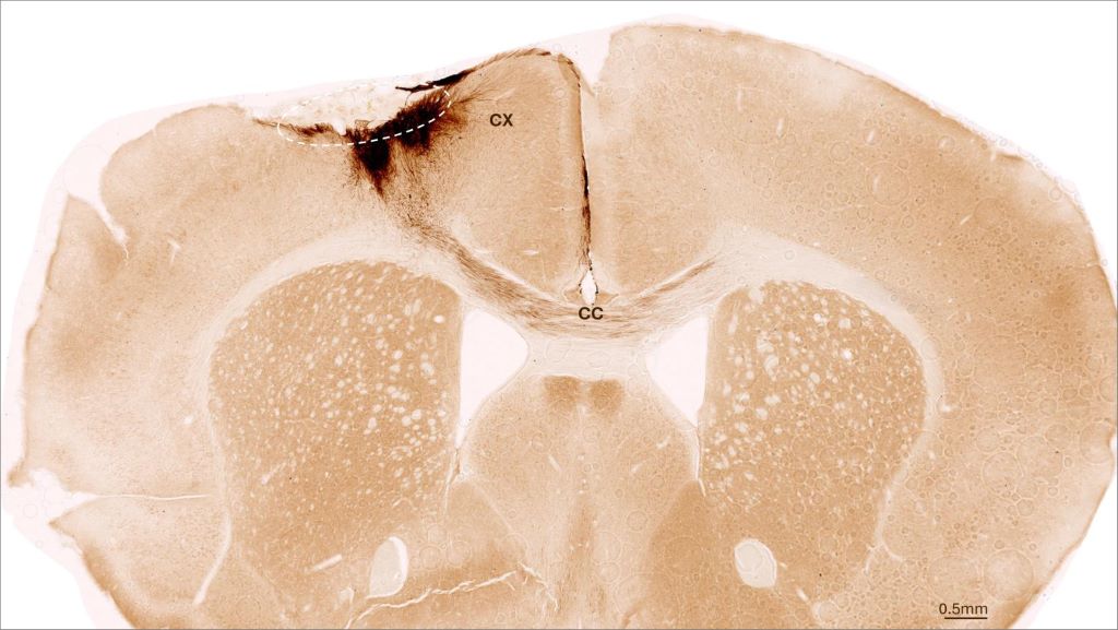
This image shows a coronal section through the mouse brain after stroke and neural stem cell transplantation. The dashed circle indicates the stroke area. The neurite projections of the transplanted human cells are stained in dark brown. Neurites extend locally into the cortex (CX) but also via the corpus callosum (CC) into the other brain hemisphere. (Image: UZH)

One in four adults suffer a stroke in their lifetime, leaving around half of them with residual damage such as paralysis or speech impairment because internal bleeding or a lack of oxygen supply kill brain cells irreversibly. No therapies currently exist to repair this kind of damage. “That’s why it is essential to pursue new therapeutic approaches to potential brain regeneration after diseases or accidents,” says Christian Tackenberg, the Scientific Head of Division in the Neurodegeneration Group at the University of Zurich (UZH) Institute for Regenerative Medicine.

Neural stem cells have the potential to regenerate brain tissue, as a team led by Tackenberg and postdoctoral researcher Rebecca Weber has now compellingly shown in two studies that were conducted in collaboration with a group headed by Ruslan Rust from the University of Southern California. “Our findings show that neural stem cells not only form new neurons, but also induce other regeneration processes,” Tackenberg says.

The first study is published in Nature Communications, the second in Science Advances.

New neurons from stem cells

The studies employed human neural stem cells, from which different cell types of the nervous system can form. The stem cells were derived from induced pluripotent stem cells, which in turn can be manufactured from normal human somatic cells. For their investigation, the researchers induced a permanent stroke in mice, the characteristics of which closely resemble manifestation of stroke in humans. The animals were genetically modified so that they would not reject the human stem cells.

One week after stroke induction, the research team transplanted neural stem cells into the injured brain region and observed subsequent developments using a variety of imaging and biochemical methods. “We found that the stem cells survived for the full analysis period of five weeks and that most of them transformed into neurons, which actually even communicated with the already existing brain cells,” Tackenberg says.

Brain regenerates itself

The researchers also found other markers of regeneration: new formation of blood vessels, an attenuation of inflammatory response processes and improved blood-brain barrier integrity. “Our analysis goes far beyond the scope of other studies, which focused on the immediate effects right after transplantation,” Tackenberg explains. Fortunately, stem cell transplantation in mice also reversed motor impairments caused by stroke. Proof of that was delivered in part by an AI-assisted mouse gait analysis.

Clinical application moving closer to reality

Human neural stem cells in culture. Cell nuclei are stained in blue, the neural stem cell-specific filament protein Nestin is shown in green, and the neural stem cell transcription factor Sox1 in red. (Image: UZH)

When he was designing the studies, Tackenberg already had his sights set on clinical applications in humans. That’s why, for example, the stem cells were manufactured without the use of reagents derived from animals. The Zurich-based research team developed a defined protocol for that purpose in collaboration with the Center for iPS Cell Research and Application (CiRA) at Kyoto University. This is important for potential therapeutic applications in humans. Another new insight discovered was that stem cell transplantation works better when it is performed not immediately after a stroke but a week later, as the second study verified. In the clinical setting, that time window could greatly facilitate therapy preparation and implementation.

Despite the encouraging results of the studies, Tackenberg warns that there is still work to be done. “We need to minimize risks and simplify a potential application in humans,” he says. Tackenberg’s group, again in collaboration with Ruslan Rust, is currently working on a kind of safety switch system that prevents uncontrolled growth of stem cells in the brain. Delivery of stem cells through endovascular injection, which would be much more practicable than a brain graft, is also under development. Initial clinical trials using induced stem cells to treat Parkinson’s disease in humans are already underway in Japan, Tackenberg reports. “Stroke could be one of the next diseases for which a clinical trial becomes possible.”

Source: University of Zurich

Sugary Drinks May Increase Risk of Metastasis in Advanced Colorectal Cancer

Photo by Breakingpic on Pexels

A new study from researchers at The University of Texas MD Anderson Cancer Center shows that the glucose-fructose mix found in sugary drinks directly fuels metastasis in preclinical models of advanced colorectal cancer. The study was published in Nature Metabolism.

A research team led by Jihye Yun, PhD, assistant professor of Genetics, studied how sugary drinks may affect late-stage colorectal cancer. Using laboratory cancer models, they compared the effects of the glucose-fructose mix found in most sugary drinks with those of glucose or fructose alone. Only the sugar mix made cancer cells more mobile, leading to faster spread to the liver – the most common site of colorectal cancer metastasis.

The sugar mix activated an enzyme called sorbitol dehydrogenase (SORD), which boosts glucose metabolism and triggers the cholesterol pathway, ultimately driving metastasis. This is the same pathway targeted by statins, common heart drugs that inhibit cholesterol production. Blocking SORD slowed metastasis, even with the sugar mix present. These findings suggest that targeting SORD could also offer an opportunity to block metastasis.

“Our findings highlight that daily diet matters not only for cancer risk but also for how the disease progresses once it has developed,” Yun said. “While these findings need further investigation, they suggest that reducing sugary drinks, targeting SORD or repurposing statins may benefit patients with colorectal cancer.”

The Yun Laboratory is interested in studying how diet affects the intestine and cancer development, and they have made important discoveries on the impacts of sugary drinks on colorectal cancer.

Sugar has long been indirectly linked to an increase in cancer risk through obesity. However, a previous study by Yun’s lab challenged that view, showing that even moderate intake of sugary drinks directly fuelled tumour growth in early-stage colorectal cancer, independent of obesity. The current study was done to determine how sugary drinks may impact later-stage disease.

While this study needs further clinical investigation, the results suggest that reducing sugary drinks and targeting the SORD enzyme may offer opportunities to reduce colorectal cancer metastasis. Additional studies are warranted to confirm these results outside of preclinical models.

Further, Yun explained it may be worthwhile to consider revisions to current dietary recommendations to reduce sugary drink consumption in this patient population. To meet nutritional needs, many patients with cancer are encouraged to have nutritional supplement drinks and concentrated juices that contain high glucose and fructose content.

Source: The University of Texas MD Anderson Cancer Center