Tag: 21/7/26

Heavy TV Watching Associated with Smaller Brain Structures, Study Finds

Reduced volume was found in areas of the brain connected to memory formation, indicating a potential link between TV watching and higher dementia risk

“Turn off that TV, it’ll rot your brain!” has been a household refrain for decades. While “rot” might be too strong a term, researchers are finding that the overall sentiment could have some merit.

study published recently in Alzheimer’s and Dementia: Journal of the Alzheimer’s Association revealed that those who reported watching TV “very often” in midlife later exhibited reduced volume in areas of the brain associated with memory, smaller frontal and occipital lobes, and areas of damage in the brain’s white matter that are associated with aging, stroke risk, cognitive decline and dementia. 

“For years we’ve focused on how much people sit. Our findings suggest we should also pay attention to what they’re doing while they’re sitting,” says David Raichlen, professor of biological sciences and anthropology at the USC Dornsife College of Letters, Arts and Sciences and a senior author of the study. 

The findings weren’t just due to TV viewing’s sedentary nature. The study found that other types of sedentary activities did not have the same associations, indicating that what one does while sitting may matter much more than previously thought.

Watching changes

The researchers analyzed data from about 1,700 adults, average age 53, who enrolled in the Atherosclerosis Risk in Communities (ARIC) Study between 1987 and 1989. ARIC is a long-running study of the U.S. population designed to investigate cardiovascular and brain health. 

Participants were asked how frequently, on a scale ranging from “never/seldom” to “very often,” they watched television during their leisure time and how much of their workday they spent sitting.

More than two decades later, participants underwent brain MRI. Compared with people who reported “never” or “seldom” watching TV, those who watched TV “very often” showed widespread structural differences across the brain.

The researchers found smaller volumes in areas associated with early signs of Alzheimer’s disease and more white matter hyperintensity volumes, an indicator of cerebral small blood vessel disease associated with cognitive decline and dementia. These participants also had smaller occipital and frontal lobes, regions associated with visual processing and executive functioning.

Differences persisted even when the researchers controlled for factors such as physical activity, diabetes, body mass index, smoking, and alcohol use.

Of note, the researchers relied on self-reported data for TV consumption, which can be less precise than timed tracking. Study participants also did not undergo a baseline MRI. Future research could begin with a baseline MRI to more concretely demonstrate changes over time.

Not all sitting is made the same

Strikingly, the sedentary element of TV watching didn’t appear to be the main driver for these changes.

Those who reported high amounts of sitting at work actually had larger frontal and occipital lobes, as well as reduced white matter hyperintensity volumes, indicating better brain health than among those who sit to watch TV. This could be due to the intellectually stimulating nature of many sit-down jobs, say the study authors. 

Men appeared to be particularly vulnerable to these changes. When the MRI scans were separated by sex, researchers found that most of the changes to the brain, both from TV watching and occupational sitting, were seen in men.

Such findings indicate there is still more research to be done on this complex topic. However, we might eventually see a different approach to health guidance around sedentary activities. Rather than just directing their patients to move more, for example, physicians might recommend they reduce television time and add cognitively engaging activities for when they do sit.  

“We frequently encourage the public not to spend too much time sitting down, but experts may want to expand that recommendation to encompass the activities done while sitting, since those seems to have distinct impacts on brain health,” says study corresponding author Natan Feter, postdoctoral scholar in the Human and Evolutionary Biology program at USC Dornsife. 

Source: University of Southern California

Researchers Reveal Iron’s Role in Allergic Airway Inflammation

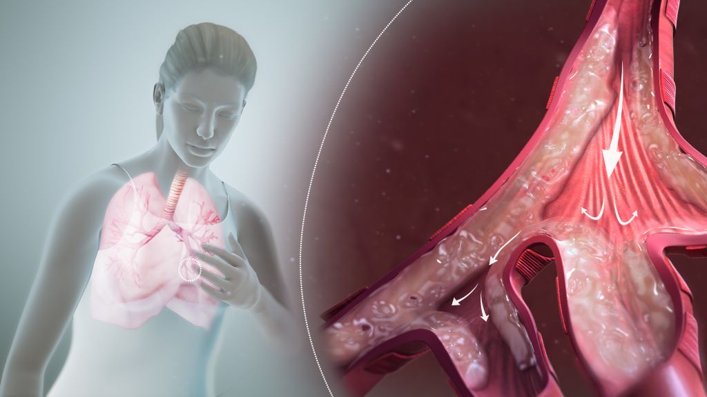
Respiratory tract. Credit: Scientific Animations CC4.0

Chinese researchers have revealed the key role of iron in initiating allergic airway inflammation. The study, which was published in Cell, was conducted by a team led by Prof SUN Bing from the Center for Excellence in Molecular Cell Science (Shanghai Institute of Biochemistry and Cell Biology) of the Chinese Academy of Sciences (CAS), along with Prof LIU Xing’s team from the Shanghai Institute of Materia and Medica of CAS.

The research showed that environmental allergens can use an iron-dependent mechanism to activate gasdermin D (GSDMD) in airway epithelial cells, thereby promoting IL-33 release and initiating allergic airway inflammation – the main pathological basis for the onset and progression of asthma.

When environmental allergens such as pollen, house dust mites, and fungal proteases enter the airway, they act on lung epithelial cells and induce the release of alarmins, including IL-33. IL-33 then activates type 2 innate lymphoid cells (ILC2s), leading to eosinophil infiltration, mucus production, and airway tissue damage.

Previous studies have shown that GSDMD is involved in IL-33 release, but how allergens activate GSDMD had remained unclear. In this study, the researchers found that allergen stimulation rapidly increased the labile iron pool in airway epithelial cells, and GSDMD was cleaved and activated through a mechanism independent of conventional proteases.

Using mouse models induced by papain or house dust mites, the researchers observed a rapid rise in lung iron levels after allergen challenge, occurring in parallel with IL-33 release. Treatment with an iron chelator markedly inhibited GSDMD cleavage and IL-33 release, whereas iron supplementation enhanced these responses. This iron-driven effect was largely abolished in GSDMD-deficient mice, indicating that the pro-inflammatory activity of iron is highly dependent on GSDMD.

Furthermore, the researchers showed that cell-surface protease-activated receptor PAR1 serves as an important entry point for allergen sensing. Papain directly cleaves PAR1, which in turn initiates NCOA4-mediated ferritinophagy and releases additional free iron. The iron chaperone PCBP2 delivers iron to the vicinity of GSDMD, where the E309/Q312 residues of GSDMD are responsible for iron binding. When these sites are mutated, GSDMD can no longer be efficiently cleaved or mediate IL-33 release.

The researchers found that this cleavage process does not depend on canonical inflammasome-associated caspases. Instead, iron delivered by PCBP2 locally triggers a Fenton reaction, generating short-range hydroxyl radicals that drive oxidative cleavage of GSDMD.

In vivo experiments showed that pretreatment with the iron chelator DFP significantly alleviates papain-induced airway inflammation, reducing eosinophil infiltration, IL-5 and IL-13 levels, and mucus secretion. Conversely, iron supplementation aggravated inflammatory responses in wild-type mice, but failed to produce the same effect in GSDMD-deficient mice.

These findings establish the iron–GSDMD–IL-33 axis as an important driver of allergen-induced type 2 immune responses, and suggest that PAR1, iron mobilisation, PCBP2, and local iron-mediated reactions may represent potential intervention points for asthma and other allergic diseases.

In summary, this study proposes a new mechanism for the initiation of allergic airway inflammation. It expands the understanding of GSDMD activation and immunological functions of iron metabolism, and provides new insight into the prevention and treatment of asthma and related allergic diseases.

Source: Chinese Academy of Sciences

Last-line Antibiotics at Risk as Children’s Resistance Rises

Photo by Ben Wicks on Unsplash

Antimicrobial resistance in children is rising globally and will worsen over the next decade, threatening the effectiveness of life‑saving antibiotics, according to a world-first monitoring platform.

The findings, led by Murdoch Children’s Research Institute (MCRI) in collaboration with the University of SydneyClinton Health Access Initiative (CHAI) and The Chinese University of Hong Kong, analysed more than 106 000 infection samples from children, aged up to 18 years, across 82 countries, discovering that antibiotic resistance increased in every region between 2004 and 2022. Babies and children in intensive care and countries with fewer healthcare resources were the most affected.

Published in JAMA Pediatrics, the research found the increase is being driven largely by Gram-negative bacteria responsible for severe infections such as sepsis and pneumonia. Resistance rose most sharply to critical ‘Watch’ and ‘Reserve’ antibiotics, which the World Health Organization (WHO) classifies to limit overuse and help preserve their effectiveness when first-line treatments fail.

MCRI Associate Professor Penelope Bryant said the findings highlighted a widening gap between recommended antibiotic use and real‑world effectiveness, reinforcing the urgent need for improved surveillance, targeted antimicrobial stewardship and better access to effective antibiotics for children.

Forecasting future antimicrobial resistance

The newly launched AMR in Kids website, created by the study team, allows clinicians, researchers and policymakers to explore antibiotic resistance by country, bacteria and antibiotic class. It also provides region and pathogen-specific forecasts to 2035, helping researchers to identify emerging threats, inform treatment decisions and help guide public health planning.

MCRI and University of Sydney Dr Yanhong Jessika Hu said by combining almost two decades of global data with forecasting, the platform could identify where resistance was likely to emerge before it becomes an even greater clinical challenge.

Antimicrobial resistance occurs when bacteria evolve to withstand antibiotic treatment. As resistant bacteria emerge and spread through communities, infections become harder to treat. Children are especially vulnerable as they experience high rates of bacterial infections and have fewer antibiotic options than adults. In 2021, about 840,000 deaths in children under five years were associated with antimicrobial resistance.

“Antimicrobial resistance is one of the biggest threats to children’s health globally, but until now we haven’t had a clear picture of how it’s changing specifically in children,” Dr Hu said. “The AMR in Kids platform addresses a major gap by developing region and pathogen-specific forecasts for the next decade.”

Forecast modelling by the platform suggests that by 2035, some of the most dangerous pathogens could become highly resistant to last‑line treatments.

CHAI Senior Clinical Director Associate Professor Joseph Harwell said, “Better data is essential, but children can’t wait for perfect data. We need to use the best available evidence to guide action now.”

The research found two types of Gram-negative bacteria were driving antimicrobial resistance globally. Acinetobacter baumannii, which commonly causes hospital acquired bloodstream infections and pneumonia, showed the highest overall resistance. Klebsiella, causing urinary tract infections and liver abscesses, recorded the fastest increase, particularly in Southeast Asia, Eastern Europe and the Western Pacific. Alarmingly, resistance to last‑line carbapenem antibiotics is projected to rise substantially by 2035, reaching 82 per cent and 35 per cent, for each superbug, respectively.

Turning data into global action

Associate Professor Bryant said despite facing unique treatment challenges, children had been overlooked in global antibiotic resistance surveillance.

“Rising resistance to first- and now second-line drugs is the clinical reality for children,” she said. “Making antibiotic resistance in children visible, through AMR in Kids, is the first step towards changing its trajectory. By understanding where resistance is emerging and how it’s changing, we can better protect children now and preserve the effectiveness of antibiotics into the future.”

But Associate Professor Bryant said more action was needed on the global stage and measures specifically targeted towards children.

“In low-income countries we need to address unregulated antibiotic use and poor sanitation,” she said. “These countries need better access to diagnostic techniques and first-line antibiotics. Encouraging, Australia’s Department of Foreign Affairs and Trade has brought together experts including from the University of Melbourne and the WHO to develop solutions in the Western Pacific.

“In high-income countries, antibiotic overuse must be tackled across farming, veterinary, primary care and hospital settings, while infection control remains critical.

“We also need child-friendly antibiotic formulations, clearer dosing guidance and funding for trials on safely reducing antibiotic use. Consumers can help by talking with their GP about the risks of overprescribing antibiotics.”

Source: Murdoch Children’s Research Institute