
Tag: 11/11/26

Experimental Treatment Promotes Repair After Heart Attack Damage in Animal Study
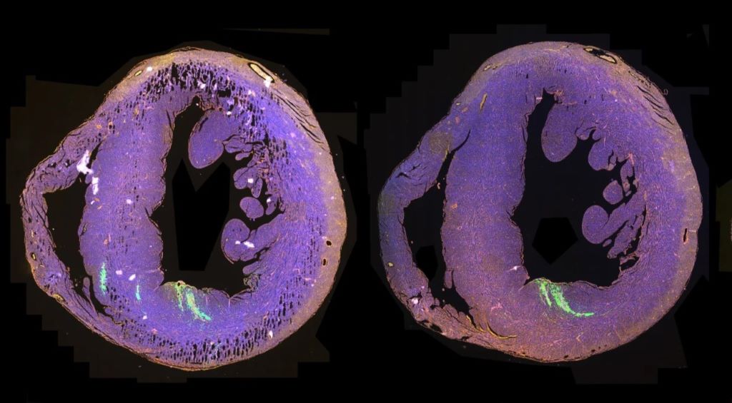
Cross sections of a whole mouse heart, following injection with the mRNA therapy. The areas glowing green mark cells that successfully took up the therapy, helping the scientists get a 3D view of where it can reach within the heart. Credit: Cambridge University

Scientists have developed a treatment that promotes the regeneration of damaged heart muscle following a heart attack, which has shown promising results in mice.

Since adult heart cells can’t repair themselves once damaged, this represents an important step towards a potential curative treatment for heart disease.

The treatment ‘reawakens’ heart muscle cells so they start to divide and help repair damaged muscle.

In a study in mice, this produced a stronger heartbeat and increased the amount of blood pumped through the heart with each heartbeat by around 10%, compared with mice that received a mock treatment, following a heart attack.

While this is not a full cure, even small improvements to heart function have been linked to lower mortality in patients and a better ability to carry out daily activities.

The treatment uses mRNA therapy to temporarily activate two proteins inside heart cells, called Myc and Cyclin T1, promoting repair of heart tissue.

This has the potential in future to be used to treat patients with heart failure. Around 200,000 people in the UK are diagnosed with heart failure each year and while current treatments can reduce symptoms and slow the progression of heart disease, they can’t regenerate heart muscle that has already been lost.

“These are really exciting results. There’s no cure for heart failure, and our treatment could potentially have a meaningful impact on the quality of life of millions of people around the world.”

Dr Catherine Wilson, Associate Professor at the University of Cambridge’s Department of Pharmacology and Gurdon Institute, co-senior author of the study.

Wilson added: “We’re not just treating the symptoms of heart failure – this approach could help the heart to repair itself.”

The team is now working to refine the treatment and has received funding from the British Heart Foundation to continue the work. Further trials in larger animal models, and then in humans, are needed to fully establish its safety and effectiveness.

The results are published today in the journal Nature Cardiovascular Research.

Following a heart attack, the mRNA therapy was injected into the hearts of the mice and its effectiveness assessed by looking at the hearts with an echocardiogram – similar to looking at a baby inside the womb.

Since the treatment is short-lived and localised, it is not expected to cause any permanent modification to the heart cells – designed to reduce the risk of overgrowth, and no adverse effects were seen in the mice.

Reassessing the mice after 28 days and after 12 weeks showed that the improvement in heart function is long-lasting.

After a heart attack, an adult human heart can lose up to one billion heart muscle cells. Unlike many other organs in the body, the adult heart can’t regenerate itself, so lost cells are never replaced. Their loss reduces the strength of the heart and can lead to scar formation, heart failure and ultimately death. 

As the world’s population grows and the prevalence of heart failure increases, the cost of patient care is anticipated to increase dramatically. The scientists believe that reawakening heart muscle cells in this way is a promising approach to treating heart failure patients.

Professor Bryan Williams, chief scientific and medical officer at the British Heart Foundation, said: “The heart is easily repaired when we are babies in the womb, but our heart muscle cells lose the ability to regenerate and fix the heart shortly after we are born. That makes it exciting when studies like this are able to reignite regeneration in heart muscle cells.

“Getting the human heart to repair itself is the holy grail for cardiologists.”

He added: “The gene therapy used by the researchers shows clear potential for heart patients. While the research is in mice, and still at an early stage, it is an important step forward towards therapies which could help repair the damage to the heart following a heart attack.”

This research was a collaboration between the University of Cambridge, QIMR Berghofer Medical Research Institute and AstraZeneca.

Reference: Boikova, A. et al: ‘A transient modified mRNA encoding Myc and Cyclin T1 improves cardiac function after myocardial injury.’ Nature Cardiovascular Research, Nov 2026. DOI: 10.1038/s44161-026-00910-7.

 The text in this work is licensed under a Creative Commons Attribution-NonCommercial-ShareAlike 4.0 International License.

By Jacqueline Garget

Source: University of Cambridge
World Spotlight on SA Laparoscopic Surgical Techniques

Skills training from Centurion to surgeons around the world

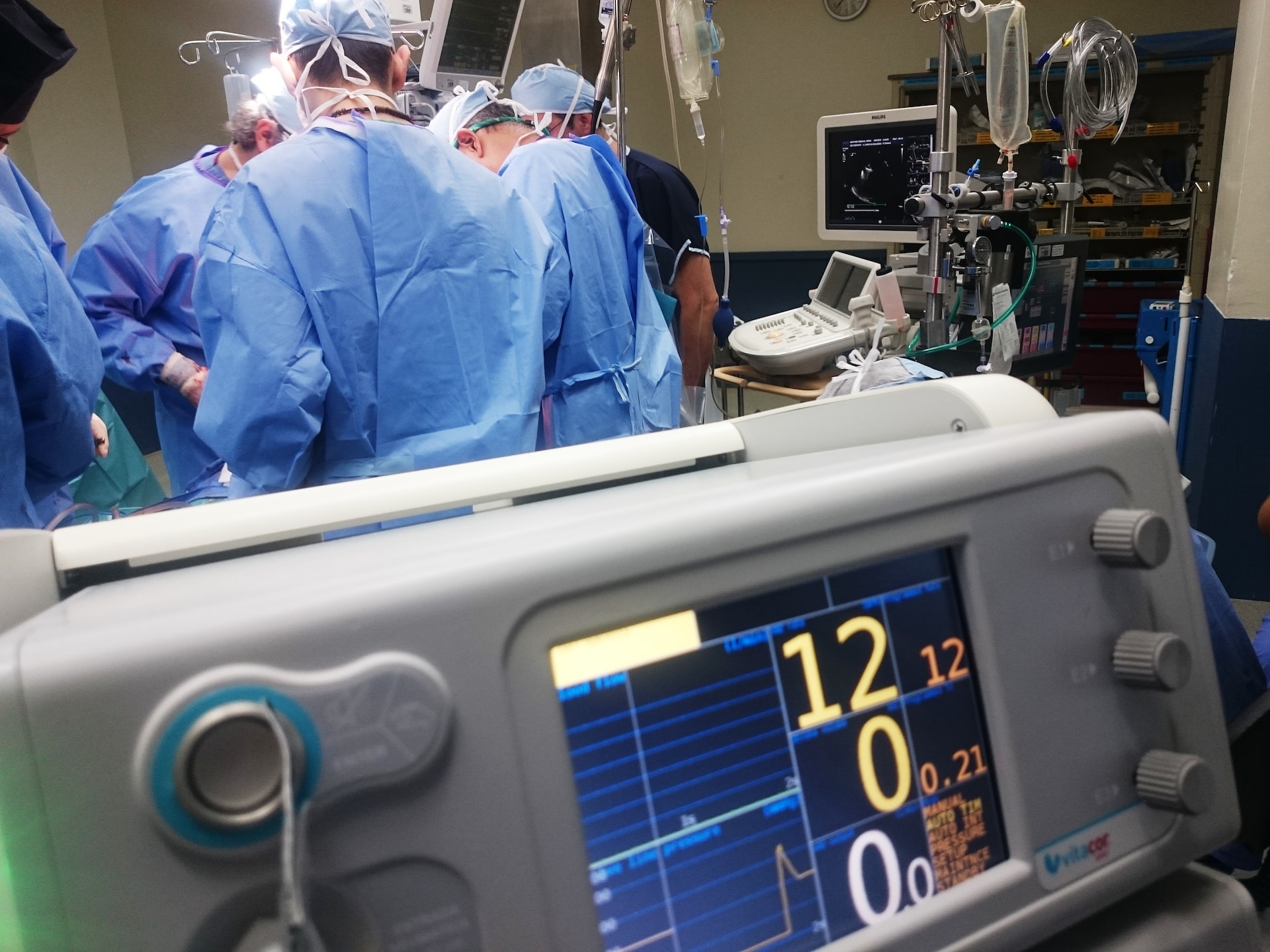
Photo by Natanael Melchor on Unsplash

Wednesday, 11 November 2026: A leading international specialist surgical training conference has once again invited veteran South African laparoscopic surgeon Professor Heine van der Walt to demonstrate his expertise on the global stage.

The Congress on Digestive System Surgery 2026, to be held on 12 and 13 November in Rome, has been described as ‘a congress built by surgeons for surgeons’ and Prof Van der Walt has demonstrated minimally invasive surgical procedures at this annual event for some 20 years. Last year, the Congress’s online streaming channels recorded over 12 million page views in addition to the 2000 healthcare professionals attending in person, illustrating the platform’s impact on surgeon training worldwide.

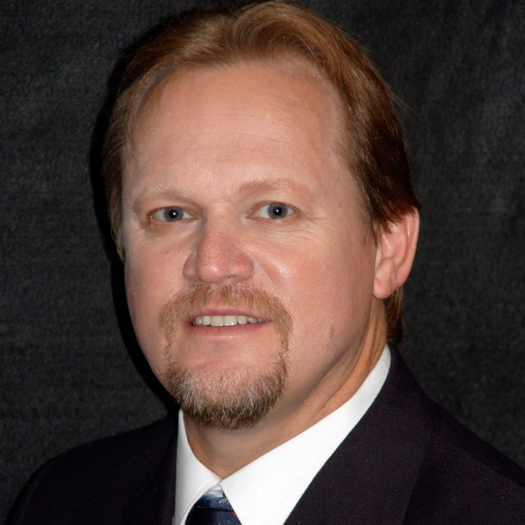
Professor Heine van der Walt

“This year, our team is performing a medley of procedures aimed at teaching foundational minimally invasive surgical techniques that are practically useful for the younger generation of surgeons to master,” says Prof Van der Walt, who will demonstrate laparoscopic techniques to delegates worldwide via video link from Netcare Unitas Hospital in Centurion.

Italian surgeon, Dr P.M. Maberti, recalls first meeting Prof Van der Walt while working in South Africa in various academic hospitals between 1985 and 1999, and the origins of the Congress. “I had the opportunity to appreciate his professionality, preparation, humanity and skill in other congresses during that period,” Dr Maberti says.

“Once back in Rome, I started collaborating with Prof Giorgio Palazzini again at the IIIth Surgical Institute of the Università La Sapienza, and Prof Palazzini had the idea of establishing the Congress on Digestive System Surgery aimed at fostering innovation and benefiting patients. Now in its 37th edition, the Congress is gaining increasing interest from the surgical community worldwide,” Dr Maberti says.

“It is now nearly two decades that Prof Van der Walt and his outstanding staff have participated in this Congress with maximum enthusiasm and skill, showing live surgery that is followed globally. The benefit is undoubted, especially for teaching young surgeons.”

Prof Van der Walt points out that the Congress has grown exponentially since communications technology enabled medical professionals to attend online, eliminating the time and expense of international travel, and widening access to the conference and the training opportunities it provides.

“I have been involved in demonstration surgeries for the international circuit since the early 1990s. I used to travel to all the different venues, operating in their hospitals with live local transmissions to the local congress venue. The transmissions started with four ISDN telephone lines, later we progressed to satellite transmissions and now 5G transmissions are negating much of the necessity for travel,” he says.

“Surgery can’t be learnt from textbooks alone. It demands practical technical skills and precision that surgeons largely pass down hand-to-hand from one generation to the next. Demonstrations such as these are a vital part of that process,” Prof Van der Walt says.

According to Dr Maberti, the great merits of the Congress are that procedures can be shared as if the delegates were in the operating theatre and the interaction it enables, which are essential for sharing surgical techniques globally.

“The Congress has become one of the largest surgical conferences, and it’s so popular because of the transfer of practical knowledge that takes place on this growing platform for the global digestive system medical community. Training is my vocation, and it is always rewarding to develop the skills that underpin better outcomes for patients,” Prof Van der Walt says. 

Netcare Unitas Hospital general manager Pieter Louw congratulated Prof Van der Walt on his extensive contribution to sharing surgical expertise through live theatre demonstrations over many years.

“By taking fellow medical professionals through a procedure as it unfolds, Prof Van der Walt shares far more than technique. He offers insight into the clinical judgement, precision and decision-making that guide each stage of surgery.

“His continued dedication to training extends the value of his experience beyond the operating theatre, helping healthcare professionals in South Africa and around the world strengthen their skills and advance patient care in their own communities. Prof Van der Walt’s participation in this world congress underscores the calibre of minimally invasive surgical expertise available to patients at Netcare Unitas Hospital,” Louw concludes.