Ketamine’s Effect on Depression is Essentially Placebo Effect

Photo by Bruce Christianson on Unsplash

Over the years, studies have demonstrated the psychoactive drug ketamine’s effect on depression, providing profound and fast relief to many people. But these studies have a critical flaw: participants usually can tell whether they have been given ketamine or a placebo. Even in blinded trials in which participants are not told which they received, ketamine’s oftentimes trippy effects are a dead giveaway.

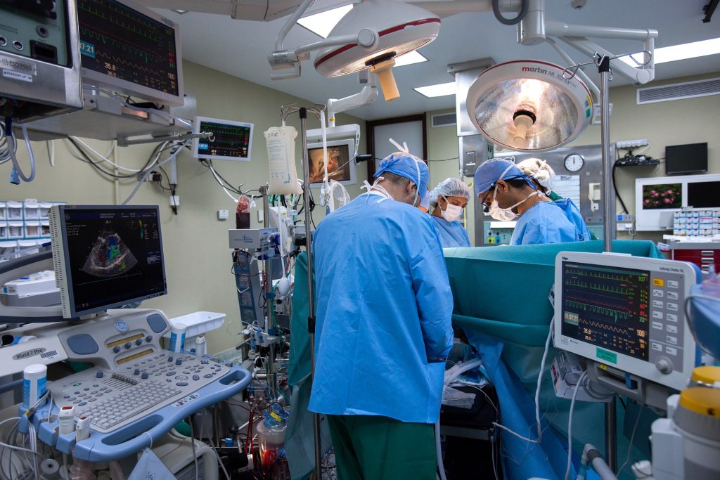
In a new study published in Nature Mental Health, Stanford Medicine researchers devised a clever workaround to hide the psychedelic, or dissociative, properties of the anesthetic first developed in 1962. They recruited 40 participants with moderate to severe depression who were also scheduled for routine surgery, then administered a single infusion of ketamine (0.5 mg kg−1) or placebo (saline) during usual anaesthesia.

All researchers and clinicians involved in the trial also were blinded to which treatment patients received. The treatments were revealed two weeks later.

The researchers were amazed to find that both groups experienced the large improvement in depression symptoms usually seen with ketamine.

“I was very surprised to see this result, especially having talked to some of those patients who said ‘My life is changed, I’ve never felt this way before,’ but they were in the placebo group,” said Boris Heifets, MD, PhD, assistant professor of anaesthesiology, perioperative and pain medicine, and senior author.

Just one day after treatment, both the ketamine and placebo groups’ scores on the Montgomery-Åsberg depression rating scale (MADRS) dropped, on average, by half. Their scores stayed roughly the same throughout the two-week follow-up.

“To put that into perspective, that brings them down to a category of mild depression from what had been debilitating levels of depression,” said Theresa Lii, MD, a postdoctoral scholar in the Heifets lab and lead author of the study.

What does it all mean?

The researchers concede that their study, having taken an unexpected turn, raises more questions than it answers.

“Now all the interpretations happen,” said Alan Schatzberg, MD, a co-author of the study. “It’s like looking at a Picasso painting.”

The researchers determined that it was unlikely the surgeries and general anaesthesia account for the improvements because studies have found that depression generally does not change after surgery; sometimes, it worsens.

A more likely interpretation, the researchers said, is that participants’ positive expectations may play a key role in ketamine’s effectiveness.

At their last follow-up visit, participants were asked to guess which intervention they had received. About a quarter said they didn’t know. Of those who ventured a guess, more than 60% guessed ketamine.

Their guesses did not correlate with their treatment – confirmation of effective blinding – but rather with how much better they felt.

Source: Stanford Medicine

Females Less Able to Recover from ACL Injuries

Photo by Andrea Piacquadio on Pexels

Injuries of the knee’s anterior cruciate ligament (ACL) are typically thought to be caused by acute traumatic events, such as sudden twists. Published in the Journal of Orthopaedic Research, new work analysing an animal model of ACLs suggests that such injuries can also occur as a result of chronic overuse, specifically due to a reduced ability to repair microtraumas associated with overuse. Importantly, the team said, females also are less able to heal from these microtraumas than males, which may explain why females are two to eight times more likely to tear their ACL ligaments than males.

“ACL tears are one of the most common injuries, affecting more than 200 000 people in the US each year, and women are known to be particularly susceptible,” said principal investigator Spencer Szczesny, associate professor of biomedical engineering and of orthopaedics and rehabilitation at Penn State. “While recent research suggests that chronic overuse can lead to ACL injuries, until now, no one had investigated the differential biological response of female and male ACLs to applied force.”

In the Penn State-led study, researchers placed ACLs from deceased male and female rabbits in a custom-made bioreactor that simulated the conditions of a living animal but allowed direct observation and measurement of the tissue. Next, they applied repetitive forces to the ACLs that mimicked those that would naturally occur during activities such as standing, walking and trotting and measured the expression of genes related to healing.

In male samples, the team found that low and moderate applied forces, such as those that would occur during standing or walking, resulted in increased expression of anabolic genes, which are related to building molecules needed for healing. By contrast, larger applied forces, such as those that would occur with repetitive trotting, decreased expression of these anabolic genes. For female samples, however, the amount of force applied did not influence the level of anabolic gene expression.

“It didn’t matter whether there was low, medium or high activity for females,” said Lauren Paschall, graduate student in biomedical engineering at Penn State and first author on the paper. “Female ACLs exposed to chronic use just didn’t heal as well as male ACLs, which may explain why women are predisposed to injuries. This supports the hypothesis that noncontact ACL injuries are attributed to microtraumas associated with chronic overuse that predispose the ACL to injury.”

According to the researchers, one explanation for the sex differences the team observed could be due to the higher amounts of oestrogen in females.

“Some studies have found that the overall effect of oestrogen on ACL injury is negative,” Paschall said. “Specifically, studies have shown that human women are more likely to tear their ACLs during the preovulatory phase, when oestrogen levels are high, than during the postovulatory phase, when oestrogen levels are low.”

She said the team plans to further investigate the role of oestrogen on ACL injury.

Szczesny noted that although the team’s study was not in humans, the findings may suggest that providing additional recovery time for women following injuries could be advantageous.

“Ultimately, this work could also help to identify targets for therapeutics to prevent ACL injuries in women,” he said.

Source: Penn State

Study Finds Urological Effects of SARS-CoV-2 Infection in Men

Photo by Help Stay on Unsplash

A study published in the Journal of Internal Medicine indicates that SARS-CoV-2 infection may worsen lower urinary tract symptoms (LUTS) in men. The study researchers found that a enlarged prostate as a result of COVID was involved.

The study included 17 986 men receiving medication for LUTS within the public healthcare system of Hong Kong in 2021–2022, half of whom had SARS-CoV-2 infection. The group with SARS-CoV-2 had significantly higher rates of retention of urine (4.55% versus 0.86%); blood in the urine (1.36% versus 0.41%); clinical urinary tract infection (4.31% versus 1.49%); bacteria in the urine (9.02% versus 1.97%); and addition of 5-alpha reductase inhibitors, which are drugs prescribed for enlarged prostate. (0.50% versus 0.02%). These urological manifestations occurred regardless of COVID severity.

The findings might relate to the presence of certain proteins targeted by SARS-CoV-2 that are known to be expressed in the prostate.

“We are excited to be the first to report the effects of COVID on complications of benign prostatic hyperplasia – or enlarged prostate – and also demonstrate the alarming extent of its urological effects,” said corresponding author Alex Qinyang Liu, MD, of Prince of Wales Hospital, in Hong Kong.

Source: Wiley

Converting Brain Immune Cells into Neurons Boosts Stroke Recovery in Mice

Source: CC0

Japanese researchers have turned microglia (brain immune cells) into neurons, successfully restoring brain function after stroke-like injury in mice. These findings, published in PNAS, suggest that replenishing neurons from immune cells could be a promising avenue for treating stroke in humans.

Recovery from stroke, where blood supply to neurons is disrupted by blockage or bleeding, is often poor, with patients suffering from severe physical disabilities and cognitive problems.

“When we get a cut or break a bone, our skin and bone cells can replicate to heal our body. But the neurons in our brain cannot easily regenerate, so the damage is often permanent,” says Professor Kinichi Nakashima, from Kyushu University’s Graduate School of Medical Sciences. “We therefore need to find new ways to replace lost neurons.”

One possible strategy is to convert other cells in the brain into neurons. Here, the researchers focused on microglia, the main immune cells in the central nervous system. Microglia are tasked with removing damaged or dead cells in the brain, so after a stroke, they move towards the site of injury and replicate quickly.

“Microglia are abundant and exactly in the place we need them, so they are an ideal target for conversion,” says first author Dr Takashi Irie, from Kyushu University Hospital.

In prior research, the team demonstrated that they could coax microglia to develop into neurons in the brains of healthy mice. Dr Irie and Professor Nakashima and colleagues, now showed that this strategy of replacing neurons also works in injured brains and contributes to brain recovery.

To conduct the study, the researchers caused a stroke-like injury in mice by temporarily blocking the right middle cerebral artery — a major blood vessel in the brain that is commonly associated with stroke in humans. A week later, the researchers examined the mice and found that they had difficulties in motor function and had a marked loss of neurons in a brain region known as the striatum. This part of the brain is involved in decision making, action planning and motor coordination.

The researchers then used a lentivirus to insert DNA into microglial cells at the site of the injury. The DNA held instructions for producing NeuroD1, a protein that induces neuronal conversion. Over the subsequent weeks, the infected cells began developing into neurons and the areas of the brain with neuron loss decreased. By eight weeks, the new induced neurons had successfully integrated into the brain’s circuits.

At only three weeks post-infection, the mice showed improved motor function in behavioural tests. These improvements were lost when the researchers removed the new induced neurons, providing strong evidence that the newly converted neurons directly contributed to recovery.

“These results are very promising. The next step is to test whether NeuroD1 is also effective at converting human microglia into neurons and confirm that our method of inserting genes into the microglial cells is safe,” says Professor Nakashima.

Furthermore, the treatment was conducted in mice in the acute phase after stroke, when microglia were migrating to and replicating at the site of injury. Therefore, the researchers also plan to see if recovery is also possible in mice at a later, chronic phase.

Source: Kyushu University

AI-based CT Scans of the Brain can Nearly Match MRI

Photo by Mart Production on Pexels

A new artificial intelligence (AI)-based method can provide as much information on subtle neurodegenerative changes in the brain captured by computed tomography (CT) as compared to magnetic resonance imaging (MRI). The method, reported in the journal Alzheimer’s & Dementia, could enhance diagnostic support, particularly in primary care, for conditions such as dementia and other brain disorders.

Compared to MRI, which requires powerful superconducting magnetics and their associated cryogenic cooling, computed tomography (CT) is a relatively inexpensive and widely available imaging technology. CT is considered inferior to MRI when it comes to reproducing subtle structural changes in the brain or flow changes in the ventricular system. Certain imaging must therefore currently be carried out by specialist departments at larger hospitals equipped with MRI.

AI trained on MRI images

Created with deep learning, a form of AI, the software has been trained to transfer interpretations from MRI images to CT images of the same brains. The new software can provide diagnostic support for radiologists and other professionals who interpret CT images.

“Our method generates diagnostically useful data from routine CT scans that, in some cases, is as good as an MRI scan performed in specialist healthcare,” says Michael Schöll, a professor at Sahlgrenska Academy who led the work involved in the study, carried out in collaboration with researchers at Karolinska Institutet, the National University of Singapore, and Lund University

“The point is that this simple, quick method can provide much more information from examinations that are already carried out on a routine basis within primary care, but also in certain specialist healthcare investigations. In its initial stage, the method can support dementia diagnosis, however, it is also likely to have other applications within neuroradiology.”

Reliable decision-making support

This is a well-validated clinical application of AI-based algorithms, and has the potential to become a fast and reliable form of decision-making support that effectively reduces the number of false negatives. The researchers believe that this solution can improve diagnostics in primary care, optimising patient flow to specialist care.

“This is a major step forward for imaging diagnosis,” says Meera Srikrishna, a postdoctor at the University of Gothenburg and lead author of the study.

“It is now possible to measure the size of different structures or regions of the brain in a similar way to advanced analysis of MRI images. The software makes it possible to segment the brain’s constituent parts in the image and to measure its volume, even though the image quality is not as high with CT.”

Applications for other brain diseases

The software was trained on images of 1117 people, all of whom underwent both CT and MRI imaging. The current study mainly involved healthy older individuals and patients with various forms of dementia. Another application that the team is now investigating is for normal pressure hydrocephalus (NPH).

With NPH, the team has obtained new results indicating that the method can be used both during diagnosis and to monitor the effects of treatment. NPH is a condition that occurs particularly in older people, whereby fluid builds up in the cerebral ventricular system and results in neurological symptoms. About two percent of all people over the age of 65 are affected. Because diagnosis can be complicated and the condition risks being confused with other diseases, many cases are likely to be missed.

“NPH is difficult to diagnose, and it can also be hard to safely evaluate the effect of shunt surgery to drain the fluid in the brain,” continues Michael. “We therefore believe that our method can make a big difference when caring for these patients.”

The software has been developed over the course of several years, and development is now continuing in cooperation with clinics in Sweden, the UK, and the US together with a company, which is a requirement for the innovation to be approved and transferred to healthcare.

Source: University of Gothenburg

No Increase in Post-surgical Pain Seen with Opioid Limits

Credit: CC0

Concerns that surgery patients would have a more difficult recovery if their doctors had to abide by a five-day limit on opioid pain medication prescriptions did not play out as expected, finds a study published in JAMA Health Forum.

Instead, the University of Michigan-led researchers found that , after the largest insurer in that US state put the limit in place, patient-reported pain levels and satisfaction didn’t change at all for adults who had their appendix or gallbladder removed, a hernia repaired, a hysterectomy or other common operations.

At the same time, the amount of opioid pain medication patients covered by that insurer received dropped immediately after the limit went into effect. On average, patients having these operations received about three fewer opioid-containing pills.

The study, which merges two statewide databases on patients covered by Blue Cross Blue Shield of Michigan, is the first large study to evaluate whether opioid prescribing limits change patient experience after surgery.

Measuring the impact of limits from patients’ perspectives

The BCBSM limit of five days’ supply, which went into effect in early 2018, is even stricter than the seven-days’ supply limit put in place a few months later by the state of Michigan.

Other major insurers and states have also implemented limits, most of which allow are seven-day limits.

Limits are designed to reduce the risk of long-term opioid use and opioid use disorder, as well as to reduce the risk of accidental overdose and the risk of unauthorized use of leftover pills.

“Opioid prescribing limits are now everywhere, so understanding their effects is crucial,” said lead author Kao-Ping Chua, MD, PhD.

“We know these limits can reduce opioid prescribing, but it hasn’t been clear until now whether they can do so without worsening patient experience.”

He noted that even the 15% of patients who had been taking opioids for other reasons before having their operations showed neither an increase in pain nor a decrease in satisfaction after the limit was put in place, even though opioid prescribing for these patients decreased.

That decrease was actually contrary to the intent of the limit, which was only designed to reduce prescribing to patients who hadn’t taken opioids recently.

How the study was done

For the new study, Chua and colleagues used data from the Michigan Surgical Quality Collaborative, which collects data on patients having common operations at 70 Michigan hospitals. The MSQC surveys patients about their pain, level of satisfaction and level of regret after their operations.

The team paired anonymized MSQC data with data on controlled substance prescription fills from Michigan state’s prescription drug monitoring programme, called MAPS.

In all, they were able to look at opioid prescribing and patient experience data from 1,323 BCBSM patients who had common operations in the 13 months before the five-day limit went into effect, and 4,722 patients who had operations in the 20 months after the limit went into effect.

About 86% of both groups were non-Hispanic white, patients’ average age was just under 49,  and just under a quarter of both groups had their operations on an emergency or urgent basis. Just under half were admitted to the hospital for at least one night.

About 27% of both groups had their gallbladders taken out laparoscopically, and a similar percentage had minor hernia repairs.

About 10% had an appendectomy done laparoscopically, and a similar percentage had laparoscopic hysterectomies.

The rest had more invasive procedures, like open hysterectomies major hernia repairs, or colon removal. 

The percentage of prescribers who prescribed opioids to their patients having these operations did not change, but the percentage of patients who filled a prescription for an opioid did, possibly because pharmacists rejected prescriptions that weren’t compliant with the BCBSM limit, Chua speculates.

Jennifer Waljee, MD, MPH, MS, senior author of the study, notes that the MSQC database doesn’t include all types of procedures, such as knee replacements and spine surgery, which typically require larger postoperative opioid prescriptions because of their associated pain.

She indicated that it’s important to understand the impact of opioid prescribing limits on the experiences of such patients, because limits have the most potential to worsen pain for these individuals. 

“Opioid prescribing limits may not worsen patient experience for common, less-invasive procedures like those we studied, because opioid prescriptions for most of these procedures were already under the maximum allowed by limits.

“But this may not be the case for painful operations where opioid prescribing was suddenly cut from an 8- to 10-day supply to a 5-day supply,” said Waljee.

She added, “The message of this study is not that we can simply go to five days’ supply across the board for operations.

“We need to understand the effects of these limits across a broad range of procedures and patients given how much pain needs vary in order to right size prescribing to patient need without resulting in additional harms.”

Source: University of Michigan

Study Reveals Great Variability in Pregnant Women’s Body Images

Photo by Anna Hecker on Unsplash

A new study published in the journal BMC Pregnancy and Childbirth has found considerable variability in the ways pregnancy affects women’s perceptions of their own body, including experiences of negative body image.

Negative body image during pregnancy is known to have serious adverse effects on both the mother and baby. Overall, average levels of body image dissatisfaction were found to be similar for pregnant women compared to the general female population, but on an individual basis, the research discovered large differences, both positive and negative.

The study is thought to be the first meta-analysis comparing pregnant and non-pregnant women, and was led by academics from Anglia Ruskin University (ARU) and the University of York.

The researchers initially screened 2017 separate academic studies, before focusing on 17 studies that provided comparable data. In total, the research included 5200 responses from women who were pregnant and 4172 responses from women who were not pregnant.

By synthesising results from multiple studies, the new research found women’s body image dissatisfaction overall was not statistically different during pregnancy compared with when not pregnant. However, when looking at the separate studies that formed part of the meta-analysis, there are significant variations on an individual level.

Body image dissatisfaction in pregnancy is made up of a combination of complex factors related to the positive and negative experiences of each woman, the researchers believe. For some, body image satisfaction will worsen during pregnancy because of ‘feeling fat’, while others describe feeling that their body is out of their control because they are aware their body will change but cannot stop it. Unrealistic portrayals of pregnant women in the media, often edited to remove uneven skin tone and stretch marks, are also believed to contribute to body image dissatisfaction.

Other pregnant women report having improved body positivity compared to when not pregnant, as they no longer compare their body to the ‘thin ideal’. Some say the improvement is because they place less attention on how their body looks and more on its functionality, focusing on the foetus’s health and their maternal role.

Understanding the causes of body image dissatisfaction in pregnant women is important since it can have physical and mental consequences for mother and baby. Many women who display body image dissatisfaction during pregnancy also exhibit depression and anxiety, both postnatally and longer term. This can lead to negative emotional, cognitive, and behavioural outcomes for the child, as well as poor quality mother-infant interactions.

In addition, body image dissatisfaction has been linked with physical illness as the expectant mother may engage in practices such as unhealthy eating, dieting, purging, and fasting. This can have unwanted negative effects on the foetus, such as low birth weight and premature birth.

Lead author Anna Crossland, University of York, said: “Due to the impact that body dissatisfaction can have on the expectant mother and foetus, it is vital to understand how body image dissatisfaction may change on an individual basis when women are pregnant. What our study has found is there is no universal experience during pregnancy, and so we shouldn’t assume how people feel. Pressures about how we look are still felt by some people during pregnancy and it is much more helpful to ask how someone is, rather than commenting on their appearance.”

Co-author Dr Elizabeth Kirk, Senior Lecturer in Psychology at Anglia Ruskin University, said: “Our earlier work found that women who didn’t feel good about their changing bodies in pregnancy reported lower feelings of bonding with their unborn baby. Therefore, it is crucial that we better understand and support women’s body image during pregnancy, to help women on an individual basis.”

Source: Anglia Ruskin University

‘Smart’ EEG Lets Paramedics Identify Stroke Type in the Ambulance

Photo by Mat Napo o Unsplash

Scientists in the Netherlands have developed a special EEG cap which can diagnose stroke in the ambulance, allowing the patient to receive appropriate treatment faster. The research is published in the journal Neurology

Every year, millions of people worldwide suffer an ischaemic stroke, the most common type of stroke, when a blood clot blocks a blood vessel of the brain, causing a part of the brain to receive no or insufficient blood. Prompt treatment is crucial to prevent permanent disability or death.  

Neurologist Jonathan Coutinho, Technical Physician Wouter Potters and professor of Radiology Henk Marquering, all from Amsterdam UMC, invented the brain-wave cap, which allows an EEG to be carried out in the back of an ambulance. This shows whether there is an ischaemic stroke and whether the blocked cerebral blood vessel is large or small.

This distinction determines the treatment: in case of a small ischaemic stroke, the patient receives a blood thinner, and in case of a large ischaemic stroke, the blood clot must be removed mechanically in a specialised hospital. “When it comes to stroke, time is literally brain. The sooner we start the right treatment, the better the outcome. If the diagnosis is already clear in the ambulance, the patient can be routed directly to the right hospital, which saves valuable time,” says Coutinho. 

Jonathan Coutinho said: “Our research shows that the brain-wave cap can recognise patients with large ischaemic stroke with great accuracy. This is very good news, because the cap can ultimately save lives by routing these patients directly to the right hospital.”

Between 2018 and 2022, the smart brain-wave cap was tested in twelve Dutch ambulances, with data collected from almost 400 patients. The study shows that the brain-wave cap can recognise patients with a large ischaemic stroke with great accuracy. “This study shows that the brain-wave cap performs well in an ambulance setting. For example, with the measurements of the cap, we can distinguish between a large or small ischaemic stroke,” adds Coutinho. 

In order to develop the brain-wave cap into a product and bring it to the market, TrianecT, an Amsterdam UMC, spin-off company was founded in 2022. In addition, a follow-up study (AI-STROKE) is currently ongoing in which even more measurements are collected in order to develop an algorithm for improved recognition of a large ischaemic stroke in the ambulance. The Dutch Heart Foundation has also recognised the importance of this research and has made 4 million euros available for large-scale research into faster treatment of ischaemic stroke.

Source: Amsterdam University Medical Centers

Multidrug-resistant E. Coli Can Take Over the Gut

Photo by CDC on Unsplash

A study published in PLOS Biology reveals that different strains of Escherichia coli can outcompete one another to take over the gut. The researchers found that a particular strain, known as MDR ST131, can readily colonise new hosts, even if those hosts are already have commensal E.coli in their healthy gut.

The international team, led by experts at the University of Birmingham, used a mouse model to help understand why strains of E.coli that live in a healthy gut are rapidly overtaken of when challenged with a multi-drug resistant strain.

Lead author Professor Alan McNally, from the Institute of Microbiology and Infection at the University of Birmingham, commented: “Antibiotic resistance has been hailed as one of the biggest health problems of our time by the World Health Organisation. There are further problems looming unless we get a better understanding of what is happening so that further drug resistance can be halted in its tracks.

“Scientists have long questioned what makes certain types of E. coli successful multi-drug resistant pathogens. It seems that extra-intestinal pathogenic E. coli, which cause urinary tract and bloodstream infections, are particularly successful when it comes to developing resistance and are therefore especially tricky to treat. Our study provides evidence that certain types of E. coli are more prone to develop antibiotic resistance than others.”

According to previous research, multi-drug resistance alone is not sufficient to drive strains to complete dominance. This most recent study demonstrates that regardless of multi-drug resistant status, certain types of E.coli will outcompete others to live in the human gut.

The work was completed in parts. First, both multi-drug resistant and non-resistant gut-dwelling E. coli were found to easily colonise a mammalian gut. In a second part of the study, the multi-drug resistant strain was found to efficiently displace an already established gut-dwelling E. coli from the mouse intestinal tract. The study provided further details to demonstrate that multidrug resistant lineages of extraintestinal E. coli have particular genetic differences that appear to give them a competitive advantage.

Successful strains of E.coli need to be able to spread between individuals or from the environment into individual hosts. The new study demonstrates that a particular strain, known as MDR ST131, can readily colonise new hosts, even if those hosts are already have E. coli in their healthy gut.

Source: EurekAlert!

Polygenic Risk Scores are not That Useful, Study Finds

Photo by Sangharsh Lohakare on Unsplash

Polygenic risk scores, which estimate a person’s disease risk based on thousands or millions of common genetic variants, perform poorly in screening and prediction of common diseases such as heart disease, according to a new study led by UCL (University College London) researchers. An extremely high number of individuals would need to be screened for each potential intervention, creating a significant burden on healthcare, while producing numerous false positive results.

It has been claimed that polygenic risk scores are set to transform the prediction and prevention of common diseases, with companies already set up to sell polygenic risk score testing services.

The new study, published in BMJ Medicine, examined 926 polygenic risk scores for 310 diseases. It found that, on average, only 11% of individuals who develop disease are identified, while at the same time 5% of people who do not develop the disease test positive. Unaffected people usually outnumber those affected which results in far more false than true positive predictions.

Lead author Professor Aroon Hingorani said: “Strong claims have been made about the potential of polygenic risk scores in medicine, but our study shows that this is not justified.

“We found that, when held to the same standards as employed for other tests in medicine, polygenic risk scores performed poorly for prediction and screening across a range of common diseases.”

For the new study, researchers looked at data available in an open-access database, the Polygenic Score Catalog, to determine what the detection rate and false positive rate of the scores would be if used in screening.

For breast cancer and coronary artery disease, the risk scores identified only 10% and 12% of eventual cases respectively, using a cut-off that resulted in 5% of unaffected individuals testing positive.

The researchers also investigated how polygenic risk scores would perform if used alongside conventional screening methods.

They found that, if used alongside conventional risk factors, several thousand people would need to have a polygenic risk score done to guide statin prescriptions to prevent one additional heart attack or stroke. The researchers noted that using age alone as a guide to statin prescription would be simpler and more effective at preventing heart attacks and strokes without the need for genetic testing.

They also found that adding polygenic risk scores as first stage screening to determine who should be prioritised for mammography would miss most women who later develop breast cancer and generate many false positives, adding to the burden on healthcare systems.

Co-author Professor Sir Nicholas Wald said: “It has been suggested that polygenic risk scores could be introduced early on to help prevent breast cancer and heart disease but, in the examples we looked at, we found that the scores contributed little, if any, health benefit while adding cost and complexity.”

In the paper, the researchers suggest regulation of commercial genetic tests based on polygenic risk scores to “protect the public from unrealistic expectations and already stretched public health systems from becoming overburdened by the management of false positive results”.

The researchers said consumers of commercial polygenic risk score tests should be informed of the detection rate and false positive rate of the polygenic risk scores as well as the absolute risk with and without a polygenic score result so they can better judge whether the test is useful.

Co-author Dr Jasmine Gratton said: “Polygenic risk scores seem attractive because genotyping is now inexpensive, the same for all diseases and is performed only once because a person’s genotype does not change. However, these features are irrelevant if the test is not useful.”

Professor Sir Nick Wald said: “Our results build on evidence that indicates that polygenic risk scores do not have a role in public health screening programmes.”

The researchers said the performance of polygenic risk scores was unlikely to change much as the variants with the strongest effect had already been identified.

Polygenic risk scores should not be confused with genetic testing for certain single gene mutations such as BRCA1 and BRCA2 which have an important role in screening for breast and ovarian cancer.

Discovering variants that are associated with a higher risk of disease is still crucial for drug development, the team emphasised, as the variants encode proteins that can be targeted with drugs that would be useful for everyone regardless of their genetic makeup.

Polygenic risk score testing is also one of the aims of the UK’s nationwide Our Future Health project.

Source: EurekAlert!