“Two-for-one” C-section and Tummy Tuck Idea Alarms Surgeons
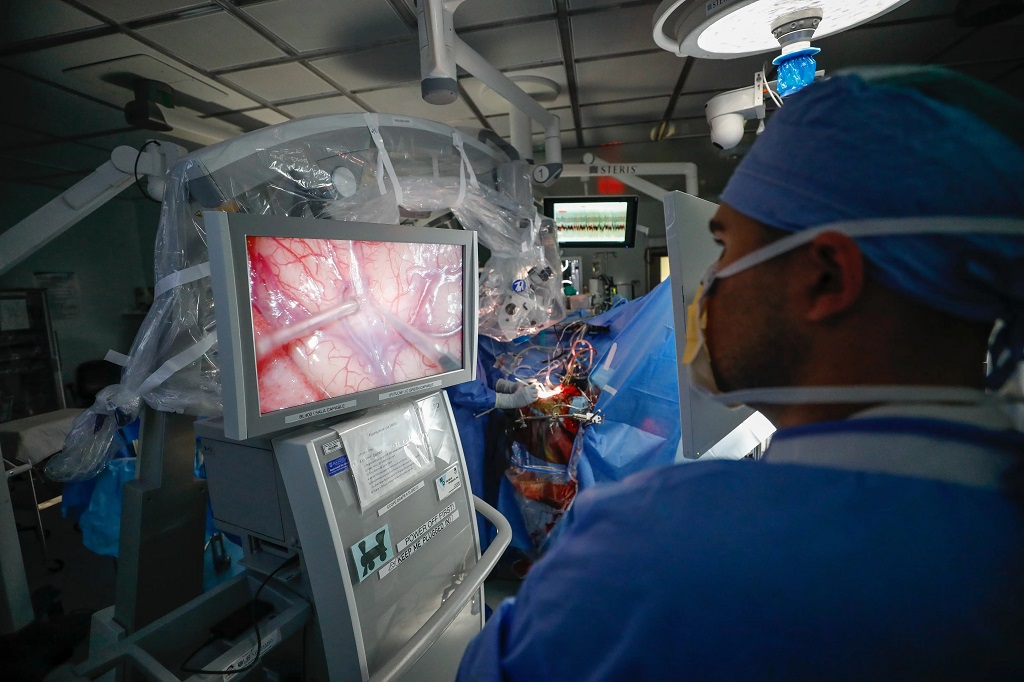
The ‘mommy makeover’ is trending, and a growing number of patients are now asking whether cosmetic procedures such as a tummy tuck, liposuction, or breast augmentation can be performed at the same time as a Caesarean section. But surgeons warn that combining elective cosmetic surgery with a C-section can sharply escalate risk during an already vulnerable period for the body.
Professor Chrysis Sofianos, a triple-board certified plastic surgeon and Academic Head of the Division of Plastic and Restorative Surgery at the University of the Witwatersrand, says procedures such as a tummy tuck should only be considered once the body has adequately recovered after childbirth – typically around six months after delivery, depending on individual healing.
“Our practice is seeing a growing number of patients ask whether body-contouring surgery can be performed while they are already in theatre for a C-section. But this reflects a dangerous misunderstanding of surgical safety and postpartum physiology.
“While the idea may appear efficient or financially attractive, pairing medically necessary obstetric surgery with elective cosmetic procedures significantly increases operative risk at a time when the patient is physiologically vulnerable.”
Combining surgeries and compounding risks
C-sections account for around 75% of private sector hospital births in South Africa. Professor Sofianos notes that because there is often an overlap between women accessing private medical care and those who may later consider elective cosmetic procedures, more patients are likely to ask whether these operations can be combined.
“However, the more important question is whether they should. And the simple answer is no,” he says. “A C-section is already a major abdominal operation. Introducing additional surgical trauma before the body has recovered would introduce excessive strain and substantially raise the risk of complications.”
Pregnancy and the immediate postpartum period are associated with a hypercoagulable state, meaning the blood has an increased tendency to clot. Postpartum women therefore face a markedly elevated risk of venous thromboembolism, particularly in the first six weeks after delivery. Prolonging operative time and increasing tissue disruption may further elevate this risk by contributing to immobility, tissue stress, and inflammatory response.
A C-section on its own carries recognised complications, including haemorrhage, infection, anaesthetic complications, and clotting risk. Adding abdominoplasty (tummy tuck) can introduce additional risks such as bleeding, fluid accumulation, wound breakdown, delayed healing, and blood clots.
Liposuction also introduces risks, such as fluid imbalance, internal injury, infection, and, in rare but serious cases, fat embolism – a potentially life-threatening condition in which fat enters the bloodstream and compromises vital organs.
The false economy of combining procedures
Professor Sofianos also notes that combining procedures rarely provides the financial or practical advantages patients may assume.
“There is a common a misconception that theatre and anaesthetic fees can be consolidated if surgeries are combined into a single session. In reality, longer operative times, greater monitoring requirements, and the potential for complications may result in far higher medical costs. More importantly, financial reasoning should never supersede patient safety.”
He adds that the combined recovery period can also be far more demanding than patients anticipate.
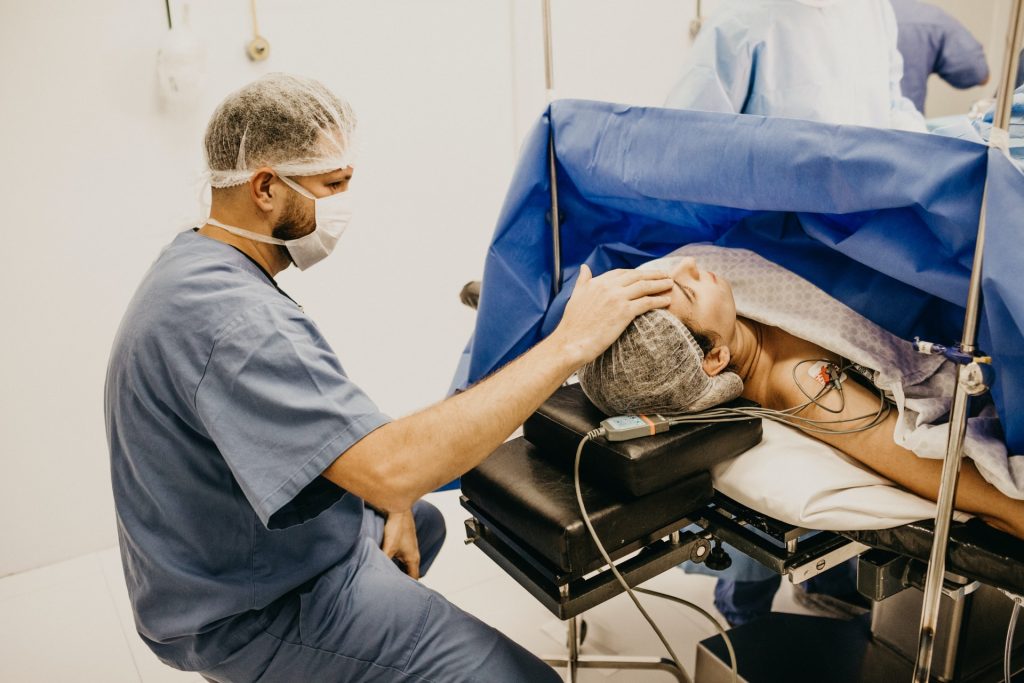
“Recovery after a C-section already places significant physical, emotional, and psychological demands on a new mother. Adding major cosmetic surgery to that recovery period can complicate mobility, wound care, and pain management at a time when the patient must also care for a newborn.
“A more intensive recovery process may further require extended postoperative care, closer medical oversight, and additional support at home, all of which can add to the existing financial burden.”
Finally, he warns that operating during the immediate postpartum period might not produce the optimal long-term aesthetic result a patient may be looking for, and could expose them to unnecessary revision surgery later.
“Medically and ethically, I do not believe combined C-section and ‘mommy makeover’ surgeries should ever be considered. No responsible surgeon should minimise the compounded risks associated with performing such procedures. Ultimately, safe, staged care remains the gold standard for medical care, or allowing the body to recover fully before elective cosmetic surgery is undertaken.”