Category: Regenerative Medicine

New Regeneration Drug for Spinal Cord Injury Passes Safety Check

Researchers in the UK have evaluated a potential drug for the treatment of spinal cord injury (SCI), which could potentially regrow damaged nerves, and found it to be safe and tolerable. The results of their Phase 1 clinical trial were published in British Journal of Clinical Pharmacology and evaluated the KCL-286 drug, which activates retinoic acid receptor beta (RARb) in the spine to promote recovery.

There are no licensed drugs that can fix the adult central nervous system’s inability to regenerate. Implants have been able to restore some function, but for most, spinal cord injuries are life-changing.

Previous studies have shown that nerve growth can be stimulated by activating the RARb2 receptor, but no drug suitable for humans has been developed. KCL-286, an RARb2 agonist, was developed by Professor Corcoran and team and used in a first in man study to test its safety in humans.

The study by the Institute of Psychiatry, Psychology & Neuroscience (IoPPN) at King’s College London, recruited 109 healthy males in a single ascending dose (SAD) adaptive design with a food interaction (FI) arm, and multiple ascending dose (MAD) arm. Participants in each arm were further divided into different dose treatments.

SAD studies are designed to establish the safe dosage range of a medicine by providing participants with small doses before gradually increasing the dose provided. Researchers look for any side effects, and measure how the medicine is processed within the body. MAD studies explore how the body interacts with repeated administration of the drug, and investigate the potential for a drug to accumulate within the body.

Researchers found that participants were able to safely take 100mg doses of KCL-286, with no severe adverse events.

Professor Jonathan Corcoran, Professor of Neuroscience and Director of the Neuroscience Drug Discovery Unit, at King’s IoPPN and the study’s senior author said, “This represents an important first step in demonstrating the viability of KCL-286 in treating spinal cord injuries. This first-in-human study has shown that a 100mg dose delivered via a pill can be safely taken by humans. Furthermore, we have also shown evidence that it engages with the correct receptor.

“Our focus can hopefully now turn to researching the effects of this intervention in people with spinal cord injuries.”

Dr Bia Goncalves, a senior scientist and project manager of the study, and the study’s first author from King’s IoPPN said, “Spinal Cord Injuries are a life changing condition that can have a huge impact on a person’s ability to carry out the most basic of tasks, and the knock-on effects on their physical and mental health are significant.

“The outcomes of this study demonstrate the potential for therapeutic interventions for SCI, and I am hopeful for what our future research will find.”

The researchers are now seeking funding for a Phase 2a trial studying the safety and tolerability of the drug in those with SCI.

Source: King’s College London
Backdoor to the Inner Ear Allows Delivery of Gene Therapy

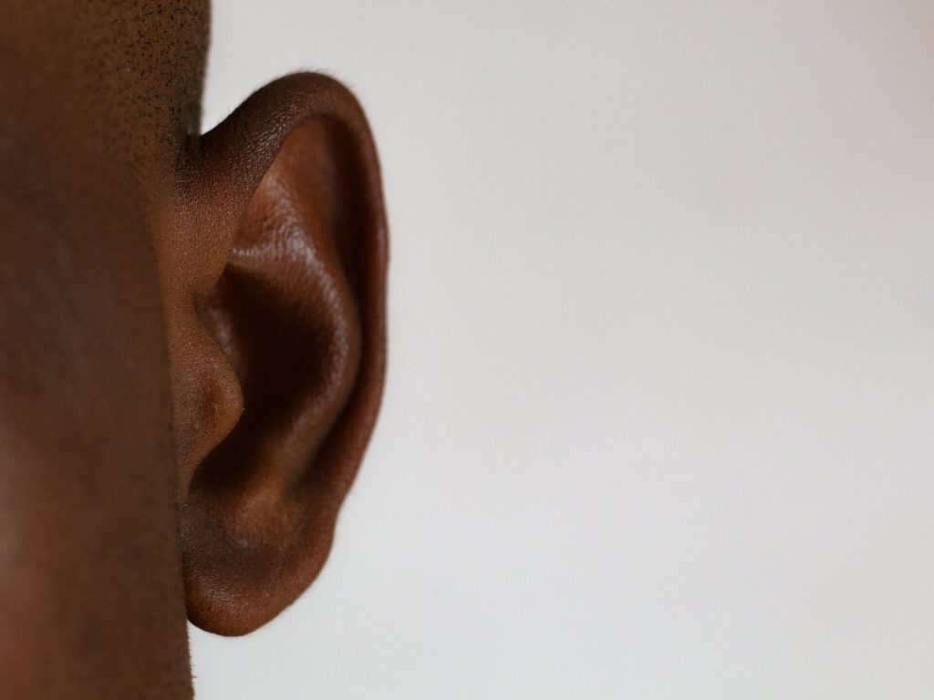
Photo by Dylann Hendricks on Unsplash

An international team of researchers has developed a new method to deliver drugs into the inner ear, according to a new study in Science Translational Medicine. The discovery was possible by harnessing the natural flow of fluids in the brain and employing a little-understood backdoor into the cochlea. When combined to deliver a gene therapy that repairs inner ear hair cells, the researchers were able to restore hearing in deaf mice.

“These findings demonstrate that cerebrospinal fluid transport comprises an accessible route for gene delivery to the adult inner ear and may represent an important step towards using gene therapy to restore hearing in humans,” says lead author Barbara Canlon, professor at Karolinska Institutet.

The number of people worldwide predicted to have mild to complete hearing loss is expected to grow to around 2.5 billion by mid-century.  The primary cause is the death or loss of function of hair cells found in the cochlea – which relay sounds to the brain – due to mutations of critical genes, aging, noise exposure, and other factors. 

While hair cells do not naturally regenerate in humans and other mammals, gene therapies have shown promise and in separate studies have successfully repaired the function of hair cells in neo-natal and very young mice.

“However, as both mice and humans age, the cochlea, already a delicate structure, becomes enclosed in the temporal bone. At this point, any effort to reach the cochlea and deliver gene therapy via surgery risks damaging this sensitive area and altering hearing,” says Barbara Canlon.

In the new study, the researchers describe a little-understood passage into the cochlea called the cochlear aqueduct. The cochlear aqueduct is a thin boney channel no larger than several strands of hair. 

Channel for spinal fluid

A new study shows that the cochlear aqueduct acts as a conduit between the cerebrospinal fluid found in the inner ear and the rest of the brain. 

Scientists are developing a clearer picture of the mechanics of the glymphatic system, the brain’s unique process of removing waste. Because the glymphatic system pumps cerebrospinal fluid deep into brain tissue to wash away toxic proteins, researchers have been eyeing it as a potential new way to deliver drugs into the brain, a major challenge in developing drugs for neurological disorders. 

The new study represented an opportunity to put the drug delivery potential of the glymphatic system to the test, while at the same time targeting a previously unreachable part of the auditory system.   

Employing several imagining and modeling technologies, the researchers were able to develop a detailed portrait of how fluid from other parts of the brain flows through the cochlear aqueduct and into the inner ear.

The team then injected an adeno-associated virus into the cisterna magna, a large reservoir of cerebrospinal fluid found at the base of the skull. 

The virus found its way into the inner ear via the cochlear aqueduct and delivered a gene therapy that expresses a protein called vesicular glutamate transporter-3, which enables the hair cells to transmit signals and rescue hearing in adult deaf mice. 

“This new delivery route into the ear may not only serve the advancement of auditory research but also prove useful when translated to humans with progressive genetic-mediated hearing loss,” says Barbara Canlon.

Source: Karolinska Institutet

Cimetidine may Aid Spinal Cord Healing by Controlling Inflammation

Researchers screening more than 1000 potential drugs for spinal cord injury treatment identified an existing one – cimetidine – that improved spinal repair in zebrafish. The results, published in the journal Theranostics, showed that the drug also helped improve recovery of movement and reduce the extent of spinal cord damage when tested in spinal-injured mice.

Healing of spinal cord injuries can be inefficient due to inflammation caused by an overreaction of the immune system. Anti-inflammatories that suppress the whole immune response also inhibit the immune cells which promote repair.

The University of Edinburgh-led study tested multiple drugs in zebrafish larvae for their ability to prevent excessive inflammation during an immune response. Scientists discovered that cimetidine acts by helping to regulate histamine levels.

The findings have enabled the team to pinpoint a specific signalling pathway that moderates the immune response after spinal injury to support repair.

The investigators say that other drugs that work in a similar way could also be tested for their ability to support recovery from spinal injury. They caution that further studies are needed to investigate their impact in human clinical trials. The researchers add that the study highlights the usefulness of zebrafish in the drug discovery process.

The research team included scientists from the University of Edinburgh, the Research Institute of the McGill University Health Centre and Technische Universität Dresden.

Source: University of Edinburgh

Researchers Identify New Compound that Could Stimulate Nerve Regeneration

Source: CC0

Research published in Nature has identified a new compound that can stimulate nerve regeneration after injury, as well as protect cardiac tissue from the sort of damage seen in heart attack. The UCL-led study identified a chemical compound, named ‘1938’, that activates the PI3K signalling pathway, and is involved in cell growth.

Results from this early research, which was done in partnership with the MRC Laboratory of Molecular Biology (MRC LMB) and AstraZeneca, showed the compound increased neuron growth in nerve cells, and in animal models, it reduced heart tissue damage after major trauma and regenerated lost motor function in a model of nerve injury.

Though further research is needed to translate these findings into the clinic, 1938 is one of just a few compounds in development that can promote nerve regeneration, for which there are currently no approved medicines.

Phosphoinositide 3-kinase (PI3K) is a type of enzyme that helps to control cell growth. It is active in various situations, such as initiating wound healing, but its functions can also be hijacked by cancer cells to allow them to proliferate. As a result, cancer drugs have been developed that inhibit PI3K to restrict tumour growth. But the clinical potential of activating the PI3K pathway remains underexplored.

Dr Roger Williams, a senior author of the study from the MRC Laboratory of Molecular Biology, said: “Kinases are ‘molecular machines’ that are key to controlling the activities of our cells, and they are targets for a wide range of drugs. Our aim was to find activators of one of these molecular machines, with the goal of making the machine work better. We found that we can directly activate a kinase with a small molecule to achieve therapeutic benefits in protecting hearts from injury and stimulating neural regeneration in animal studies.”

In this study, researchers from UCL and MRC LMB worked with researchers from AstraZeneca to screen thousands of molecules from its chemical compound library to create one that could activate the PI3K signalling pathway. They found that the compound named 1938 was able to activate PI3K reliably and its biological effect were assessed through experiments on cardiac tissue and nerve cells.

Researchers at UCL’s Hatter Cardiovascular Institute found that administering 1938 during the first 15 minutes of blood flow restoration following a heart attack provided substantial tissue protection in a preclinical model. Ordinarily, areas of dead tissue form when blood flow is restored that can lead to heart problems later in life.

When 1938 was added to lab-grown nerve cells, neuron growth was significantly increased. A rat model with a sciatic nerve injury was also tested, with delivery of 1938 to the injured nerve resulting in increased recovery in the hind leg muscle, indicative of nerve regeneration.

Senior author Professor James Phillips said: “There are currently no approved medicines to regenerate nerves, which can be damaged as a result of injury or disease, so there’s a huge unmet need. Our results show that there’s potential for drugs that activate PI3K to accelerate nerve regeneration and, crucially, localised delivery methods could avoid issues with off-target effects that have seen other compounds fail.”

Given the positive findings, the group is now working to develop new therapies for peripheral nerve damage, such as those sustained in serious hand and arm injuries. They are also exploring whether PI3K activators could be used to help treat damage in the central nervous system, for example due to spinal cord injury, stroke or neurodegenerative disease.

Source: University College London

Cultured Cells may Restore Vision Lost to Photoreceptor Damage

A preclinical study that produced progenitor photoreceptor cells and transplanted them into experimental models of damaged retinas has resulted in significant vision recovery. This finding marks a first step towards potentially restoring vision in eye diseases characterised by photoreceptor loss.

“Our laboratory has developed a novel method that enables the production of photoreceptor progenitor cells resembling those in human embryos,” said Assistant Professor Tay Hwee Goon, first author of the study published in Molecular Therapy. “Transplantation of these cells into experimental models has yielded partial restoration of the retinal function.”

The degeneration of photoreceptors in the eye is a significant cause of declining vision that can eventually lead to blindness and for which there is currently no effective treatment. Photoreceptor degeneration occurs in a variety of inherited retinal diseases, such as retinitis pigmentosa, a rare eye disease that breaks down cells in the retina over time and eventually causes vision loss, and age-related macular degeneration, a leading cause of vision impairment worldwide.

Asst Prof Tay and her team from Duke-NUS Medical School, the Singapore Eye Research Institute and the Karolinska Institute in Sweden, developed a procedure to grow human embryonic stem cells in the presence of purified laminin proteins. These proteins are involved in normal development of human retinas, and in their presence, stem cells could be directed to differentiate into photoreceptor progenitor cells responsible for converting light into signals that are sent to the brain.

When these cells were transplanted into damaged retinas, the preclinical models showed significant recovery of vision. A diagnostic test called electroretinogram also identified significant recovery in the retinas via electrical activity in the retina in response to a light stimulus. The transplanted cells established connections with surrounding retinal cells and nerves in the inner retina. They also survived and functioned for many weeks after transplantation.

Moving forward, the team hopes to refine their method to make it simpler and achieve more consistent results than earlier attempts to explore stem cell therapy for photoreceptor cell replacement.

“It is exciting to find these results, which suggest a promising route towards using stem cells to treat those forms of visual deterioration and blindness caused by the loss of photoreceptors,” said Dr Helder Andre, Head of Molecular and Cellular Research from Karolinska Institute’s Department of Clinical Neuroscience and a senior author of the study.

Associate Professor Enrico Petretto, Director of the Centre for Computational Biology at Duke-NUS and the study’s bioinformatics analysis lead, added: “Our method may also be useful for understanding the molecular and cellular pathways that drive the progression of macular degeneration, perhaps leading to the development of other therapeutic approaches.”

The next challenge for the researchers is to explore the efficacy of their method in models of photoreceptor degeneration that more closely match the human condition.

“If we get promising results in our future studies, we hope to move to clinical trials in patients,” said Professor Karl Tryggvason, from Duke-NUS’ Cardiovascular and Metabolic Disorders Programme, and the corresponding author of the study. “That would be an important step towards for being able to reverse damage of the retina and restore vision.”

Source: Duke-NUS Medical School

Can Progressive Hearing Loss be Reversed?

Photo by Dylann Hendricks on Unsplash

In humans, hearing loss from exposure to loud noises is progressive because the primary cells which detect sound, cochlear hair cells, cannot regenerate if damaged or lost. People who have repeated exposure to loud noises, like military personnel, construction workers, and musicians, are most at risk for this type of hearing loss, though it can happen to anyone over time.

On the other hand, birds and fish can regenerate these hair cells, and now researchers report their advances in promoting this effect in mammals. Their work is published in Frontiers in Cellular Neuroscience.

“We know from our previous work that expression of an active growth gene, called ERBB2, was able to activate the growth of new hair cells (in mammals), but we didn’t fully understand why,” said Patricia White, PhD, professor of Neuroscience and Otolaryngology at the University of Rochester Medical Center. The 2018 study led by Jingyuan Zhang, PhD, a postdoctoral fellow in the White lab at the time, found that activating the growth gene ERBB2 pathway triggered a cascading series of cellular events by which cochlear support cells began to multiply and activate other neighbouring stem cells to become new sensory hair cells.

“This new study tells us how that activation is happening – a significant advance toward the ultimate goal of generating new cochlear hair cells in mammals,” said White.

Using single-cell RNA sequencing in mice, researchers compared cells with an overactive growth gene (ERBB2 signalling) with similar cells that lacked such signalling. They found the growth gene, ERBB2, promoted stem cell-like development by initiating the expression of multiple proteins – including SPP1, a protein that signals through the CD44 receptor. The CD44 receptor is known to be present in cochlear-supporting cells. This increase in cellular response promoted mitosis in the supporting cells, a key event for regeneration.

“When we checked this process in adult mice, we were able to show that ERBB2 expression drove the protein expression of SPP1 that is necessary to activate CD44 and grow new hair cells,” said Dorota Piekna-Przybylska, PhD, a staff scientist in the White Lab and first author of the study. “This discovery has made it clear that regeneration is not only restricted to the early stages of development. We believe we can use these findings to drive regeneration in adults.”

“We plan to further investigation of this phenomenon from a mechanistic perspective to determine whether it can improve auditory function after damage in mammals. That is the ultimate goal,” said White.

Source: University of Rochester Medical Center

Transplanted Hair Follicles Successfully Reduced Scars

Photo by Diana Polekhina on Unsplash

By treating skin scars in three volunteers with hair follicle transplants, researchers found that the scarred skin began to behave more like uninjured skin. According to the results published in Nature Regenerative Medicine, the scarred skin harboured new cells and blood vessels, remodelled collagen to restore healthy patterns, and even expressed genes found in healthy unscarred skin.

The findings could lead to better treatments for scarring both on the skin and inside the body, leading to hope for patients with extensive scarring, which can impair organ function and cause disability.

Lead author Dr Claire Higgins, of Imperial’s Department of Bioengineering, said: “After scarring, the skin never truly regains its pre-wound functions, and until now all efforts to remodel scars have yielded poor results. Our findings lay the foundation for exciting new therapies that can rejuvenate even mature scars and restore the function of healthy skin.”

Hope in hair

Scar tissue in the skin lacks hair, sweat glands, blood vessels and nerves, impairing temperature regulation and sensation. Scarring can also hinder movement as well as potentially causing discomfort and emotional distress.

Compared to scar tissue, healthy skin undergoes constant remodelling by the hair follicle. Hairy skin heals faster and scars less than non-hairy skin- and hair transplants had previously been shown to aid wound healing. Inspired by this, the researchers hypothesised that transplanting growing hair follicles into scar tissue might induce scars to remodel themselves.

To test their hypothesis, Imperial researchers worked with Dr Francisco Jiménez, lead hair transplant surgeon at the Mediteknia Clinic and Associate Research Professor at University Fernando Pessoa Canarias, in Gran Canaria, Spain. They transplanted hair follicles into the mature scars on the scalp of three participants in 2017. The researchers selected the most common type of scar, called normotrophic scars, which usually form after surgery.

They took and microscope imaged 3mm-thick biopsies of the scars just before transplantation, and then again at two, four, and six months afterwards.

The researchers found that the follicles inspired profound architectural and genetic shifts in the scars towards a profile of healthy, uninjured skin.

Dr Jiménez said: “Around 100 million people per year acquire scars in high-income countries alone, primarily as a result of surgeries. The global incidence of scars is much higher and includes extensive scarring formed after burn and traumatic injuries. Our work opens new avenues for treating scars and could even change our approach to preventing them.”

Architects of skin

After transplantation, the follicles continued to produce hair and induced restoration across skin layers.

Scarring causes the epidermis to thin out, leaving it vulnerable to tears. At six months post-transplant, the epidermis had doubled in thickness alongside increased cell growth, bringing it to around the same thickness as uninjured skin.

The next skin layer down, the dermis, is populated with connective tissue, blood vessels, sweat glands, nerves, and hair follicles. Scar maturation leaves the dermis with fewer cells and blood vessels, but after transplantation the number of cells had doubled at six months, and the number of vessels had reached nearly healthy-skin levels by four months. This demonstrated that the follicles inspired the growth of new cells and blood vessels in the scars, which are unable to do this unaided.

Scarring also increases the density of collagen fibres, causing them to align and make the scar stiffer. The hair transplants reduced the fibre density, allowing them to form a healthier, ‘basket weave’ pattern, which reduced stiffness – a key factor in tears and discomfort.

The authors also found that after transplantation, the scars expressed 719 genes differently to before. Genes that promote cell and blood vessel growth were expressed more, while genes that promote scar-forming processes were expressed less.

Underling mechanism still unknown

It is not known how exactly the transplants brought about the change. Having of a hair follicle in the scar was cosmetically acceptable for the participants as the scars were on the scalp. The researchers are now working to uncover the underlying mechanisms so they can develop therapies that remodel scar tissue towards healthy skin, without the hair follicle transplant. They can then test their findings on non-hairy skin, or on organs like the heart, which can suffer scarring after heart attacks, and the liver, which can suffer scarring through fatty liver disease and cirrhosis.

Dr Higgins said: “This work has obvious applications in restoring people’s confidence, but our approach goes beyond the cosmetic as scar tissue can cause problems in all our organs.

“While current treatments for scars like growth factors focus on single contributors to scarring, our new approach tackles multiple aspects, as the hair follicle likely delivers multiple growth factors all at once that remodel scar tissue. This lends further support to the use of treatments like hair transplantation that alter the very architecture and genetic expression of scars to restore function.”

Source: Imperial College London

Scientists Coax CNS Axons into Regenerating

Source: Pixabay CC0

Severed axons are unable to regenerate, which means that central nervous system (CNS) injuries such as to the spinal cord, can result in permanent loss of sensory and motor function. Presently, there are very limited options to help these patients regain their motor abilities. In mice, researchers have found that deleting a certain gene can cause axons to regrow. The results have recently been published in the scientific journal Neuron.

In a study using mice, a research team led by Associate Professor Kai Liu found that the deletion of PTPN2, a phosphatase-coding gene, in neurons can prompt axons to regrow. Combination with the type II interferon IFNγ, can accelerate the process and increase the number of axons regenerated.

Unlike the CNS, peripheral nerves have a greater ability to regrow and repair by themselves after injury. Scientists have yet to fully understand the relationship between this self-repair and the intrinsic immune mechanism of the nervous system. Thus, the team aimed to resolve how immune-related signalling pathways affected neurons after injury, and whether they could enhance axonal regeneration directly.

This study investigated whether the signalling pathway IFNγ-cGAS-STING had any role in the regeneration process of peripheral nerves. Researchers found that peripheral axons could directly modulate the immune response in their injured environment to promote self-repair after injury.

In previous research, Prof Liu’s team had already demonstrated that elevating the neuronal activity and regulating the neuronal glycerolipid metabolism pathway could  boost axon regenerative capacity. The current study is providing further insights into the search of treatment solutions for challenging conditions such as spinal cord injuries, with one possible option being the joining of several types of different signalling pathways.

Source: EurekAlert!

Scientists Discover that Leprosy has an Organ Regeneration Secret

Photo by Aldo Hernandez on Unsplash

Researchers say that leprosy may hold to the key to safe and effective organ regeneration, after discovering that leprosy can double the size of livers in armadillos by stimulating normal, healthy growth.

Their findings, published in the journal Cell Reports, reveal a previously unknown interaction of the leprosy bacterium with its host, in this study, an armadillo – the only one known one besides humans that the disease may manifest in.

The researchers found that leprosy appears to rewind the developmental clock of liver cells, effectively reprogramming them to be in an ‘adolescent’ state.

Regenerative medicine aims for ‘grown to order’ organs to replace those damaged by disease or age, but organ development is an extremely complex process which takes place in vivo and so far only limited progress has been made using in vitro models. The liver, a highly resilient organ, stops regenerating once it reaches its original size, making it difficult to study regeneration pathways.

Leprosy, also referred to as Hansen disease, is a chronic granulomatous infection generally caused by Mycobacterium leprae and Mycobacterium lepromatosis, both of which primarily affect the skin and peripheral nerves. It also has the ability to convert body tissues from one type to another.

Researchers infected four cloned armadillos with the bacteria, and observed the growth of their livers. The bacteria enlarged the liver, basically give themselves more room – and this was accomplished in a way that left the livers perfectly functional and healthy.

The researchers suggest that, as with other body tissues, the bacteria-induced partial reprogramming also works in adult liver in vivo, turning hepatocytes into liver progenitor-like cells leading to proliferation and subsequent re-differentiation in the microenvironment created by the bacteria.

Prof Anura Rambukkana, from the University of Edinburgh’s centre for regenerative medicine described the discover as “completely unexpected”.

“It is kind of mind-blowing,” Prof Rambukkana told the BBC. “How do they do that? There is no cell therapy that can do that.”

Fixing Spinal Cord Injuries with Stem Cell Grafts and Rehabilitation

Photo by Cottonbro on Pexels

In recent years, researchers have made strides in promoting tissue regeneration in spinal cord injuries (SCI) through implanted neural stem cells or grafts in animal models. Separate efforts have shown that intensive physical rehabilitation can improve function after SCI by promoting greater or new roles for undamaged cells and neural circuits.

University of California San Diego researchers tested whether rehabilitation can pair with pro-regenerative therapies, such as stem cell grafting. They published their findings in in JCI Insight, 

The researchers induced a cervical lesion in rats that impaired the animals’ ability to grasp with its forelimbs. The animals were divided into four groups: animals who underwent the lesion alone; animals who received a subsequent grafting of neural stem cells designed to grow and connect with existing nerves; animals who received rehabilitation only; and animals who received both stem cell therapy and rehabilitation.

Rehabilitation therapy for some animals began one month after initial injury, a time point that approximates when most human patients are admitted to SCI rehabilitation centers. Rehabilitation consisted of daily activities that rewarded them with food pellets if they performed grasping skills.

The researchers found that rehabilitation enhanced regeneration of injured corticospinal axons at the lesion site in rats, and that a combination of rehabilitation and grafting produced significant recovery in forelimb grasping when both treatments occurred one month after injury.

“These new findings indicate that rehabilitation plays a critically important role in amplifying functional recovery when combined with a pro-regenerative therapy, such as a neural stem cell transplant,” said first author Paul Lu, PhD, associate adjunct professor of neuroscience at UC San Diego School of Medicine and research health science specialist at the Veterans Administration San Diego Healthcare System.

“Indeed, we found a surprisingly potent benefit of intensive physical rehabilitation when administered as a daily regimen that substantially exceeds what humans are now provided after SCI.”

Senior author Mark H. Tuszynski, MD, PhD, professor of neurosciences and director of the Translational Neuroscience Institute at UC San Diego School of Medicine, and colleagues have long worked to address the complex challenges of repairing SCIs and restoring function.

In 2020, for example, they reported on the observed benefits of neural stem cell grafts in mice and in 2019, described 3D-printed implantable scaffolding that would promote nerve cell growth.

“There is a great unmet need to improve regenerative therapies after SCI,” said Tuszynski. “We hope that our findings point the way to a new potential combination treatment consisting of neural stem cell grafts plus rehabilitation, a strategy that we hope to move to human clinical trials over the next two years.”

Source: University of California – San Diego