Deep within either hemisphere of the brain is the “claustrum complex”, which contributes to consciousness and awareness. Many diseases known to be related to higher cognitive function, such as Alzheimer’s, schizophrenia, and ADD/ADHD, are also closely linked to abnormal function of this particular part of the brain. But how the different parts of the claustrum complex work or how its circuits and communication system are organised is not fully understood.
Researchers at Aarhus University have now uncovered this, and their results identify, down to the cellular level, which part of the claustrum complex controls our ability to discriminate familiar and novel things.
“Our study focuses on an area of the claustrum called the ‘endopiriform,’ which is a relatively unknown brain structure despite its unique brain network and cellular properties,” explains Asami Tanimura, an associate professor and the lead researcher of the study appearing as a preprint in eLife.
“For the first time, we have dissected the circuit of endopiriform to the hippocampus, and demonstrated how this pathway is crucial for recognition memory.”
In mouse models, researchers were able to observe how the mice’s behaviour changed when they respectively ‘turned on’ and ‘turned off’ the activity in this specific cell group.
Asami explains: “We observed that the cells in the endopiriform were active when the mice interacted with new conspecifics or objects, and when we inhibited this cell group, it reduced the mice’s ability to distinguish novel mouse or object from familiar ones.”
Based on this, the researchers concluded that this specific cell group in the claustrum seems to play a key role in sending memory-guided attention signal to the hippocampus.
“This is entirely new knowledge about this small but important part of the brain, and it gives us a unique understanding of the special circuit involved in recognition memory,” explains Asami.
What this knowledge might mean, and whether it could lead to the development of new treatment methods targeted at disorders in this part of the brain, remains to be seen. However, Asami and her colleagues are optimistic:
“To develop effective treatment methods, a very detailed understanding of the cells’ circuits is required. With our study, we have at least opened a door that has previously been closed in terms of specific role of the endopiriform-hippocampal circuit on higher cognitive function.”
A study published in Nature reveals the functional relevance of tumour-neuron interactions that regulate the growth of ependymoma brain tumours, one of the most common types in children. The study, conducted by researchers at Baylor College of Medicine and St. Jude Children’s Research Hospital, highlights how neuronal signalling, modifications in DNA-associated proteins and developmental programs are intertwined to drive malignancy in brain cancer.
“Ependymomas are the third most common type of paediatric brain tumours,” said co-corresponding author, Dr Benjamin Deneen professor in the Department of Neurosurgery. “These tumours are aggressive, resistant to chemotherapy and lack tumour-specific therapies, leading to poor survival.”
“We have not made an impact on patient survival in the last three decades. A major factor has been a poor understanding of the disease. The motivation of our collaborative work with the Deneen lab is to dissect the biology of these tumours as a basis for developing new therapies,” said co-corresponding author Dr Stephen Mack, associate member at St. Jude Children’s Research Hospital and member of the Department of Neurobiology, Neurobiology and Brain Tumor Program and Center of Excellence in Neuro-Oncology Sciences.
Previous studies have shown in other types of brain tumours that brain activity surrounding the tumour can influence its growth. “In the current study, we investigated whether brain activity played a role in ependymoma growth, specifically in a very aggressive type driven by a protein called ZFTA-RELA,” said first author Hsiao-Chi Chen, a graduate student in the Deneen lab. “In collaboration with the Mack lab, we developed an animal model to study this rare paediatric brain tumour and validated these findings in human tumour samples.”
The researchers discovered evidence of abnormal neuronal activity in ependymoma’s environment and investigated whether it affected ependymoma growth. They found that while hyperactivity of some neural circuits promoted tumour growth, hyperactivity of other neural circuits surprisingly reduced tumour growth, which had not been described before. Their study revealed a novel chain of events at play that regulates tumour growth, which may hold therapeutic applications.
“First, we found that normal neurons located in the brain region called dorsa raphe nucleus (dRN) project towards the cortex, where ependymoma grows. These neurons secrete serotonin, a brain chemical that carries messages between nerve cells, which surprisingly slows tumour growth,” Chen said.
Interestingly, ependymoma cells carry a serotonin transporter, a molecule that imports serotonin within the cell. “We were surprised to discover that serotonin enters ependymoma cells and binds to histone H3, a protein that is tightly associated with DNA,” Chen said. “Histone serotonylation, the addition of serotonin to histone, regulated tumour growth. Promoting it enhanced tumour growth while preventing it slowed down ependymoma growth in animal models.”
“Discovering histone serotonylation in ependymoma piqued our interest because a previous study from our lab had revealed that adding serotonin to histones affects which genes the cell turns on,” Deneen said.
The team discovered that histone serotonylation in ependymoma increases the expression of transcription factors, genes that regulate the expression of other genes,” Chen said. “We focused on transcription factor ETV5 whose overexpression accelerated tumour growth. But how does it do it?”
The next experiments showed that ETV5 expression triggers changes in the 3D structure of chromatin, the combination of DNA and proteins that forms chromosomes. The 3D changes prevent the activation of genes encoding neurotransmitters, molecules that mediate neural activity. The team focused on a neurotransmitter called neuropeptide Y (NPY) and found that growing tumours have little NPY. Restoring the levels of NPY in tumours slowed down tumour progression and tumour-associated neural hyperactivity through the remodeling of surrounding synapses or neuron-to-neuron communication.
“We knew that brain tumours release factors that remodel synapses towards hyperactivity. Here we found the opposite also can happen, that ependymoma tumours can release factors that suppress excitatory synaptic remodeling and that repressing this mechanism is essential for tumour progression,” Deneen said.
“I am excited that this work has redefined our understanding of how brain tumour cells grow, and how they take advantage of factors in their surrounding environment to initiate tumours,” Mack said. “I am equally excited that this work has revealed many new avenues for research that may in the future lead to new therapies, which is desperately needed for this devastating disease.”
More than half of us are carriers of chronic herpesvirus infections. But even though the herpes simplex virus can infect nerve cells, it rarely causes serious infection of the brain. Researchers from Aarhus University have now discovered a key element of the explanation.
The researchers have discovered a previously unknown defence mechanism in the body that is the reason why herpes infection causes a serious and potentially fatal brain inflammation in only one out of 250 000 cases. The study has recently been published in the scientific journal Nature.
“The study has exciting perspectives because it gives us a better understanding of how the brain defends itself against viral infections,” says Professor Søren Riis Paludan from the Department of Biomedicine at Aarhus University. He is the article’s last author, a Lundbeck Foundation Professor and centre director of the Excellence Centre CiViA.
“We’ve discovered how our body prevents herpesvirus from entering into the brain, even though 50–80% of us are chronically infected with this particular virus. The idea behind CiViA is that we want to understand how the body fights infections without harming itself at the same time. The mechanism we’ve found doesn’t cause inflammatory reactions,” he says.
The answer lies in the protective TMEFF1 gene.
The brain uses a novel mechanism to keep the virus out
Many years of experimenting with the genome-wide CRISPR screening technology and development of mice that lacked the critical gene have finally convinced the researchers that TMEFF1 produces a protein that prevents herpesvirus from entering into nerve cells.
The study in Nature is accompanied by another article describing two patients with brain inflammation caused by herpesvirus infection, called herpes encephalitic. In a collaborative study led by researchers in New York, the research group in Aarhus discovered that two children who developed herpes encephalitis were carrying a genetic defect that disabled the protective TMEFF1 gene.
“The new study is groundbreaking because it updates the basic understanding of immunity against viral infections,” explains Søren Riis Paludan.
“This is interesting for immunologists because it illustrates that there are still many immunological mechanisms in the brain that we don’t know about. “The study is also relevant for neuroscience because it sheds light on how the brain, so to say, prevents unwanted visitors from intruding without causing harm to the brain itself, i.e. the neuronal cells,” he says.
May provide a better understanding of Alzheimer’s
Søren Riis Paludan hopes that the study is the first step towards revealing a completely new range of brain defence mechanisms. One of the tracks that the researchers will now investigate is what the discovery may mean for the development of dementia.
Research has already demonstrated a correlation between infection with herpesviruses and later development of Alzheimer’s disease.
“Perhaps our discovery of a new antiviral mechanism in the brain can help to clarify whether individual differences in this particular mechanism or similar mechanisms can give the virus access to the brain and accelerate neurodegenerative processes,” says Søren Riis Paludan.
New research published in Developmental Medicine & Child Neurology reveals that children born preterm are more likely to screen positive for autism than full-term children.
For the study, 9725 toddlers were screened at 15-, 18-, or 24-month well child visits using a test called the Modified Checklist for Autism in Toddlers, Revised.
Screening results that were positive for autism were most common among children born extremely preterm (51.35%) and least common among those born full-term (6.95%). Subsequent evaluations after positive screening revealed the following rates of autism diagnoses: 16.05% of extremely preterm, 2.00% of very preterm, 2.89% of moderately preterm, and 1.49% of full-term births.
Utilising the screening test at ages unadjusted for early birth was effective for identifying autism, as only a small number of preterm children (1.90%) who screened positive with the test did not receive a diagnosis of autism or other developmental delay following evaluation.
“With this research, we are hoping to help dissipate doubts that clinicians might have about the utility of screening for autism in toddlers born preterm,” said corresponding author Georgina Perez Liz, MD, of the AJ Drexel Autism Institute. “Low-cost, universal public health strategies such as screening can lead to less disparity in autism detection and help children on the spectrum start specific intervention and supports earlier in life.”
Enhancing prefrontal cortex (PFC) cognitive functions requires identifying suitable exercises that increase cerebral blood flow. A recent study using functional near-infrared spectroscopy found that short-duration, low-intensity physical exercises, except static stretching with monotonous movements, increased oxygenated haemoglobin (oxy-Hb) levels, thereby activating the PFC and enhancing blood flow in children. This study marks a significant step toward improving both the physical and mental health of children.
Cognitive functions, also known as intellectual functions, encompass thinking, understanding, memory, language, computation, and judgment, and are performed in the cerebrum. The prefrontal cortex (PFC), located in the frontal lobe of the cerebral cortex, handles these functions. Studies have shown that exercise improves cognitive function through mechanisms such as enhanced cerebral blood flow, structural changes in the brain, and promotion of neurogenesis. However, 81% of children globally do not engage in enough physical activity, leading to high levels of sedentary behavior and insufficient exercise. This lack of physical activity raises concerns about its negative impact on children’s healthy brain development and cognitive function.
A recent study from Waseda University published in Scientific Reports, by doctoral student Takashi Naito from the Graduate School of Sport Sciences, along with Professors Kaori Ishii and Koichiro Oka from the Faculty of Sport Sciences, offers insights into potential solutions. The study investigated the effects of short-duration and light-intensity exercise on increasing cerebral blood flow in children. “Our goal is to develop a light-intensity exercise program that is accessible to everyone, aiming to enhance brain function and reduce children’s sedentary behaviour. We hope to promote and implement this program in schools through collaborative efforts,” says Naito.
To enhance cognitive performance, it is essential to develop exercise programs that increase cerebral blood flow. While previous studies have established the benefits of moderate-to-vigorous exercise on cognitive functions, changes in cerebral blood flow during light-intensity exercise, particularly in children, is yet to be investigated. To address this gap, the team conducted an experimental study to examine the effects of short-term, light-intensity exercises on prefrontal cortex (PFC) hemodynamics. They focused on exercises that can be easily performed on the spot without special equipment, such as stretching. Functional near-infrared spectroscopy (fNIRS), an imaging technique that measures changes in cerebral blood flow through oxy-Hb concentrations, was used for this purpose.
The study enrolled 41 healthy children ranging from fifth-grade elementary to third-year junior high school students. The children were taught seven different types of low-intensity exercises along with associated safety measures. These exercises included Upward Stretch, Shoulder Stretch, Elbow Circles, Trunk Twist, Washing Hands, Thumb and Pinky, and Single-leg Balance. The exercises were performed while seated except Single-leg Balance, with movement patterns lasting for 10 and 20 seconds. Researchers recorded and compared oxy-Hb levels at rest and during exercise.
The study’s results were highly promising, showing a significant increase in oxy-Hb levels in multiple regions of the PFC during all forms of exercise compared to the resting state. However, no significant change in oxy-Hb levels was observed during static stretching with movement in one direction. “By combining the types of exercise that easily increase blood flow in the PFC identified in this study, it is possible to develop an exercise program that everyone can easily engage in to improve children’s executive functions. It may also be used in the future to prevent cognitive decline in adults and the elderly,” explains Naito optimistically.
In conclusion, this groundbreaking study represents a significant step forward in combating sedentary lifestyles and activating brain functions in children, thereby supporting their physical and mental growth. Although this study demonstrated that even short-duration, low-intensity exercise can increase cerebral blood flow in the prefrontal cortex, future research is needed to confirm whether such exercises actually lead to improved cognitive function.
The placebo effect is very real. This we’ve known for decades, as seen in real-life observations and the best double-blinded randomised clinical trials researchers have devised for many diseases and conditions, especially pain. And yet, how and why the placebo effect occurs has remained a mystery. Now, neuroscientists have discovered a key piece of the placebo effect puzzle, reporting it in Nature.
Researchers at the University of North Carolina School of Medicine – with colleagues from Stanford, the Howard Hughes Medical Institute, and the Allen Institute for Brain Science – discovered a pain control pathway that links the cingulate cortex in the front of the brain, through the pons region of the brainstem, to cerebellum in the back of the brain.
The researchers, led by Greg Scherrer, PharmD, PhD, associate professor in the UNC Department of Cell Biology and Physiology, the UNC Neuroscience Center, and the UNC Department of Pharmacology, then showed that certain neurons and synapses along this pathway are highly activated when mice expect pain relief and experience pain relief, even when there is no medication involved.
“That neurons in our cerebral cortex communicate with the pons and cerebellum to adjust pain thresholds based on our expectations is both completely unexpected, given our previous understanding of the pain circuitry, and incredibly exciting,” said Scherrer. “Our results do open the possibility of activating this pathway through other therapeutic means, such as drugs or neurostimulation methods to treat pain.”
Scherrer and colleagues said research provides a new framework for investigating the brain pathways underlying other mind-body interactions and placebo effects beyond the ones involved in pain.
The Placebo Paradox
In conjunction with millennia of evolution, our brains can search for ways to alleviate the sensation of pain, in some cases quantifiably as with released chemicals, and less quantifiably through positive thinking and even prayer which have some documented benefit. And then there is the placebo effect.
In clinical research, the placebo effect is often seen in the “sham” treatment group that receives a fake pill or intervention that is supposed to be inert; no benefit is expected. Except that the brain is so powerful and individuals so desire to feel better that some experience a marked improvement in their symptoms. Some placebo effects are so strong that individuals are convinced they received a real treatment meant to help them.
In fact, it’s thought that some individuals in the “actual” treatment group also derive benefit from the placebo effect, complicating experimental design and driving larger sample sizes. One way to help scientists account for this is to first understand what precisely is happening in the brain of someone experiencing the placebo effect.
Enter the Scherrer lab
The scientific community’s understanding of the biological underpinnings of pain relief through placebo analgesia came from human brain imaging studies, which showed activity in certain brain regions. Those imaging studies did not have enough precision to show what was actually happening in those brain regions. So Scherrer’s team designed a set of meticulous, complementary, and time-consuming experiments to learn in more detail, with single nerve cell precision, what was happening in those regions.
First, the researchers created an assay that generates in mice the expectation of pain relief and then very real placebo effect of pain relief. Then the researchers used a series of experimental methods to study the intricacies of the anterior cingulate cortex (ACC), which had been previously associated with the pain placebo effect. While mice were experiencing the effect, the scientists used genetic tagging of neurons in the ACC, imaging of calcium in neurons of freely behaving mice, single-cell RNA sequencing techniques, electrophysiological recordings, and optogenetics – the use of light and fluorescent-tagged genes to manipulate cells.
These experiments helped them see and study the intricate neurobiology of the placebo effect down to the brain circuits, neurons, and synapses throughout the brain.
The scientists found that when mice expected pain relief, the rostral anterior cingulate cortex neurons projected their signals to the pontine nucleus, which had no previously established function in pain or pain relief. And they found that expectation of pain relief boosted signals along this pathway.
“There is an extraordinary abundance of opioid receptors here, supporting a role in pain modulation,” Scherrer said. “When we inhibited activity in this pathway, we realised we were disrupting placebo analgesia and decreasing pain thresholds. And then, in the absence of placebo conditioning, when we activated this pathway, we caused pain relief.
Lastly, the scientists found that Purkinje cells – a distinct class of large branch-like cells of the cerebellum – showed activity patterns similar to those of the ACC neurons during pain relief expectation. Scherrer and first author Chong Chen, MD, PhD, a postdoctoral research associate in the Scherrer lab, said that this is cellular-level evidence for the cerebellum’s role in cognitive pain modulation.
“We all know we need better ways to treat chronic pain, particularly treatments without harmful side effects and addictive properties,” Scherrer said. “We think our findings open the door to targeting this novel neural pain pathway to treat people in a different but potentially more effective way.”
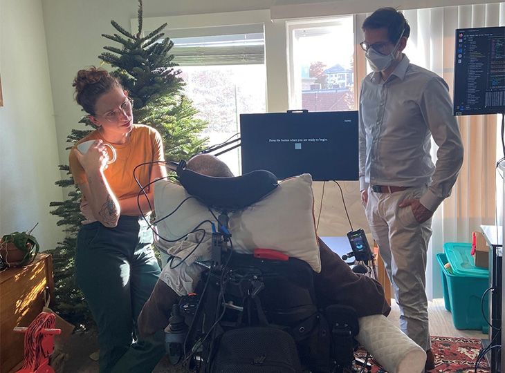
A new brain-computer interface (BCI) developed at UC Davis Health translates brain signals into speech with up to 97% accuracy – the most accurate system of its kind. The researchers implanted sensors in the brain of a man with severely impaired speech due to amyotrophic lateral sclerosis (ALS). The man was able to communicate his intended speech within minutes of activating the system.
ALS, also known as Lou Gehrig’s disease, affects the nerve cells that control movement throughout the body. The disease leads to a gradual loss of the ability to stand, walk and use one’s hands. It can also cause a person to lose control of the muscles used to speak, leading to a loss of understandable speech.
The new technology is being developed to restore communication for people who can’t speak due to paralysis or neurological conditions like ALS. It can interpret brain signals when the user tries to speak and turns them into text that is ‘spoken’ aloud by the computer.
“Our BCI technology helped a man with paralysis to communicate with friends, families and caregivers,” said UC Davis neurosurgeon David Brandman. “Our paper demonstrates the most accurate speech neuroprosthesis (device) ever reported.”
When someone tries to speak, the new BCI device transforms their brain activity into text on a computer screen. The computer can then read the text out loud.
To develop the system, the team enrolled Casey Harrell, a 45-year-old man with ALS, in the BrainGate clinical trial. At the time of his enrolment, Harrell had weakness in his arms and legs (tetraparesis). His speech was very hard to understand (dysarthria) and required others to help interpret for him.
In July 2023, Brandman implanted the investigational BCI device. He placed four microelectrode arrays into the left precentral gyrus, a brain region responsible for coordinating speech. The arrays are designed to record the brain activity from 256 cortical electrodes.
“We’re really detecting their attempt to move their muscles and talk,” explained neuroscientist Sergey Stavisky. Stavisky is an assistant professor in the Department of Neurological Surgery. He is the co-director of the UC Davis Neuroprosthetics Lab and co-principal investigator of the study. “We are recording from the part of the brain that’s trying to send these commands to the muscles. And we are basically listening into that, and we’re translating those patterns of brain activity into a phoneme – like a syllable or the unit of speech – and then the words they’re trying to say.”
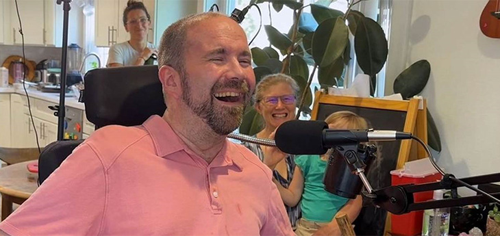
Casey Harrell with his personal assistant Emma Alaimo and UC Davis neuroscientist Sergey Stavisky
Faster training, better results
Despite recent advances in BCI technology, efforts to enable communication have been slow and prone to errors. This is because the machine-learning programs that interpreted brain signals required a large amount of time and data to perform.
“Previous speech BCI systems had frequent word errors. This made it difficult for the user to be understood consistently and was a barrier to communication,” Brandman explained. “Our objective was to develop a system that empowered someone to be understood whenever they wanted to speak.”
Harrell used the system in both prompted and spontaneous conversational settings. In both cases, speech decoding happened in real time, with continuous system updates to keep it working accurately.
The decoded words were shown on a screen. Amazingly, they were read aloud in a voice that sounded like Harrell’s before he had ALS. The voice was composed using software trained with existing audio samples of his pre-ALS voice.
At the first speech data training session, the system took 30 minutes to achieve 99.6% word accuracy with a 50-word vocabulary.
“The first time we tried the system, he cried with joy as the words he was trying to say correctly appeared on-screen. We all did,” Stavisky said.
In the second session, the size of the potential vocabulary increased to 125 000 words. With just an additional 1.4 hours of training data, the BCI achieved a 90.2% word accuracy with this greatly expanded vocabulary. After continued data collection, the BCI has maintained 97.5% accuracy.
“At this point, we can decode what Casey is trying to say correctly about 97% of the time, which is better than many commercially available smartphone applications that try to interpret a person’s voice,” Brandman said. “This technology is transformative because it provides hope for people who want to speak but can’t. I hope that technology like this speech BCI will help future patients speak with their family and friends.”
The study reports on 84 data collection sessions over 32 weeks. In total, Harrell used the speech BCI in self-paced conversations for over 248 hours to communicate in person and over video chat.
“Not being able to communicate is so frustrating and demoralising. It is like you are trapped,” Harrell said. “Something like this technology will help people back into life and society.”
“It has been immensely rewarding to see Casey regain his ability to speak with his family and friends through this technology,” said the study’s lead author, Nicholas Card. Card is a postdoctoral scholar in the UC Davis Department of Neurological Surgery.
“Casey and our other BrainGate participants are truly extraordinary. They deserve tremendous credit for joining these early clinical trials. They do this not because they’re hoping to gain any personal benefit, but to help us develop a system that will restore communication and mobility for other people with paralysis,” said co-author and BrainGate trial sponsor-investigator Leigh Hochberg. Hochberg is a neurologist and neuroscientist at Massachusetts General Hospital, Brown University and the VA Providence Healthcare System.
Brandman is the site-responsible principal investigator of the BrainGate2 clinical trial. The trial is enrolling participants. To learn more about the study, visit braingate.org or contact braingate@ucdavis.edu.
New research published in the journal iSCIENCE has revealed new insights into early sensorimotor features and cognitive abilities of toddlers who are later diagnosed with Autism Spectrum Disorder (ASD). The research, led by Kristina Denisova, a professor of Psychology and Neuroscience at the CUNY Graduate Center and Queens College, takes an important step toward better understanding ASD so that more precise, individually tailored interventions can be developed.
ASD, typically diagnosed around the ages of 4 to 5 years, is a neurodevelopmental disorder with complex and varied presentations, including atypical communication and restrictive and repetitive patterns of behaviour. Moreover, cognitive abilities are often lower in individuals with ASD. Despite the established link between lower intelligence quotient (IQ) in infancy and a future diagnosis of ASD, not all children with ASD exhibit lower cognitive abilities during infancy. The study addresses the critical gap in knowledge regarding the early features that differentiate children with varying cognitive abilities who later develop ASD.
The research team investigated the relationship between movement and cognitive abilities in toddlers before their ASD diagnosis, both during sleep and wakefulness. The study posed two key questions: Do ASD children with lower IQ exhibit altered movement during sleep compared to children with higher IQ? Additionally, are lower motor skills during wakefulness characteristic of lower-IQ children with ASD compared to those of higher-IQ ASD toddlers?
The research was conducted in two stages. In the first sample, the team examined sensorimotor features obtained from sleep functional magnetic resonance imaging (fMRI) in 111 toddlers with ASD. In the second, independent sample, they analysed sensorimotor functioning during wakefulness in over 1000 toddlers with ASD, categorised by lower vs higher cognitive abilities.
The findings revealed that toddlers with ASD and lower IQs have significantly altered sensorimotor features compared to toddlers with ASD and higher IQs. Interestingly, the sensorimotor features of higher-IQ ASD toddlers were nearly indistinguishable from typically developing (TD) toddlers. This suggests that a higher IQ may confer resilience to atypical sensorimotor functioning, and conversely, that poor sensorimotor functioning may be a key marker for lower IQ in childhood autism.
Moreover, the study found that lower-IQ ASD toddlers consistently exhibited lower gross motor skills across various age milestones (6, 12, 18, 24, and 30 months). This disruption in early sensorimotor learning during critical developmental periods indicates a potential vulnerability in the brain’s motor control circuitry, associated with lower cognitive abilities in toddlers who later receive an ASD diagnosis.
“The implications of these findings are far-reaching,” said Denisova. “They underscore the need for more precise, tailored interventions for children with ASD, particularly those with lower cognitive abilities. Interventions for lower-IQ autistic children may need to focus on enhancing both sensorimotor and cognitive skills, while interventions for higher-IQ autistic children might prioritise leveraging their strengths to mitigate potential mental health consequences.”
Denisova emphasised the importance of future research in this area, particularly involving underserved families who face barriers in accessing early intervention services.
The lines on this diagram of the brain represent connections between various areas of the cerebral cortex involved in language processing. When we read, the neurons in these areas fire in precise synchronicity, a phenomenon known as “co-rippling.” Photo credit: UC San Diego Health Sciences
Researchers at University of California San Diego School of Medicine have brought us closer to solving how the brain processes information from specialised areas into a whole. By delving into the brain with intracranial electroencephalography, they observed how neurons synchronise across the human brain while reading. The findings are published in Nature Human Behaviour and are also the basis of a thesis by UC San Diego School of Medicine doctoral candidate Jacob Garrett.
“How the activity of the brain relates to the subjective experience of consciousness is one of the fundamental unanswered questions in modern neuroscience,” said study senior author Eric Halgren, Ph.D., professor in the Departments of Neurosciences and Radiology at UC San Diego School of Medicine. “If you think about what happens when you read text, something in the brain has to turn that series of lines into a word and then associate it with an idea or an object. Our findings support the theory that this is accomplished by many different areas of the brain activating in sync.”
This synchronisation of different brain areas, called “co-rippling” is thought to be essential for binding different pieces of information together to form a coherent whole. In rodents, co-rippling has been observed in the hippocampus, the part of the brain that encodes memories. In humans, Halgren and his colleagues previously observed that co-rippling also occurs across the entire cerebral cortex.
To examine co-rippling at the mechanistic level, Ilya Verzhbinsky, an MD/PhD candidate completing his research in Halgren’s lab, led a study published in PNAS that looked at what happens to single neurons firing in different cortical areas during ripples. The present study looks at the phenomenon with a wider lens, asking how the many billions of neurons in the cortex are able to coordinate this firing to process information.
“There are 16 billion neurons in the cortex – double the number of people on Earth,” said Halgren. “In the same way a large chorus needs to be organised to sound as a single entity, our brain neurons need to be coordinated to produce a single thought or action. Co-rippling is like neurons singing on pitch and in rhythm, allowing us to integrate information and make sense of the world. Unless they’re co-rippling, these neurons have virtually no effect on the other, but once ripples are present about two thirds of neuron pairs in the cortex become synchronised. We were surprised by how powerful the effect was.”
Co-rippling in the cortex has been difficult to observe in humans due to limitations of noninvasive brain scanning. To work around this problem, the researchers used an approach called intracranial electroencephalography (EEG) scanning, which measures the electrical activity of the brain from inside the skull. The team studied a group of 13 patients with drug-resistant epilepsy who were already undergoing EEG monitoring as part of their care.
Participants were shown a series of animal names interspersed with strings of random consonants or nonsense fonts and then asked to press a button to indicate the animal whose name they saw. The researchers observed three stages of cognition during these tests: an initial hierarchical phase in visual areas of the cortex in which the participant could see the word without conscious understanding of it; a second stage in which this information was “seeded” with co-ripples into other areas of the cortex involved in more complex cognitive functions; and a final phase, again with co-ripples, where the information across the cortex is integrated into conscious knowledge and a behavioural response – pressing the button.
The researchers found that throughout the exercise, co-rippling (~100ms-long ~90Hz oscillations) occurred between the various parts of the brain engaged in these cognitive stages, but the rippling was stronger when the participants were reading real words.
The study’s findings have potential long-term implications for the treatment of neurological and psychiatric disorders, such as schizophrenia, which are characterised by disruptions in these information integration processes.
“It will be easier to find ways to reintegrate the mind in people with these disorders if we can better understand how minds are integrated in typical, healthy cases,” added Halgren.
More broadly, the study’s findings have significant implications for our understanding of the link between brain function and human experience.
“This is a fundamental question of human existence and gets at the heart of the relationship between mind and brain,” said Halgren. “By understanding how our brain’s neurons work together, we can gain new insights into the nature of consciousness itself.”
The general anaesthetic propofol may hold the keys to developing new treatment strategies for epilepsy and other neurological disorders, according to a study led by researchers at Weill Cornell Medicine and Linköping University in Sweden.
In their study, published in Nature, the researchers determined the high-resolution structural details of how propofol inhibits the activity of HCN1, an ion channel protein found on many types of neurons. Drug developers consider inhibiting HCN1 a promising strategy for treating neurologic disorders including epilepsy and chronic pain. The researchers also found, to their surprise, that when HCN1 contains either of two epilepsy-associated mutations, propofol binds to it in a way that restores its functionality.
“We might be able to exploit propofol’s unique way of binding to HCN1 for the treatment of these drug-resistant epilepsies and other HCN1-linked disorders, either by directly repurposing propofol or by designing new, more selective drugs that have the same mechanism of action,” said study co-senior author Dr Crina Nimigean, professor of physiology and biophysics in anaesthesiology at Weill Cornell Medicine.
The study’s first author was Dr Elizabeth Kim, a postdoctoral research associate in the Nimigean laboratory.
HCN ion channels in humans come in four basic forms, HCN1 to HCN4, and are found especially on cells in the heart and nervous system. They work as switches to control the electrical voltage across the cell membrane, opening to admit an inward flow of positively charged potassium and sodium ions – thus “depolarising” the cell – when the voltage reaches a certain threshold. This function underpins much of the rhythmic activity of brain and heart muscle cells, which is why HCN channels are also called pacemaker channels.
In the study, the researchers used cryo-electron microscopy and other methods to determine, at near-atomic scale, how propofol reduces HCN1 activity – which it does with selectivity for HCN1 over other HCNs. They found that the drug inhibits HCN1 by binding within a groove between two elements of the channel protein’s central pore structure, making it harder for the pore to open.
As they investigated propofol’s action on HCN1, the researchers examined how the drug affects different known mutants of the channel, including mutants that leave it excessively open and are associated with hard-to-treat epilepsy syndromes such as early infantile epileptic encephalopathy (EIEE). The researchers were surprised to find that for two different HCN1 mutations that cause EIEE, propofol restores the mutant channels to normal or near-normal function.
From their experiments, the researchers derived a model in which the mutations decouple HCN1’s voltage-sensing and pore mechanisms, while propofol effectively recouples them, allowing membrane voltage to control ion flow again.
The results suggest at least two possibilities for translation to therapies. One is simply to use propofol, an existing, approved drug, to treat these HCN1-mutation epilepsies and potentially other HCN1-linked disorders. Propofol is a potent anesthetic that requires careful monitoring by anaesthesiologists, but it might be able to restore HCN1 function at doses below those used for general anaesthesia.
The other possibility, the researchers said, is to use the new structural data on propofol’s binding to design modified, non-anesthetic versions of propofol, or even completely different compounds, that bind to HCN1 with a similar effect but much more selectively—in other words, without binding to other channels, including other HCNs, in the body and thereby potentially causing unwanted side effects.
“For that we will need a better understanding of how propofol inhibits HCN1 better than other HCN channels,” Dr Kim said.