Category: Implants and Prostheses

Heart Valve Which ‘Grows’ with Young Children Undergoing Preclinical Testing

The Iris Valve, a transcatheter, growth-accommodating pulmonary valve designed for very young children, was developed at UC Irvine and is currently progressing toward FDA clinical approval. Arash Kheradvar

Researchers at the University of California, Irvine have successfully performed preclinical laboratory testing of a replacement heart valve intended for toddlers and young children with congenital cardiac defects, a key step toward obtaining approval for human use. The results of their study were published recently in the Journal of the American Heart Association.

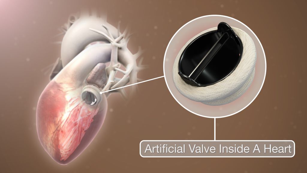
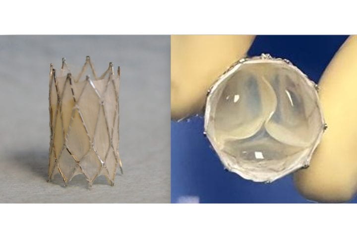
The management of patients with congenital heart disease who require surgical pulmonary valve replacement typically occurs between the ages of 2 and 10. To be eligible for a minimally invasive transcatheter pulmonary valve procedure, patients currently must weigh at least 20.4kg. For children to receive minimally invasive treatment, they must be large enough so that their veins can accommodate the size of a crimped replacement valve. The Iris Valve designed and developed by the UC Irvine team can be implanted in children weighing as little as 7.7 to 10kg and gradually expanded to an adult diameter as they grow.

Research and development of the Iris Valve has been supported by the Eunice Kennedy Shriver National Institute of Child Health and Human Development; the National Heart, Lung, and Blood Institute; and the National Science Foundation.

This funding has enabled benchtop fracture testing, which demonstrated the valve’s ability to be crimped down to a 3mm diameter for transcatheter delivery and subsequently enlarged to 20mm without damage, as well as six-month animal studies that confirmed successful device integration within the pulmonary valve annulus, showing valve integrity and a favourable tissue response.

“We are pleased to see the Iris Valve performing as we expected in laboratory bench tests and as implants in Yucatan mini pigs, a crucial measure of the device’s feasibility,” said lead author Arash Kheradvar, UC Irvine professor of biomedical engineering. “This work represents the result of longstanding collaboration between our team at UC Irvine and Dr Michael Recto at Children’s Hospital of Orange County built over several years of joint research and development.”

Congenital heart defects affect about 1% of children born in the United States and Europe, with over 1 million cases in the US alone. These conditions often necessitate surgical interventions early in life, with additional procedures required to address a leaky pulmonary valve and prevent right ventricular failure as children grow.

The Iris Valve can be implanted via a minimally invasive catheter through the patient’s femoral vein. The Kheradvar group employed origami folding techniques to compress the device into a 12-French transcatheter system, reducing its diameter to no more than 3mm. Over time, the valve can be balloon-expanded up to its full 20mm diameter.

This implantation method, along with the ability to begin treatment earlier in very young patients, helps mitigate the risk of complications from delayed care and reduces the need for multiple surgeries in this vulnerable population.

“Once the Iris Valve comes to fruition, it will save hundreds of children at least one operation – if not two – throughout the course of their lives,” said Recto, an interventional paediatric cardiologist at CHOC who’s also a clinical professor of paediatrics at UC Irvine. “It will save them from having to undergo surgical pulmonary valve placement, as the Iris Valve is delivered via a small catheter in the vein and can be serially dilated to an adult diameter and also facilitate the future placement of larger transcatheter pulmonary valves – with sizes greater than 20 millimetres, like the Melody, Harmony and Sapien devices – if needed.”

Source: University of California, Irvine

First-of-its-kind Technology Helps Man with ALS ‘Speak’ in Real Time

An investigational brain-computer interface (BCI) allows the study participant to communicate through a computer. Credit: UC Davis

Researchers at the University of California, Davis, have developed an investigational brain-computer interface that holds promise for restoring the ability to hold real-time conversations to people who have lost the ability to speak due to neurological conditions.

In a new study published in the scientific journal Nature, the researchers demonstrate how this new technology can instantaneously translate brain activity into voice as a person tries to speak – effectively creating a digital vocal tract with no detectable delay.

The system allowed the study participant, who has amyotrophic lateral sclerosis (ALS), to “speak” through a computer with his family in real time, change his intonation and “sing” simple melodies.

“Translating neural activity into text, which is how our previous speech brain-computer interface works, is akin to text messaging. It’s a big improvement compared to standard assistive technologies, but it still leads to delayed conversation. By comparison, this new real-time voice synthesis is more like a voice call,” said Sergey Stavisky, senior author of the paper and an assistant professor in the UC Davis Department of Neurological Surgery. Stavisky co-directs the UC Davis Neuroprosthetics Lab.

“With instantaneous voice synthesis, neuroprosthesis users will be able to be more included in a conversation. For example, they can interrupt, and people are less likely to interrupt them accidentally,” Stavisky said.

Decoding brain signals at heart of new technology

The man is enrolled in the BrainGate2 clinical trial at UC Davis Health. His ability to communicate through a computer has been made possible with an investigational brain-computer interface (BCI). It consists of four microelectrode arrays surgically implanted into the region of the brain responsible for producing speech.

These devices record the activity of neurons in the brain and send it to computers that interpret the signals to reconstruct voice.

“The main barrier to synthesising voice in real-time was not knowing exactly when and how the person with speech loss is trying to speak,” said Maitreyee Wairagkar, first author of the study and project scientist in the Neuroprosthetics Lab at UC Davis. “Our algorithms map neural activity to intended sounds at each moment of time. This makes it possible to synthesise nuances in speech and give the participant control over the cadence of his BCI-voice.”

Instantaneous, expressive speech with BCI shows promise

The brain-computer interface was able to translate the study participant’s neural signals into audible speech played through a speaker very quickly – one-fortieth of a second. This short delay is similar to the delay a person experiences when they speak and hear the sound of their own voice.

The technology also allowed the participant to say new words (words not already known to the system) and to make interjections. He was able to modulate the intonation of his generated computer voice to ask a question or emphasize specific words in a sentence.

The participant also took steps toward varying pitch by singing simple, short melodies.

His BCI-synthesized voice was often intelligible: Listeners could understand almost 60% of the synthesized words correctly (as opposed to 4% when he was not using the BCI).

Real-time speech helped by algorithms

The process of instantaneously translating brain activity into synthesized speech is helped by advanced artificial intelligence algorithms.

The algorithms for the new system were trained with data collected while the participant was asked to try to speak sentences shown to him on a computer screen. This gave the researchers information about what he was trying to say.

The electrodes measured the firing patterns of hundreds of neurons. The researchers aligned those patterns with the speech sounds the participant was trying to produce at that moment in time. This helped the algorithm learn to accurately reconstruct the participant’s voice from just his neural signals.

Clinical trial offers hope

“Our voice is part of what makes us who we are. Losing the ability to speak is devastating for people living with neurological conditions,” said David Brandman, co-director of the UC Davis Neuroprosthetics Lab and the neurosurgeon who performed the participant’s implant.

“The results of this research provide hope for people who want to talk but can’t. We showed how a paralyzed man was empowered to speak with a synthesized version of his voice. This kind of technology could be transformative for people living with paralysis.”

Brandman is an assistant professor in the Department of Neurological Surgery and is the site-responsible principal investigator of the BrainGate2 clinical trial.

Limitations

The researchers note that although the findings are promising, brain-to-voice neuroprostheses remain in an early phase. A key limitation is that the research was performed with a single participant with ALS. It will be crucial to replicate these results with more participants, including those who have speech loss from other causes, such as stroke.

Unlocking New Areas of the Brain for Stimulation in Parkinson’s

Deep brain stimulation illustration. Credit: NIH

People with Parkinson’s disease increasingly lose their mobility over time and are eventually unable to walk. Hope for these patients rests on deep brain stimulation. In a recent study, researchers at Ruhr University Bochum and Philipps-Universität Marburg, Germany, investigated whether and how stimulation of a certain region of the brain can have a positive impact on ambulatory ability and provide patients with a better quality of life. To do so, the researchers used a technique in which the nerve cells are activated and deactivated via light. Their report appeared in the journal Scientific Reports.

Improving ambulatory ability

If medication is no longer sufficient in alleviating restricted mobility in the advanced stage of Parkinson’s disease, one alternative is deep brain stimulation. An electrical pulse emitter is implanted within the brain, such as in the subthalamic nucleus, which is functionally part of the basal ganglia system. 

The group under Dr Liana Melo-Thomas from Philipps-Universität Marburg was able to show in previous studies on rats that stimulation of the inferior colliculus, chiefly known for processing auditory input, can be used to overcome mobility restrictions. “There are indications that stimulation of this region of the brain leads to activation of the mesencephalic locomotor region, or MLR,” says Melo-Thomas.

Interestingly, the colliculus inferior – unlike the basal ganglia –is not affected by Parkinson’s disease. However, the research group under Melo-Thomas discovered that its stimulation activates alternative motor pathways and can improve patients’ mobility.

The current study aimed to further investigate this activating influence of the inferior colliculus on the MLR. “We suspected that this would have a positive effect on ambulatory ability,” says Melo-Thomas.

Optically influencing nerve cells

The Marburg group led by Professor Rainer Schwarting sought support by Dr Wolfgang Kruse from the Department of General Zoology and Neurobiology at Ruhr University Bochum. The team in Bochum led by Professor Stefan Herlitze played a significant role in co-developing the methods of optogenetics.

While doing so, the researchers ensure that the nerve cells of genetically modified test animals produce a light-sensitive protein in interesting regions of the brain. Light that reaches these nerve cells via small, implanted optical fibres allows the researchers to activate or inhibit them specifically. “This method is thus much more precise than electrical stimulation, which always affects the area around the cells as well,” says Kruse.

For the first time, the effect of the stimulation was directly documented with electrophysiological measurements of neuronal activity in the target structures. A multi-electrode system originally developed at Philipps-Universität Marburg was used for this purpose. By combining these methods, the researchers were able to directly understand the effect of the stimulation. Parallel measurement with up to four electrodes is also highly efficient, allowing minimisation of the number of animals used. Behavioural effects that can be triggered by the stimulation were monitored in conscious animals.

Stimulation of the inferior colliculus provides the desired effect

Optogenetic stimulation in the inferior colliculus predominantly triggered the expected increase in neuronal activity within it. “Simultaneous measurements in the deeper MLR region showed increased activity in the majority of cells, although nearly one quarter of the cells were inhibited by the additional activity in the inferior colliculus,” reports Kruse. The activation of individual nerve cells occurred with an average delay of 4.7 milliseconds, indicating a functional synaptic interconnection between the inferior colliculus and MLR.

Foundations for new types of therapy

Investigating circuits outside of the basal ganglia that are affected by Parkinson’s disease is a promising step in the search for a new therapeutic approach to alleviating motor deficits resulting from the disease. Such is the case with the connection between the inferior colliculus and the MLR that was investigated for this study.

“Even if the path toward new therapeutic approaches to alleviating the symptoms of Parkinson’s disease still appears long, such foundational research is immensely important,” emphasises Kruse. The exact mechanisms that lead to the observed relief of symptoms with deep brain stimulation in the basal ganglia are not fully understood. Further investigation of the underlying interconnections may provide new insight that could optimise therapy in the long term.

Source: Ruhr-University Bochum

New Auditory Brainstem Implant Shows Early Promise

A new study co-led by Mass General Brigham researchers points to a promising new type of auditory brainstem implant (ABI) that could benefit people who are deaf due to Neurofibromatosis type 2 (NF2) and other severe inner ear abnormalities that prevent them from receiving cochlear implants. With further tests and trials, researchers hope it will provide a more effective treatment alternative than what is currently used.

In the new research, published in Nature Biomedical Engineering, scientists at Mass Eye and Ear, a member of the Mass General Brigham healthcare system, collaborated with scientists at the École Polytechnique Fédérale de Lausanne (EPFL) in Geneva, Switzerland, to report on a new class of soft, flexible ABIs that were designed to address the limitations of those currently used. These implants bypass damaged auditory structures and directly stimulate the brainstem’s sound-processing region to restore auditory function.

The new ABI was borne out of a decade-long collaboration between Mass Eye and Ear and EPFL scientists. It features an elastic, multilayer construct that includes ultra-thin platinum electrodes and silicone, a novel design that allows it to conform closely to the brainstem’s curved surface.

Conventional ABIs that are sometimes used in patients with NF2 rely on stiff electrodes that struggle to conform to the curved surface of the cochlear nucleus in the brainstem. That limits their effectiveness to modest benefits, typically providing only basic sound awareness to aid lip reading. The design can also cause side effects like discomfort that discourages long-term use.

The novel, soft electrode design was developed using advanced thin-film processing techniques, allowing for closer contact and more precise stimulation. In preclinical tests conducted in Switzerland, two macaques received the implants and underwent several months of behavioural testing. Results showed the animals could consistently distinguish between different patterns of stimulation – which indicated high-resolution auditory perception, a promising sign for eventual human use.

“While cochlear implants are life-changing for many, there remains a group of patients for whom current technology falls short,” said study co-senior author Daniel J. Lee, MD, FACS, Ansin Foundation Chair in Otolaryngology at Mass Eye and Ear. “Our research lays the groundwork for a future auditory brainstem implant that could improve hearing outcomes and reduce side effects in patients who are deaf and do not benefit from the cochlear implant.”

Source: Mass Eye and Ear

Overlooked Factor Key to Good Recovery After Hip Replacement Surgery

Photo by DanR. CC BY-NC-SA-2.0

Hip replacement surgery, or total hip arthroplasty (THA), can lessen pain and improve function in individuals with hip osteoarthritis. Some patients, however, continue to experience long-term physical deficits, including muscle weakness, decreased functional mobility, and increased fall risk, after the procedure. New research published in the Journal of Orthopaedic Research reveals that a patient’s muscle quality before THA may predict their risk of such suboptimal recovery after surgery.

In the study, 10 people undergoing THA underwent imaging tests before surgery. Patients whose imaging results indicated poor muscle quality were more likely to perform poorly on movement tasks after surgery, compared with those with good muscle quality. The severity of patients’ osteoarthritis before surgery (as indicated by the imaging tests performed) was not linked to their functional abilities after surgery.

“The findings from this study indicate that hip muscle quality may be an important predictor of post-operative biomechanical recovery following hip replacement. Muscle quality is often overlooked, and magnetic resonance imaging is needed to visualise muscle composition, which is not routinely collected for hip replacement patients,” said corresponding author Jeannie F. Bailey, PhD, of the University of California, San Francisco. “Future studies will seek to understand possible implications for poor hip muscle quality on long-term functional outcomes.”

Source: Wiley

Study Finds that Titanium Particles are Common Around Dental Implants

Photo by Tima Miroshnichenko on Pexels

Titanium micro-particles in the oral mucosa around dental implants are common. This is shown in a new study from the University of Gothenburg, which also identified 14 genes that may be affected by these particles.

According to the researchers, there is no reason for concern, but more knowledge is needed.

“Titanium is a well-studied material that has been used for decades. It is biocompatible and safe, but our findings show that we need to better understand what happens to the micro-particles over time. Do they remain in the tissue or spread elsewhere in the body?” says Tord Berglundh, senior professor of periodontology at Sahlgrenska Academy, University of Gothenburg.

Found at all implants

Previous research has shown that titanium particles may occur in inflamed tissues around dental implants. The new study, published in Communications Medicine, showed that titanium micro-particles were consistently found at all examined implants—even those without signs of inflammation.

The researchers analysed tissue samples from 21 patients with multiple adjacent implants. Samples were taken both at healthy implants and at implants affected by peri-implantitis, an inflammatory disease in the tissue around the implant. Each patient thus served as their own control. The density of particles varied between patients, but not between sites with and without peri-implantitis within the same patient. The analyses were conducted in collaboration with Uppsala University, where researchers used an advanced method called µ-PIXE to map the distribution of titanium particles in the tissue samples.

Affected genes

Peri-implantitis is a microbial biofilm-associated inflammatory disease around dental implants, with features similar to those of periodontitis around teeth. The inflammatory process is complex and the resulting destruction of supporting bone in peri-implantitis may lead to loss of the implant. 

“We observed that tissue samples with higher concentrations of titanium particles had an altered gene expression, especially genes related to inflammation and wound healing. We identified 14 such genes, but it is unclear whether the particles influence the local immune response or if the difference in gene expression reflects inter-individual variability in inflammatory conditions,” says Carlotta Dionigi, specialist in periodontology and researcher at the Department of Periodontology, Sahlgrenska Academy, University of Gothenburg.

The researchers suspect that titanium particles are released during the surgical installation procedure, when the screw-shaped implant is inserted into the prepared canal in the alveolar bone. In this context, the observation on differences in micro-particle densities between various implant systems deserves attention, since the surface structure of the implant may influence the deposition of micro-particles. This is now an important topic for continued research.

Source: University of Gothenburg

Prosthetic Hand ‘Knows’ What it’s Touching, Grasps Like a Human

Sriramana Sankar/Johns Hopkins University.

Johns Hopkins University engineers have developed a pioneering prosthetic hand that can grip plush toys, water bottles, and other everyday objects like a human, carefully conforming and adjusting its grasp to avoid damaging or mishandling whatever it holds.

The system’s hybrid design is a first for robotic hands, which have typically been too rigid or too soft to replicate a human’s touch when handling objects of varying textures and materials. The innovation offers a promising solution for people with hand loss and could improve how robotic arms interact with their environment.

Details about the device appear in Science Advances.

“The goal from the beginning has been to create a prosthetic hand that we model based on the human hand’s physical and sensing capabilities—a more natural prosthetic that functions and feels like a lost limb,” said Sriramana Sankar, a Johns Hopkins PhD student in biomedical engineering who led the work. “We want to give people with upper-limb loss the ability to safely and freely interact with their environment, to feel and hold their loved ones without concern of hurting them.”

The device, developed by the same Neuroengineering and Biomedical Instrumentations Lab that in 2018 created the world’s first electronic “skin” with a humanlike sense of pain, features a multifinger system with rubberlike polymers and a rigid 3D-printed internal skeleton. Its three layers of tactile sensors, inspired by the layers of human skin, allow it to grasp and distinguish objects of various shapes and surface textures, rather than just detect touch. Each of its soft air-filled finger joints can be controlled with the forearm’s muscles, and machine learning algorithms focus the signals from the artificial touch receptors to create a realistic sense of touch, Sankar said.

“The sensory information from its fingers is translated into the language of nerves to provide naturalistic sensory feedback through electrical nerve stimulation,” Sankar said.

In the lab, the hand identified and manipulated 15 everyday objects, including delicate stuffed toys, dish sponges, and cardboard boxes, as well as pineapples, metal water bottles, and other sturdier items. In the experiments, the device achieved the best performance compared with the alternatives, successfully handling objects with 99.69% accuracy and adjusting its grip as needed to prevent mishaps. The best example was when it nimbly picked up a thin, fragile plastic cup filled with water, using only three fingers without denting it.

“We’re combining the strengths of both rigid and soft robotics to mimic the human hand,” Sankar said. “The human hand isn’t completely rigid or purely soft—it’s a hybrid system, with bones, soft joints, and tissue working together. That’s what we want our prosthetic hand to achieve. This is new territory for robotics and prosthetics, which haven’t fully embraced this hybrid technology before. It’s being able to give a firm handshake or pick up a soft object without fear of crushing it.”

To help amputees regain the ability to feel objects while grasping, prostheses will need three key components: sensors to detect the environment, a system to translate that data into nerve-like signals, and a way to stimulate nerves so the person can feel the sensation, said Nitish Thakor, a Johns Hopkins biomedical engineering professor who directed the work.

“The goal from the beginning has been to create a prosthetic hand that we model based on the human hand’s physical and sensing capabilities—a more natural prosthetic that functions and feels like a lost limb.”

Sriramana Sankar

PhD student, Biomedial engineering

The bioinspired technology allows the hand to function this way, using muscle signals from the forearm, like most hand prostheses. These signals bridge the brain and nerves, allowing the hand to flex, release, or react based on its sense of touch. The result is a robotic hand that intuitively “knows” what it’s touching, much like the nervous system does, Thakor said.

“If you’re holding a cup of coffee, how do you know you’re about to drop it? Your palm and fingertips send signals to your brain that the cup is slipping,” Thakor said. “Our system is neurally inspired—it models the hand’s touch receptors to produce nervelike messages so the prosthetics’ ‘brain,’ or its computer, understands if something is hot or cold, soft or hard, or slipping from the grip.”

While the research is an early breakthrough for hybrid robotic technology that could transform both prosthetics and robotics, more work is needed to refine the system, Thakor said. Future improvements could include stronger grip forces, additional sensors, and industrial-grade materials.

“This hybrid dexterity isn’t just essential for next-generation prostheses,” Thakor said. “It’s what the robotic hands of the future need because they won’t just be handling large, heavy objects. They’ll need to work with delicate materials such as glass, fabric, or soft toys. That’s why a hybrid robot, designed like the human hand, is so valuable—it combines soft and rigid structures, just like our skin, tissue, and bones.”

Össur South Africa Launches ‘What’s Your Epic?’ to Further Empower Those Living with Limb Loss

Transform Lives, Break Barriers, Redefine Possibilities

Dane Wilson, Michael Stevens and Reuben van Niekerk of Jumping Kids, a nonprofit that supports young amputees.

Ahead of this year’s Cape Epic, Össur South Africa has announced the launch of its ‘What’s Your Epic?’ initiative. In partnership with Aramex, this campaign supports three nonprofit organisations (NPOs) – all of which provide hope, mobility, and independence to individuals with limited movement. ‘What’s Your Epic?’ aims to further empower amputees to overcome barriers, move freely, and live life to the fullest.

“Movement is a fundamental right. While not everyone may be an elite athlete, everyone deserves the freedom to move,” says Blignaut Knoetze, MD of Össur South Africa, a global provider of non-invasive orthopaedics. “We are committed to improving people’s mobility so that they can live their life without limitations. Our efforts and expertise are focused on helping those living with limb loss to be confident, safe and mobile, regardless of injuries or conditions that could compromise their quality of life.”

To help drive awareness for these NPOs and the valuable work that they do, so closely aligned with its own mission, Össur South Africa has entered three teams into the prestigious Cape Epic mountain bike race, with each team representing and raising funds for one of the selected NPOs. These teams not only showcase the resilience of their riders but also shine a light on the important and essential work carried out by these organisations.

The NPOs and Riders Making a Difference

Rejuvenate SA
After an elective amputation in 2020, Travis Warwick-Oliver turned to adaptive sports and co-founded Rejuvenate SA with prosthetist Luvan Cass. Their nonprofit provides mobility aids and vocational training to underprivileged individuals, particularly in rural KwaZulu-Natal. As they gear up for another Cape Epic, they see it as more than just a race – it’s an opportunity to raise awareness and inspire involvement. “We’re not just trying to get people moving; we’re trying to create a better future and give them the opportunity to fend for themselves,” explains Cass. Their journey embodies resilience, community, and the belief that movement is the key to opportunity and dignity.

Jumping Kids
Led by director Michael Stevens, Jumping Kids supports young amputees by providing prosthetics, education, and sporting opportunities – equipping children with limb loss with the prosthetic technology they need to run, play, and chase their dreams. Ambassadors Reuben van Niekerk and Dane Wilson (both amputees) advocate for mobility solutions, emphasising that the ‘What’s Your Epic?’ campaign is about more than just sports—it’s about redefining possibilities. “Whether through donations, raising awareness, or inspiring others, every action helps build a future where children with disabilities can thrive,” says Stevens.

Zimele NPC
Rentia Retief lost her leg in a 2023 cycling accident. Just a year later, she is set to compete in the Cape Epic alongside teammate Jackie Church. Supporting Zimele NPC (‘independence’ in Xhosa), a nonprofit dedicated to empowering adult amputees to lead independent lives, Rentia is proving that disability does not define potential. “Being part of this experience is truly inspiring,” says Church, an Össur South Africa employee. “Rentia is showing others what’s possible and breaking barriers for amputees everywhere.”

“These three NPOs are lifelines for those who often lack essential resources or healthcare,” says Knoetze, Össur South Africa aims to support them by raising vital funds, thereby helping them to expand their impact and, in turn, help ensure that more amputees have the freedom to move, dream, and live fully.

“’What’s Your Epic?’ is more than a fundraising initiative—it’s a movement to shift perceptions, raise awareness, and advocate for individuals with limb loss,” adds Knoetze. “Movement changes lives. Together, we can empower amputees to overcome challenges, dream boldly, and achieve the extraordinary.”

What’s your Epic?

To support these heroes taking part in the upcoming Cape Epic, and help to extend the impact of these three NPOs (Rejuvenate SA, Jumping Kids and Zimele), please visit GivenGain:  https://www.givengain.com/event/ossur-sa-giving-back.

Mechanical Heart Valve Replacements have Better Long-term Survival

Artificial heart valve. Credit: Scientific Animations CC4.0

Patients aged between 50 to 70 years with a mechanical heart valve replacement had better long-term survival compared to those with a biological valve, new research led by the University of Bristol has found. The study is published in the European Journal of Cardio-Thoracic Surgery.

The last two decades have seen an increase in the use of biological over mechanical heart valve replacements. However, while short-term clinical outcomes are known to be the same, long-term outcomes are still under debate.

Existing guidelines support the use of mechanical valves made of synthetic materials in patients below the age of 50, while biological valves made of animal tissue are favoured for those above the age of 65 or 70. The guidelines leave the choice to the decision of surgeons and patients who are 50 to 70 years old.

The research team wanted to find out the clinical outcomes for patients aged between 50 to 70 years undergoing elective and urgent heart valve replacement at the Bristol Heart Institute (BHI) over a 27 year period [1996 to 2023].

The researchers also sought to investigate trends, early outcomes and long-term survival rates, the incidence of repeat valve interventions and patient prosthesis mismatch (PPM).

A total of 1708 (61% male) patients with an average age of 63 years were included with 1191 (69.7%) receiving a biological valve replacement.

The research found there were no short-term differences when comparing patients receiving biological and mechanical valves. However, patients who received mechanical valves had better long-term survival up to 13 years after having surgery. 

Patients with a size 19mm biological valve replacement (a fairly small valve commonly used in females) had the worse long-term survival. Patients with a size 21mm mechanical valve had better survival compared to both size 19 and 21mm biological valves.  The study confirmed that severe PPM is a significant risk factor for poor long-term survival.

Gianni Angelini, BHF Professor of Cardiac Surgery at the Bristol Medical School: Translational Health Sciences (THS), Director of the Bristol Heart Institute and corresponding author, said: “Our study has implications for decision-making in surgical heart valve replacements for patients aged between 50 and 70 years old. The evidence supporting better long-term survival in patients receiving a mechanical heart valve suggests the current trend favouring biological valves in this age bracket should be urgently reconsidered. The survival benefit is especially clear in smaller sized valves.”

The research team recommends the evaluation of the long-term benefits associated with mechanical valves, especially in smaller sizes, despite long-term blood thinners not being needed with biological valves.

Study limitations

The single-institution design, retrospective collection of data, and absence of randomisation make the study open to bias. The lack of echocardiographic information could potentially underestimate the incidence of structural valve failure. In terms of repeat valve interventions, only patients who underwent re-do surgical aortic valve replacement or valve in valve transcatheter aortic valve implantation (TAVI) at the BHI were included.

As the BHI is a supra-regional centre, it is very unlikely that many patients might have undergone reintervention in other institutions. The cause of  death (cardiovascular/non cardiovascular) was not available.

Source: University of Bristol

Progress and Challenges in the Development of Brain Implants

Pixabay CC0

In a paper recently published in The Lancet Digital Health, a scientific team led by Stanisa Raspopovic from MedUni Vienna looks at the progress and challenges in the research and development of brain implants. New achievements in the field of this technology are seen as a source of hope for many patients with neurological disorders and have been making headlines recently. As neural implants have an effect not only on a physical but also on a psychological level, researchers are calling for particular ethical and scientific care when conducting clinical trials.

The research and development of neuroprostheses has entered a phase in which experiments on animal models are being followed by tests on humans. Only recently, reports of a paraplegic patient in the USA who was implanted with a brain chip as part of a clinical trial caused a stir. With the help of the implant, the man can control his wheelchair, operate the keyboard on his computer and use the cursor in such a way that he can even play chess. About a month after the implantation, however, the patient realised that the precision of the cursor control was decreasing and the time between his thoughts and the computer actions was delayed.

“The problem could be partially, but not completely, resolved – and illustrates just one of the potential challenges for research into this technology,” explains study author Stanisa Raspopovic from MedUni Vienna’s Center for Medical Physics and Biomedical Engineering, who published the paper together with Marcello Ienca (Technical University of Munich) and Giacomo Valle (ETH Zurich). “The questions of who will take care of the technical maintenance after the end of the study and whether the device will still be available to the patient at all after the study has been cancelled or completed are among the many aspects that need to be clarified in advance in neuroprosthesis research and development, which is predominantly industry-led.”

Protection of highly sensitive data

Neuroprostheses establish a direct connection between the nervous system and external devices and are considered a promising approach in the treatment of neurological impairments such as paraplegia, chronic pain, Parkinson’s disease and epilepsy. The implants can restore mobility, alleviate pain or improve sensory functions. However, as they form an interface to the human nervous system, they also have an effect on a psychological level: “They can influence consciousness, cognition and affective states and even free will. This means that conventional approaches to safety and efficacy assessment, such as those used in clinical drug trials, are not suitable for researching these complex systems. New models are needed to comprehensively evaluate the subjective patient experience and protect the psychological privacy of the test subjects,” Raspopovic points out.

The special technological features of neuroimplants, in particular the ability to collect and process neuronal data, pose further challenges for clinical validation and ethical oversight. Neural data is considered particularly sensitive and requires an even higher level of protection than other health information. Unsecured data transmission, inadequate data protection guidelines and the risk of hacker attacks are just some of the potential vulnerabilities that require special precautions in this context. “The use of neural implants cannot be reduced to medical risks,” summarises Stanisa Raspopovic. “We are only in the initial phase of clinical studies on these technological innovations. But questions of ethical and scientific diligence in dealing with this highly sensitive topic should be clarified now and not only after problems have arisen in test subjects or patients.”

Source: Medical University of Vienna