Skipping Breakfast may Weaken Immune Cells

Fasting may be detrimental to fighting off infection, and could lead to an increased risk of heart disease, suggests a new study published in Immunity. The research in mouse models, shows that skipping meals triggers a response in the brain that negatively affects immune cells. The results could lead to a better understanding of how chronic fasting may affect the body long term.
“There is a growing awareness that fasting is healthy, and there is indeed abundant evidence for the benefits of fasting. Our study provides a word of caution as it suggests that there may also be a cost to fasting that carries a health risk,” says lead author Filip Swirski, PhD, Director of the Cardiovascular Research Institute at Icahn Mount Sinai. “This is a mechanistic study delving into some of the fundamental biology relevant to fasting. The study shows that there is a conversation between the nervous and immune systems.”
As part of a larger study on different fasting times affect the immune systems, researchers focused on the role of breakfast. They fed one group of mice breakfast right after waking up (breakfast is their largest meal of the day), and the other group had no breakfast. Researchers collected blood samples in both groups when mice woke up (baseline), then four hours later, and eight hours later.
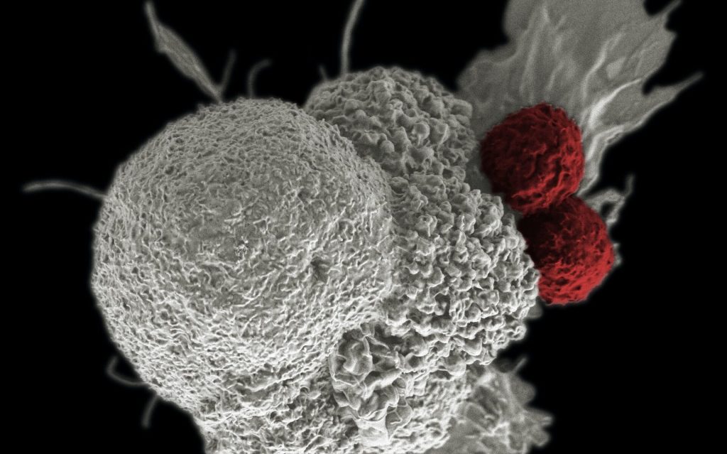
Analysing the blood work, researchers saw a difference in the number of monocytes, which are white blood cells that are made in the bone marrow and travel through the body, where they play many critical roles, from fighting infections, to heart disease, to cancer.
At baseline, all mice had the same amount of monocytes. But after four hours, monocytes in mice from the fasting group were dramatically affected. Researchers found that 90% of these cells disappeared from the bloodstream, and the number further declined at eight hours. Meanwhile monocytes in the non-fasting group were unaffected.
In fasting mice, researchers discovered the monocytes travelled back to the bone marrow to hibernate. At the same time, production of new cells in the bone marrow diminished. The normally short-lived monocytes in the bone marrow survived longer as a consequence of staying in the bone marrow, and aged differently than the monocytes that stayed in the blood.
The researchers continued to fast mice for up to 24 hours, and then reintroduced food. The cells hiding in the bone marrow surged back into the bloodstream within a few hours, provoking heightened level of inflammation. Instead of protecting against infection, these altered monocytes were more inflammatory, making the body less resistant to fighting infection.
Researchers found that specific regions in the brain controlled the monocyte response during fasting. This study demonstrated that fasting elicits a stress response in the brain (feeling ‘hangry’) and this instantly triggers a large-scale migration of these white blood cells from the blood to the bone marrow, and then back to the bloodstream shortly after food is reintroduced.
Dr Swirski emphasised that while there is also evidence of the metabolic benefits of fasting, this new study is a useful advance in the full understanding of the body’s mechanisms.
“The study shows that, on the one hand, fasting reduces the number of circulating monocytes, which one might think is a good thing, as these cells are important components of inflammation. On the other hand, reintroduction of food creates a surge of monocytes flooding back to the blood, which can be problematic. Fasting, therefore regulates this pool in ways that are not always beneficial to the body’s capacity to respond to a challenge such as an infection,” explains Dr Swirski. “Because these cells are so important to other diseases like heart disease or cancer, understanding how their function is controlled is critical.”
Source: The Mount Sinai Hospital / Mount Sinai School of Medicine