Category: Immune System

Research Shines a Light into Sex Differences in Diseases

Photo by Daniil Onischenko on Unsplash

Many diseases affect men and women differently. Asthma tends to strike men earlier in life, yet more women develop asthma as they get older. Parkinson’s is more common in men, but Alzheimer’s is more common in women. 

The differences are even more stark when it comes to autoimmune disease. Women are around two and a half times more likely than men to develop multiple sclerosis and nine times more likely to develop lupus.

Why would some diseases strike one sex more than another? And why do some tissues, such as the lungs and brain, seem especially vulnerable to these sex-based differences?

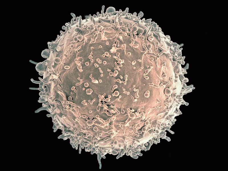
To answer these questions, scientists at La Jolla Institute for Immunology (LJI) are leading new research into how our immune cells defend specific parts of the body.

In a new Science review, LJI Professor, President & CEO Erica Ollmann Saphire, PhD, MBA, and LJI Associate Professor Sonia Sharma, PhD, examine how genetics, sex hormones, and environmental factors come together to shape the immune system.

“In just the last two years, LJI scientists have uncovered a whole new body of information about how the immune systems of men and women are very different,” says Saphire. “We’re looking at what is genetically encoded in our XX or XY chromosomes, and how hormones like oestrogen and testosterone affect what is genetically programmed into our immune cells.”

In the paper, the researchers define biological sex (in an immunology context) as the presence of XX chromosomes in females and XY chromosomes in males. “Every cell in your body is either XX or XY,” says Saphire. “That X chromosome has many, many immune-related genes. Women have two copies of each. That gives them, in a sense, twice the palette of colours to paint from in formulating an immune response. It can also give them a stronger immune response for those genes that are doubly active – active in both copies simultaneously.

Sex hormones are important for much more than reproductive function. Immune cells can also sense hormones such as oestrogen and testosterone and use them to determine which genes to turn on or off and which ones to turn on more brightly or dim. This means similar immune cells can do different things, depending on whether that cell is from a male or a female. 

Further, female cells vary in which of their two copies of X is “turned on.” As a result, women have organs with a collage, or mosaic, of immune cells that work differently in different tissues. This innate “variety” of immune cells appears to be an effective way to ward off infectious disease (women are better than men at fighting off pathogens such as SARS-CoV-2). 

But scientists have also found that having more genes from X chromosomes may predispose women to autoimmune disease. This increased X chromosome “dosage” is closely linked to a higher risk for autoimmune diseases such as Sjögren’s syndrome and scleroderma.

New research into sex-based immune system differences is also critical for developing new cancer immunotherapies, Sharma explains.

“We’re increasingly understanding how sex-based differences affect disease outcomes. When it comes to medicine, one size doesn’t fit everybody,” says Sharma, who directs LJI’s Center for Sex-Based Differences in the Immune System. “This is leading to new research, particularly in the cancer field, toward precision medicine. We’re asking how a person’s individual immune system is contributing to controlling that cancer through immunotherapy.”

Saphire and Sharma also highlight environmental factors, such as nutrition and chemical exposures, that may add to the complex interplay of chromosomes and sex hormones. Men and women also appear to have some signature differences in their skin and gut microbiomes.

The researchers hope these foundational discoveries can lead to medical advances for all, and they’re working with collaborators across the country to move this research forward. “It takes a team to translate these findings,” says Sharma.

The new review, titled “Sex differences in tissue-specific immunity and immunology,” includes co-author Alicia Gibbons of LJI and UC San Diego.

Source: La Jolla Institute for Immunology

Decoding How Immune Cells Communicate in Autoimmune Disease and Cancer

Squamous cancer cell being attacked by cytotoxic T cells. Image by National Cancer Institute on Unsplash

By measuring interactions between cells, the method offers insights into how the human body fights viral infections, how malfunctions can lead to autoimmune diseases and why immunotherapies work for some people but not others.    

A healthy immune system is trained to detect and destroy infections and cancer cells. This defence is based on a complex communication system at cellular level, in which different immune cells each perform a specialised task: recognising infectious agents, alerting other immune cells, and eliminating harmful cells or pathogens. Problems arise when communication between different cell types is disrupted, potentially leading to a variety of diseases.  

For example, cancer cells often develop strategies to specifically disrupt or circumvent the exchange of information in the immune system – this allows them to evade immune surveillance and grow unhindered. “Modern immunotherapies have fundamentally changed the treatment of certain types of cancer by restoring or specifically strengthening communication between immune cells,” explains Prof Simon Haas, one of the leaders of the study.  

Dr Daniel Hübschmann, also head of the study, adds: “However, not all patients respond equally well to these therapies and reliable methods for predicting which patients will benefit most are still lacking.”   

Decoding immune cell communication for personalised cancer therapies    

Scientists have now developed a technology that overcomes many of these hurdles through a better understanding of immune cell communication. With this method, made possible by interdisciplinary collaboration, millions of cell-cell interactions can be measured quickly and cost-effectively, both in research laboratories and in the clinic.   

The scientists are using the newly developed technology to investigate the behaviour and kinetics of immunotherapies and to gain insights into how these therapies work at the level of cell-cell interactions. They were able to show that the approach enables the prediction of individual therapy responses and can thus create a central basis for personalised immunotherapies and targeted therapy decisions.   

In addition, the researchers were able to use their new technology to visualise, in high resolution, how cells of the immune system interact with each other during viral infections and autoimmune diseases. The results allow them to develop dynamic maps of immune cell networks, illustrating for the first time how the immune defence is coordinated in different tissues.   

Together with clinical partners, the team is now working on translating these findings from research into practice, for example to better predict treatment success and utilise immunotherapies in a more personalised manner.   

The study was published in Nature Methods.  

Source: Queen Mary University London

Study Reveals Trained Immunity May Cause Lung Damage

Discovery could help explain why some people are more vulnerable to lung damage during severe inflammation

Photo by Anna Shvets

Trained immunity – a process being explored in vaccine and therapy development to boost immune defences – appears to be counterproductive in certain contexts, researchers at McGill University and the Research Institute of the McGill University Health Centre (The Institute) have found.

Trained immunity is when the body’s first line of defence remembers past threats and becomes more reactive, responding more strongly to future infections even if they are different, by changing immune cells’ behaviour.

In an earlier study, the researchers had determined beta-glucan, a molecule found in the cell walls of fungi like yeast and mushrooms, can reduce lung damage during influenza infection. That study had focused on beta-glucan’s impact on neutrophils.

However, in a new study, published in the journal eLifethe team found exposure to beta-glucan can reprogram alveolar macrophages in a way that worsens lung damage during severe inflammation caused by viral or bacterial products. These cells help keep the lungs clean by clearing out dust, debris and pathogens.

“To date, most trained immunity research has focused on circulating immune cells that arise from the bone marrow,” said lead author Renaud Prével, a postdoctoral fellow at the Meakins-Christie Laboratories at The Institute. “We wanted to explore whether beta-glucan could induce trained immunity in alveolar macrophages, and whether that might be helpful or harmful.”

The researchers exposed mice to beta-glucan, which is known to trigger trained immunity and is found in some health supplements. A week later, the mice were exposed to signals that mimic severe viral or bacterial infections with sepsis-like phenotype. Using high-resolution microCT scans and fluid analysis, they found that mice given beta-glucan developed significantly more severe lung damage compared to the untreated control group.

To confirm the immune cells were causing the damage, researchers removed them from the mice, and the inflammation went away. When they put trained alveolar macrophages into other mice, the inflammation came back. The cells showed signs of immune training, but surprisingly, this didn’t happen through the usual immune pathways. It needed signals from infections and help from other immune cells.

“Our study shows that immune memory in the lungs is more dynamic than previously thought,” said senior author Maziar Divangahi, Professor of Medicine at McGill and Associate Director of the Meakins-Christie Laboratories. “This could help explain why some individuals develop more severe lung inflammation, especially in settings like sepsis.”

Source: McGill University

Prenatal Exposure to PFAS ‘Forever Chemicals’ Shapes Baby Immunity

PFAS lurks in numerous consumer products – from nonstick cookware and food packaging to stain-resistant fabrics and personal care items. Photo by Cooker King on Unsplash

New research reveals that tiny amounts of per- and polyfluoroalkyl substances (PFAS; widely known as “forever chemicals”) cross the placenta and breast milk to alter infants’ developing immune systems, potentially leaving lasting imprints on their ability to fight disease.

University of Rochester Medical Center (URMC) researchers tracked 200 local healthy mother–baby pairs, measuring common PFAS compounds in maternal blood during pregnancy and then profiling infants’ key T‑cell populations at birth, six months, and one year. By age 12 months, babies whose mothers had higher prenatal PFAS exposure exhibited significantly fewer T follicular helper (Tfh) cells – vital coaches that help B cells produce strong, long‑lasting antibodies – and disproportionately more Th2, Th1, and regulatory T cells (Tregs), each linked to allergies, autoimmunity, or immune suppression when out of balance.

“This is the first study to identify changes in specific immune cells that are in the process of developing at the time of PFAS exposure,” said Kristin Scheible, MD, an associate professor of Pediatrics and Microbiology & Immunology at URMC and lead author of the study, which appears in the journal Environmental Health Perspectives. “Identification of these particular cells and pathways opens up the potential for early monitoring or mitigation strategies for the effects of PFAS exposure, in order to prevent lifelong diseases.”

Implications for vaccines, allergies, and autoimmunity

Tfh cell depletion helps explain previous findings that higher PFAS levels in children correlate with weaker vaccine responses to tetanus, measles, and other routine immunisations. Conversely, the uptick in Th2 and Treg cells can predispose to allergic inflammation or dampened defences, while excess Th1 activity raises concerns about future autoimmune conditions such as juvenile arthritis or type 1 diabetes.

“The cells impacted by PFAS exposure play important roles in fighting infections and establishing long-term memory to vaccines,” said Darline Castro Meléndez, PhD, a researcher in Scheible’s lab and first author of the study. “An imbalance at a time when the immune system is learning how and when to respond can lead to a higher risk of recurrent infections with more severe symptoms that could carry on through their lifetime.”

Minimising PFAS exposure

Although Rochester’s drinking water meets current safety standards, PFAS lurks in numerous consumer products – from nonstick cookware and food packaging to stain-resistant fabrics and personal care items. The study’s mothers had relatively low PFAS blood levels compared to other regions, yet the immune shifts were pronounced even in this small sample.

While not all environmental exposures can be avoided, families can reduce PFAS contact during critical windows of foetal and infant immune development. “Use water filters, minimise cooking in damaged nonstick pans, switch to alternatives like stainless steel or cast iron, and store food in glass or ceramic containers,” said Scheible. “Small steps can help lower the cumulative burden of exposure.”

The team plans a longer follow-up to determine whether these early T‑cell imbalances persist into toddlerhood and whether they translate into more infections, allergies, or autoimmune diseases. Measuring PFAS in infants directly and unravelling the molecular underpinnings of these immune disruptions are key objectives for future research.

Source: University of Rochester

Researchers Identify New Protein Target to Control Chronic Inflammation

A woman with Systemic Lupus Erythematosus. Source: Wikimedia CC0

Chronic inflammation occurs when the immune system is stuck in attack mode, sending cell after cell to defend and repair the body for months or even years. Diseases associated with chronic inflammation, like arthritis or cancer or autoimmune disorders, weigh heavily on human health – and their incidence is expected to rise. A new study by investigators from Mass General Brigham identified a protein called WSTF that could be targeted to block chronic inflammation. Crucially, this strategy would not interfere with acute inflammation, allowing the immune system to continue responding appropriately to short-term threats, such as infection by a pathogen. Results are published in Nature.

“Chronic inflammatory diseases cause a great deal of suffering and death, but we still have much to learn about what drives chronic inflammation and how to treat it,” said senior author Zhixun Dou, PhD, of the Center for Regenerative Medicine and Krantz Family Center for Cancer Research at Massachusetts General Hospital. “Our findings help us separate chronic and acute inflammation, as well as identify a new target for stopping chronic inflammation that results from aging and disease.”

Using chronically inflamed human cells, the researchers found that WSTF interacts with other proteins inside cell nuclei, which prompts its excretion and degradation. Since WSTF is responsible for concealing pro-inflammatory genes, this nucleus-eviction reveals those genes and, in turn, amplifies inflammation. They confirmed that WSTF loss could promote inflammation in mouse models of aging and cancer. They also found, using human cells, that WSTF loss only occurred in chronic inflammation, not acute. Using these findings, the researchers designed a WSTF-restoring therapeutic to suppress chronic inflammation and observed preliminary success in mouse models of aging, metabolic dysfunction-associated steatohepatitis (MASH), and osteoarthritis.

The researchers went further to examine tissue samples from patients with MASH or osteoarthritis. They found that WSTF is lost in the livers of patients with MASH, but not in the livers of healthy donors. Using cells from the knees of osteoarthritis patients undergoing joint replacement surgery, they showed that WSTF-restoring therapeutic reduces chronic inflammation from the inflamed knee cells. These findings highlight the potential of developing new treatments targeting WSTF to combat chronic inflammatory diseases.

Further research is needed to validate the therapeutic potential of WSTF restoration in broader settings and to develop specific strategies to target WSTF. Additionally, the findings suggest other similar proteins may be involved in chronic inflammation, opening a promising new avenue for studying and treating inflammation in the future.

Source: Mass General Brigham

Multiple Sclerosis Drug Ocrelizumab Works by Reshaping the Immune System

Myelin sheath damage. Credit: Scientific Animations CC4.0

When ocrelizumab became the first FDA-approved treatment for early forms of multiple sclerosis (MS) in 2017, it offered patients immense hope. The long-awaited drug is a monoclonal antibody that depletes B cells – the immune cells that drive MS progression. Exactly how ocrelizumab does this, however, remains unclear.

In a new study published in The Journal of Clinical Investigation, Yale scientists begin to answer this question. By using single-cell RNA sequencing, a technique that provides a window into the gene expression in individual cells, the researchers laid out a detailed view of how ocrelizumab achieves its therapeutic effects.

“The surprise was that the drug doesn’t work at all the way we thought it was working,” says David A. Hafler, MD, Professor of Neurology at Yale School of Medicine, who led the study. “We knew what the end result was and that the drug was enormously effective in patients. But what’s driving the drug’s action is a type of white blood cell in the central nervous system. No one would ever hypothesise that.”

The roles of T cells and B cells in multiple sclerosis

B and T cells have closely intertwined roles in the immune system. B cells are critical cells that recognise foreign objects, bind them, and present them to T cells, which then signal other immune cells to take action. But this relationship goes awry in disease.

Scanning electron micrograph of a B cell. Credit: NIH

In MS, abnormally active B cells trigger T cells to attack the myelin sheath, the protective layer of nerve fibres, leading to neurological symptoms, such as loss of vision, muscle weakness, and cognitive impairment. More than two decades ago, Hafler and his team discovered this was due to defects in regulatory T cells, which normally put the brake on immune responses, but when defective, unleash immune cells that mistakenly target the body’s own tissues.

In the early stages of MS, both B and T cells are deemed to be the drivers of the disease. Once the disease progresses to a neurodegenerative stage, other inflammatory processes become more prominent.

“Once you enter the neurodegenerative phase of the disease, it is much more difficult to stop the process,” Hafler says. “What we’ve learned is that the earlier you treat the disease, the better the outcome.”

Ocrelizumab binds to the surface of B cells, leading to their destruction. And especially for people in the early stages of MS, it can be quite effective. “The drug works incredibly well,” Hafler says. But Hafler and his team found that ocrelizumab was doing far more than just controlling B cells.

In the new study, the researchers analysed the blood and cerebrospinal fluid of 18 patients, all of whom had an early-onset form of multiple sclerosis in which patients cycle between periods of disease remission and relapse. The scientists measured the cell type-specific changes in protein expression before and after the patients received six months of ocrelizumab, in an effort to identify immune molecules that might change in response to the drug.

They discovered that the reduction in B cells driven by ocrelizumab led to an increase in the pro-inflammatory molecule TNF-α. This was unexpected because TNF-α has been shown to trigger the immune system and exacerbate inflammation in certain diseases. In fact, medications that block the activity of TNF-α are typically used for treating various autoimmune diseases such as rheumatoid arthritis and inflammatory bowel disease.

As they looked further, the researchers found that by inducing TNF-α, ocrelizumab led to an increase in a specific type of regulatory T cell. This, in turn, curbed the circulation of T cells that attack the myelin.

“This unpredicted increase in TNF-α shows that ocrelizumab works in a paradoxical way,” says Hafler.

Understanding the cause of multiple sclerosis

One of the current working models of MS suggests that the disease originates from the Epstein-Barr virus. “How the Epstein-Barr virus triggers the disease is a point that we don’t yet understand,” Hafler says. However, there is a strong body of evidence to show that the virus infects B cells. Therefore, understanding how a B cell-depleting drug affects T cell activity may lead to further explanations.

The current finding also explains why a fifth of the genes linked to MS risk involve the TNF pathway and why many of those genetic changes are protective in other diseases, such as inflammatory bowel diseases.

“This shows that biology has a richness to it,” Hafler says. “When these molecules are made, where they’re made, and what cell they’re working on have very different effects.”

Hafler suspects that ocrelizumab might be acting through other mechanisms as well, an inkling that motivates his lab to continue their investigation. “For something to work that well, there must be other things going on,” he says.

The team is now beginning to study the pathogenesis of MS in a large cohort of women who have at least one parent with the disease. By following the genetic evolution of the disease, the scientists are hoping to better understand how B cells change the immune landscape in real time.

“This study is only one piece of the puzzle,” Hafler says. “We’ll continue to look for other pieces.”

Source: Yale School of Medicine

Autoimmune Disease Linked to Doubling in Depression, Anxiety, Bipolar Risks

Risks higher in women than in men with the same condition
Chronic exposure to systemic inflammation may explain associations, say researchers

Photo by Sydney Sims on Unsplash

Living with an autoimmune disease is linked to a near doubling in the risk of persistent mental health issues, such as depression, generalised anxiety, and bipolar disorder, with these risks higher in women than in men, finds a large population-based UK study, published in the open access journal BMJ Mental Health.

Chronic exposure to the systemic inflammation caused by the autoimmune disease may explain the associations found, say the researchers.

A growing body of evidence suggests that inflammation is linked to mental ill health, but many of the published studies have relied on small sample sizes, limiting their statistical power, note the researchers.

In a bid to overcome this, they drew on data from 1.5 million participants in the recently established Our Future Health dataset from across the UK. Participants’ average age was 53; just over half (57%) were women; and 90% identified as White.

On recruitment to Our Future Health, participants completed a baseline questionnaire to provide personal, social, demographic, health and lifestyle information.

Health information included lifetime diagnoses–including for their biological parents–for a wide range of disorders, including autoimmune and psychiatric conditions.

Six autoimmune conditions were included in the study: rheumatoid arthritis; Graves’ syndrome (thyroid hormone disorder); inflammatory bowel disease; lupus, multiple sclerosis; and psoriasis.

The mental health conditions of interest were self-reported diagnoses of affective disorders, defined as depression, bipolar, or anxiety disorder.

In all, 37 808 participants reported autoimmune conditions and 1 525 347 didn’t. Those with autoimmune conditions were more likely to be women (74.5% vs 56.5%) and more likely to report lifetime diagnoses of affective disorders for their biological parents:  8% vs 5.5% for fathers; 15.5% vs 11% for mothers.

Chronic and pathogenic immune system activation—including the presence of markers of inflammation—is a hallmark of many autoimmune conditions. And in the absence of direct measurements of inflammatory biomarkers, an autoimmune condition was regarded as a proxy for chronic inflammation in this study.

The lifetime prevalence of any diagnosed affective disorder was significantly higher among people with an autoimmune disorder than it was among the general population: 29% vs 18%.

Similar associations in lifetime prevalence emerged for depression and anxiety: 25.5% vs just over 15% for depression; and just over 21% vs 12.5% for anxiety.

While the overall prevalence of bipolar disorder was much lower, it was still significantly higher among those with an autoimmune disorder than it was among the general population:  just under 1% compared with 0.5%.

The prevalence of current depression and anxiety was also higher among people with autoimmune conditions.

And the prevalence of affective disorders was significantly and consistently higher among women than it was among men with the same physical health conditions: 32% compared to 21% among participants with any autoimmune disorder.

The reasons for this aren’t clear, say the researchers, but “theories suggest that sex hormones, chromosomal factors, and differences in circulating antibodies may partly explain these sex differences,” they write.

“Women (but not men) with depression exhibit increased concentrations of circulating cytokines and acute phase reactants compared with non-depressed counterparts. It is therefore possible that women may experience the compounding challenges of increased occurrence of autoimmunity and stronger effects of immune responses on mental health, resulting in the substantially higher prevalence of affective disorders observed in this study,” they add.

Overall, the risk for each of the affective disorders was nearly twice as high—87-97% higher—in people with autoimmune conditions, and remained high even after adjusting for potentially influential factors, including age, household income, and parental psychiatric history.

No information was available on the time or duration of illness, making it impossible to determine whether autoimmune conditions preceded, co-occurred with, or followed, affective disorders, note the researchers.

No direct measurements of inflammation were made either, and it was therefore impossible to establish the presence, nature, timing or severity of inflammation, they add.

“Although the observational design of this study does not allow for direct inference of causal mechanisms, this analysis of a large national dataset suggests that chronic exposure to systemic inflammation may be linked to a greater risk for affective disorder,” they conclude.

“Future studies should seek to determine whether putative biological, psychological, and social factors—for example, chronic pain, fatigue, sleep or circadian disruptions and social isolation—may represent potentially modifiable mechanisms linking autoimmune conditions and affective disorders.”

And they suggest that it may be worth regularly screening people diagnosed with autoimmune disease for mental health conditions, especially women, to provide them with tailored treatment early on.

Source: BMJ

Daytime Gives a Boost to the Immune System, Scientists Find

Photo by Julian Jagtenberg on Pexels

A breakthrough study, led by scientists at Waipapa Taumata Rau, University of Auckland, has uncovered how daylight can boost the immune system’s ability to fight infections.

The circadian clock is a 2.5-billion-year-old cellular timekeeper that allows organisms to adapt to the rhythms of day and night. Evidence has shown that disruption of our internal body clock through the likes of shift work or jet lag makes people more susceptible to infections, so the researchers wanted to find out what in the body contributed to that susceptibility.

“In earlier studies, we had observed that immune responses to infection peaked in the morning, during the animals’ early active phase,” says lead researcher Associate Professor Christopher Hall, from the Department of Molecular Medicine and Pathology.

“We think this represents an evolutionary response such that during daylight hours the host is more active so more likely to encounter bacterial infections,” says Hall.

However, the scientists wanted to find out how the immune response was being synchronised with daylight.

They focused on ‘neutrophils’ the most abundant immune cells in our bodies. These cells move quickly to the site of an infection and kill invading bacteria.

The scientists used zebrafish, a small freshwater fish, as a model organism, because its genetic make-up is similar to ours and they can be bred to have transparent bodies, making it easy to observe biological processes in real time.

With this new study, published in Science Immunology, neutrophils were found to possess a circadian clock that alerted them to daytime, and boosted their ability to kill bacteria.

Circadian clocks are present in almost every cell and tissue in the body, telling them what is going on in the outside world and coordinating physiological processes like metabolism, hormone release, and sleep-wake cycles.

Light has the biggest influence on resetting these circadian clocks.

“Given that neutrophils are the first immune cells to be recruited to sites of inflammation, our discovery has very broad implications for therapeutic benefit in many inflammatory diseases,” Hall says.

“This finding paves the way for development of drugs that target the circadian clock in neutrophils to boost their ability to fight infections.”

Source: University of Auckland

Cytokines Also Act on the Brain, Inducing Anxiety or Sociability

Photo by Andrea Piacquadio on Pexels

Immune molecules called cytokines play important roles in the body’s defence against infection, helping to control inflammation and coordinating the responses of other immune cells. A growing body of evidence suggests that some of these molecules also influence the brain, leading to behavioural changes during illness.

Two new studies from MIT and Harvard Medical School, focused on a cytokine called IL-17, now add to that evidence. The researchers found that IL-17 acts on two distinct brain regions — the amygdala and the somatosensory cortex — to exert two divergent effects. In the amygdala, IL-17 can elicit feelings of anxiety, while in the cortex it promotes sociable behaviour.

These findings suggest that the immune and nervous systems are tightly interconnected, says Gloria Choi, an associate professor of brain and cognitive sciences, a member of MIT’s Picower Institute for Learning and Memory, and one of the senior authors of the studies.

“If you’re sick, there’s so many more things that are happening to your internal states, your mood, and your behavioural states, and that’s not simply you being fatigued physically. It has something to do with the brain,” she says.

Jun Huh, an associate professor of immunology at Harvard Medical School, is also a senior author of both studies, which appear today in CellOne of the papers was led by research scientists Byeongjun Lee and Jeong-Tae Kwon, and the other was led by postdocs Yunjin Lee and Tomoe Ishikawa.

Behavioral effects

Choi and Huh became interested in IL-17 several years ago, when they found it was involved in a phenomenon known as the fever effect. Large-scale studies of autistic children have found that for many of them, their behavioural symptoms temporarily diminish when they have a fever.

In a 2019 study in mice, Choi and Huh showed that in some cases of infection, IL-17 is released and suppresses a small region of the brain’s cortex known as S1DZ. Overactivation of neurons in this region can lead to autism-like behavioral symptoms in mice, including repetitive behaviours and reduced sociability.

“This molecule became a link that connects immune system activation, manifested as a fever, to changes in brain function and changes in the animals’ behaviour,” Choi says.

IL-17 comes in six different forms, and there are five different receptors that can bind to it. In their two new papers, the researchers set out to map which of these receptors are expressed in different parts of the brain. This mapping revealed that a pair of receptors known as IL-17RA and IL-17RB is found in the cortex, including in the S1DZ region that the researchers had previously identified. The receptors are located in a population of neurons that receive proprioceptive input and are involved in controlling behaviour.

When a type of IL-17 known as IL-17E binds to these receptors, the neurons become less excitable, which leads to the behavioural effects seen in the 2019 study.

“IL-17E, which we’ve shown to be necessary for behavioural mitigation, actually does act almost exactly like a neuromodulator in that it will immediately reduce these neurons’ excitability,” Choi says. “So, there is an immune molecule that’s acting as a neuromodulator in the brain, and its main function is to regulate excitability of neurons.”

Choi hypothesises that IL-17 may have originally evolved as a neuromodulator, and later on was appropriated by the immune system to play a role in promoting inflammation. That idea is consistent with previous work showing that in the worm C. elegans, IL-17 has no role in the immune system but instead acts on neurons. Among its effects in worms, IL-17 promotes aggregation, a form of social behaviour. Additionally, in mammals, IL-17E is actually made by neurons in the cortex, including S1DZ.

“There’s a possibility that a couple of forms of IL-17 perhaps evolved first and foremost to act as a neuromodulator in the brain, and maybe later were hijacked by the immune system also to act as immune modulators,” Choi says.

Provoking anxiety

In the other Cell paper, the researchers explored another brain location where they found IL-17 receptors — the amygdala. This almond-shaped structure plays an important role in processing emotions, including fear and anxiety.

That study revealed that in a region known as the basolateral amygdala (BLA), the IL-17RA and IL-17RE receptors, which work as a pair, are expressed in a discrete population of neurons. When these receptors bind to IL-17A and IL-17C, the neurons become more excitable, leading to an increase in anxiety.

The researchers also found that, counterintuitively, if animals are treated with antibodies that block IL-17 receptors, it actually increases the amount of IL-17C circulating in the body. This finding may help to explain unexpected outcomes observed in a clinical trial of a drug targeting the IL-17-RA receptor for psoriasis treatment, particularly regarding its potential adverse effects on mental health.

“We hypothesise that there’s a possibility that the IL-17 ligand that is upregulated in this patient cohort might act on the brain to induce suicide ideation, while in animals there is an anxiogenic phenotype,” Choi says.

During infections, this anxiety may be a beneficial response, keeping the sick individual away from others to whom the infection could spread, Choi hypothesises.

“Other than its main function of fighting pathogens, one of the ways that the immune system works is to control the host behaviour, to protect the host itself and also protect the community the host belongs to,” she says. “One of the ways the immune system is doing that is to use cytokines, secreted factors, to go to the brain as communication tools.”

The researchers found that the same BLA neurons that have receptors for IL-17 also have receptors for IL-10, a cytokine that suppresses inflammation. This molecule counteracts the excitability generated by IL-17, giving the body a way to shut off anxiety once it’s no longer useful.

Distinctive behaviours

Together, the two studies suggest that the immune system, and even a single family of cytokines, can exert a variety of effects in the brain.

“We have now different combinations of IL-17 receptors being expressed in different populations of neurons, in two different brain regions, that regulate very distinct behaviours. One is actually somewhat positive and enhances social behaviours, and another is somewhat negative and induces anxiogenic phenotypes,” Choi says.

Her lab is now working on additional mapping of IL-17 receptor locations, as well as the IL-17 molecules that bind to them, focusing on the S1DZ region. Eventually, a better understanding of these neuro-immune interactions may help researchers develop new treatments for neurological conditions such as autism or depression.

“The fact that these molecules are made by the immune system gives us a novel approach to influence brain function as means of therapeutics,” Choi says. “Instead of thinking about directly going for the brain, can we think about doing something to the immune system?”

Source: Massachusetts Institute of Technology

Findings on T Cell Exhaustion: The Body Prepares Early for a Long Fight

Scanning electron micrograph of a T cell lymphocyte. Credit: NIH / NIAID

When an infection is prolonged and severe, T cell exhaustion comes into play to prevent to reign in the immune system and prevent damage to the body. A study published in Nature reveals that right from the beginning of mild illness, the body also produces these special T cells previously known only from chronic, severe infections and tumours.

There are different types of T cells in the body, all of which play a crucial role in the immune system. They fight pathogens and control the immune response. However, some subtypes become less effective or even cease their activity altogether as the disease progresses. This has a protective function: in persisting infections, it would harm the body if the immune system continued to fight the pathogens aggressively. But in cancer treatment, T cell exhaustion means that therapeutic measures may no longer be effective.

Until now, it was assumed that the body only produces such T cells in severe and persisting infections. The results of the researchers, from Helmholtz Munich and the Technical University of Munich, show that this is not the case. “We were able to show that the body prepares T cell subtypes that are predisposed to exhaustion even in early infection phases of moderate diseases,” says Dietmar Zehn, Professor of Animal Physiology and Immunology at TUM and last study author.

Different T Cells for Different Purposes

The team deduces from the discovery that the body assembles a range of different T cells early on at the onset of the disease to arm itself for different disease progressions. Depending on the course of the disease, it then has cells at its disposal to make the immune response more aggressive or more gentle — and in some circumstances, even to abort it.

“Our results expand the classic idea of the development of T cell exhaustion,” says Dietmar Zehn. “We therefore assume that our observations will help to further decipher the mechanisms behind T cell exhaustion.” A better understanding of these processes could help in the future to control the immune response in a targeted manner — for example, to strengthen the immune system in cancer patients or to weaken excessive defences, as is typical in severe cases of COVID-19, for instance.

Source: Technical University of Munich (TUM)