Excessive amounts of visceral fat, the hidden fat surrounding organs, is linked with faster ageing of the heart, a new study has found. Ageing is the biggest risk factor for heart disease but why some people age faster than others isn’t fully understood.
The scientists leading the research, which is published in the European Heart Journal, say that visceral body fat could play an important role in accelerating ageing of the heart and blood vessels. This type of fat is known to be harmful to health and this study now links it to faster heart ageing.
Sex differences
The study, led by scientists from the Medical Research Council (MRC) Laboratory of Medical Sciences, also found differences between men and women. They discovered that fat around the hips and thighs could potentially slow heart ageing in women. The scientists analysed data from 21 241 participants in UK Biobank, which includes whole body imaging to map the amount of fat and where it is located in the body. The study was
Determining an individual’s ‘heart age’
The UK Biobank data also includes detailed imaging of the heart and blood vessels. Artificial intelligence (AI) was used to analyse these images to capture signs of organ ageing such as tissues becoming stiff and inflamed. An individual was given a ‘heart age’ which can be compared to their actual age at the time of the scan.
The risks of ‘hidden’ fat
The researchers found that faster heart ageing was linked to having more visceral adipose tissue. Visceral adipose tissue is fat found deep inside the abdomen around organs such as the stomach, intestines, and liver. This type of fat cannot be seen from the outside, and some people can have large amounts of visceral fat despite having a healthy weight.
Premature ageing
The researchers found signs on blood tests that visceral fat is linked to increased inflammation in the body, which is a potential cause of premature ageing. They also found differences between the sexes. Male-type fat distribution, which is fat around the belly and often called ‘apple-shaped’, was particularly predictive of early ageing in men.
The role of hormones
In contrast, a genetic predisposition to female-type fat, primarily fat on the hips and thighs, often called ‘pear-shaped’, was protective against heart ageing in women. The researchers also found a link between higher oestrogen levels in pre-menopausal women and a slowing of heart ageing. They suggested that this could indicate a role for hormones in protecting against heart ageing.
Increasing healthy lifespan
Professor Declan O’Regan, who led the research at the MRC Laboratory of Medical Sciences and Imperial College London, said:
We have known about the apple and pear distinction in body fat, but it hasn’t been clear how it leads to poor health outcomes. Our research shows that ‘bad’ fat, hidden deep around the organs, accelerates ageing of the heart.
But some types of fat could protect against ageing, specifically fat around the hips and thighs in women.
We also showed that body mass index wasn’t a good way of predicting heart age which underscores the importance of knowing where fat is stored in the body and not just total body weight.
The goal of our research is to find ways to increase healthy lifespan. While being active is important, we found that hidden fat could still be harmful even in fit people.
In the future, we plan to investigate how drug therapies, such as GLP-1 inhibitors (for example, Ozempic) could improve not just diabetes and obesity but target the ageing effects of hidden visceral fat.
Disposable syringe of Clexane (enoxaparin), a low molecular weight heparin (LMWH). CC0
Researchers at McMaster University have discovered that a rare but dangerous reaction to heparin is caused by a single antibody – overturning decades of medical misunderstanding and opening the door to more precise ways of diagnosing and treating this medical complication.
The study, published in the New England Journal of Medicine, focused on heparin-induced thrombocytopenia (HIT), a serious immune complication that affects approximately one per cent of hospitalised patients treated with the blood thinner heparin. Nearly half of those who develop HIT experience life-threatening blood clots, which can lead to strokes, heart attacks, amputations, and even death, making early detection and treatment critically important.
Until now, scientists believed that the immune response causing HIT involved many different types of antibodies working together. But this research has revealed something surprising: in every patient studied, only one antibody was causing the disease, while the rest created what the researchers describe as a kind of “smokescreen,” making it harder to identify the true culprit. This discovery helps pinpoint the exact source of the problem, opening the door to more accurate diagnosis and better-targeted treatments.
“This study not only challenges our existing understanding of HIT but also revolutionises how we think about the immune response overall,” said Ishac Nazy, senior author of the study and scientific director of the McMaster Platelet Immunology Laboratory and co-director of the Michael G. DeGroote Center for Transfusion Research (MCTR).
“This work corrects decades of misunderstanding in HIT. This status quo was a key reason behind the high rate of false-positive test results and frequent misdiagnoses in HIT, which can lead to severe consequences for patients, including unnecessary treatment or avoidable complications. Our findings lay the groundwork for more accurate diagnostics and targeted treatments,” said Nazy, a professor in the Department of Medicine and the Department of Biochemistry and Biomedical Sciences at McMaster.
The research team was comprised of scientists from the McMaster Platelet Immunology Laboratory (MPIL) within MCTR as well as collaborators from the University of Massachusetts Amherst.
They analysed blood samples from nine patients diagnosed with HIT. In each case, they found that the antibodies targeting platelet factor 4 (PF4) – a protein involved in blood clotting – were monoclonal. This suggests that HIT may be driven by a highly specific immune reaction, rather than a generalised one.
“This discovery could reshape how we diagnose HIT and eventually how we treat it,” said Jared Treverton, first author of the study and a PhD candidate at McMaster. “Knowing that HIT is caused by a monoclonal antibody will allow us to develop improved tests specific to patients with this disorder and design better targeted therapies. This is a major step towards making diagnostics more accurate and treatments much safer.”
The findings are expected to have immediate relevance for haematologists, laboratory specialists, and researchers in immunology, as well as for patients receiving heparin in hospitals across Canada and around the world.
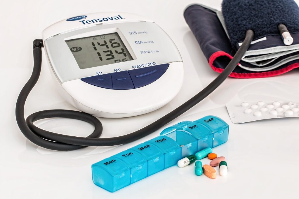
Preventing and managing high blood pressure with healthy lifestyle behaviours combined with early treatment with medication to lower blood pressure if necessary are recommended to reduce the risk of heart attack, stroke, heart failure, kidney disease, cognitive decline and dementia, according to a new clinical guideline published in the American Heart Association’s peer-reviewed journals Circulation and Hypertension, and in JACC, the flagship journal of the American College of Cardiology.
The “2025 AHA / ACC / AANP / AAPA / ABC / ACCP / ACPM / AGS / AMA / ASPC / NMA / PCNA / SGIM Guideline for the Prevention, Detection, Evaluation, and Management of High Blood Pressure in Adults” replaces the 2017 guideline and includes new or updated recommendations for blood pressure management based on the latest scientific evidence to achieve the best health outcomes for patients.
The new guideline reflects several major changes since 2017, including use of the American Heart Association’s PREVENTTM (Predicting Risk of cardiovascular disease EVENTs) risk calculator to estimate cardiovascular disease risk. It also provides updated guidance on medication options, including the early treatment for high blood pressure to reduce the risk of cognitive decline and dementia; use of specific medications including the possible addition of newer therapies such as GLP-1 medications for some patients with high blood pressure and overweight or obesity, and recommendations for managing high blood pressure before, during and after pregnancy.
High blood pressure (including stage 1 or stage 2 hypertension) affects nearly half (46.7%) of all adults in the U.S., is the leading cause of death in the U.S. and around the world. The blood pressure criteria remain the same as the 2017 guideline:
normal blood pressure is less than 120/80 mm Hg;
elevated blood pressure is 120-129 mm Hg and <80 mm Hg;
stage 1 hypertension is 130-139 mm Hg or 80-89 mm Hg; and
stage 2 hypertension is ≥140 mm Hg or ≥90 mm Hg.
“High blood pressure is the most common and most modifiable risk factor for heart disease,” said Chair of the guideline writing committee Daniel W. Jones, M.D., FAHA, dean and professor emeritus of the University of Mississippi School of Medicine in Jackson, Mississippi, and was a member of the writing committee for the 2017 high blood pressure guideline. “By addressing individual risks earlier and offering more tailored strategies across the lifespan, the 2025 guideline aims to aid clinicians in helping more people manage their blood pressure and reduce the toll of heart disease, kidney disease, Type 2 diabetes and dementia.”
“This updated guideline is designed to support health care professionals – from primary care teams to specialists, and to all clinicians across health systems – with the diagnosis and care of people with high blood pressure. It also empowers patients with practical tools that can support their individual health needs as they manage their blood pressure, whether through lifestyle changes, medications or both,” Jones said.
Importance of healthy lifestyle
The new guideline reaffirms the critical role healthy lifestyle behaviours play in preventing and managing high blood pressure, and it encourages health care professionals to work with patients to set realistic, achievable goals. Healthy behaviours such as those in Life’s Essential 8, the American Heart Association’s metrics for heart health, remain the first line of care for all adults.
Specific blood pressure-related guidance includes:
limiting sodium intake to less than 2,300 mg per day, moving toward an ideal limit of 1,500 mg per day by checking food labels (most adults in the U.S. get their sodium from eating packaged and restaurant foods, not the salt shaker);
ideally, consuming no alcohol or for those who choose to drink, consuming no more than two drinks per day for men and no more than one drink per day for women;
managing stress with exercise, as well as incorporating stress-reduction techniques like meditation, breathing control or yoga;
maintaining or achieving a healthy weight, with a goal of at least a 5% reduction in body weight in adults who have overweight or obesity;
following a heart healthy eating pattern, for example the DASH eating plan, which emphasizes reduced sodium intake and a diet high in vegetables, fruits, whole grains, legumes, nuts and seeds, and low-fat or nonfat dairy, and includes lean meats and poultry, fish and non-tropical oils;
increasing physical activity to at least 75-150 minutes each week including aerobic exercise (such as cardio) and/or resistance training (such as weight training); and
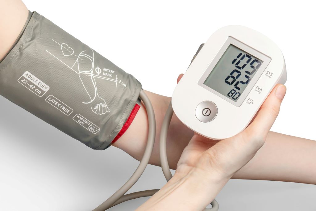
home blood pressure monitoring is recommended for patients to help confirm office diagnosis of high blood pressure and to monitor, track progress and tailor care as part of an integrated care plan.
Addressing each of these lifestyle factors is especially important for people with high blood pressure and other major risk factors for cardiovascular disease because it may prevent, delay or treat elevated or high blood pressure.
New risk calculator and earlier intervention
The new guideline recommends that health care professionals use thePREVENTTM risk calculator to estimate a person’s risk of a heart attack, stroke or heart failure. Developed by the American Heart Association in 2023, PREVENTTM is a tool to estimate 10- and 30-year risk of cardiovascular disease in people ages 30-79 years. It includes variables such as age, sex, blood pressure, cholesterol levels and other health indicators, including zip code as a proxy for social drivers of health. It is the first risk calculator that combines measures of cardiovascular, kidney and metabolic health to estimate risk for cardiovascular disease. More precise risk estimates can help guide treatment decisionspersonalized for each individual.
In addition to the use of the PREVENTTM risk assessment tool, the new guideline recommends two important changes to laboratory testing for initial evaluation.
The ratio of urine albumin and creatinine (a test that assesses kidney health) is now recommended for all patients with high blood pressure. It was recommended as an optional test in the 2017 guideline.
The guideline also expands the indication for use of the plasma aldosterone-to-renin ratio test as a screening tool for primary aldosteronism in more patients including those with obstructive sleep apnea. (Primary aldosteronism is a condition that occurs when the adrenal glands make too much aldosterone, leading to high blood pressure and low potassium levels.)
Screening for primary aldosteronism may also be considered in adults with stage 2 hypertension to increase rates of detection, diagnosis and targeted treatment.
Association of high blood pressure with cognitive decline and dementia
While high blood pressure is a leading cause of heart attack and stroke, the new guideline highlights other serious risks. More recent research confirms that blood pressure affects brain health, including cognitive function and dementia. High blood pressure can damage small blood vessels in the brain, which is linked to memory problems and long-term cognitive decline. The guideline recommends early treatment for people diagnosed with high blood pressure with a goal of systolic blood pressure (top number) goal of <130 mm Hg for adults with high blood pressure to prevent cognitive impairment and dementia.
Tailored approaches to medication for high blood pressure
For many people with high blood pressure, especially those who have Type 2 diabetes, obesity or kidney disease, more than one medication may be needed to lower blood pressure to meet the <130/80 mm Hg criteria. The guideline highlights several types of blood pressure medications to initiate treatment, including angiotensin-converting enzyme (ACE) inhibitors, angiotensin II receptor blockers (ARBs), long-acting dihydropyridine calcium channel blockers and thiazide-type diuretics. If blood pressure remains high after one medication, clinicians may individualize treatment to either increase the dose or add a second medication from a different medication class.
The guideline maintains the recommendation to begin treatment with two medications at once – preferably in a single combination pill – for people with blood pressure levels 140/90 mm Hg or higher (stage 2 hypertension). The guideline also suggests possible addition of newer therapies such as GLP-1 medications for some patients with high blood pressure and overweight or obesity.
High blood pressure and pregnancy
High blood pressure during pregnancy can have lasting effects on the mother’s health, including an increased risk of future high blood pressure and cardiovascular conditions. Without treatment, high blood pressure during pregnancy can lead to serious complications, such as preeclampsia, eclampsia, stroke, kidney problems and/or premature delivery. Women with high blood pressure who are planning a pregnancy or are pregnant should be counselled about the potential benefits of low-dose aspirin (81 mg/day) to reduce the risk of preeclampsia.
For pregnant women with chronic hypertension (high blood pressure before pregnancy or diagnosed before 20 weeks of pregnancy), the new guideline recommends treatment with certain medications when systolic blood pressure reaches 140 mm Hg or higher and/or diastolic blood pressure reaches 90 mm Hg or higher. This change reflects growing evidence that tighter blood pressure control for some individuals during pregnancy may help to reduce the risk of serious complications.
In addition, postpartum care is especially important because high blood pressure can begin or persist after delivery. The guideline urges continued blood pressure monitoring and timely treatment during the postpartum period to help prevent complications. Patients with a history of pregnancy-associated high blood pressure are encouraged to have their blood pressure measured at least annually.
“It is important for people to be aware of the recommended blood pressure goals and understand how healthy lifestyle behaviours and appropriate medication use can help them achieve and maintain optimal blood pressure. Prevention, early detection and management of high blood pressure are critical to long-term heart and brain health, which means longer, healthier lives,” said Jones.
Blood pressure matters at all ages. Children with higher blood pressure at age 7 may be at an increased risk of dying of cardiovascular disease by their mid-50s, according to preliminary research presented at the American Heart Association’s Hypertension Scientific Sessions 2025. The study is simultaneously published in JAMA.
“We were surprised to find that high blood pressure in childhood was linked to serious health conditions many years later. Specifically, having hypertension or elevated blood pressure as a child may increase the risk of death by 40% to 50% over the next five decades of an individual’s life,” said Alexa Freedman, Ph.D., lead author of the study and an assistant professor in the department of preventive medicine at the Northwestern University’s Feinberg School of Medicine in Chicago. “Our results highlight the importance of screening for blood pressure in childhood and focusing on strategies to promote optimal cardiovascular health beginning in childhood.”
Previous research has shown that childhood blood pressure is associated with an increased risk of cardiovascular disease in adulthood, and a 2022 study found that elevated blood pressure in older children (average age of 12 years) increased the risk of cardiovascular death by middle age (average age of 46 years). The current study is the first to investigate the impact of both systolic (top number) and diastolic (bottom number) blood pressure in childhood on long-term cardiovascular death risk in a diverse group of children. Clinical practice guidelines from the American Academy of Pediatrics recommend checking blood pressure at annual well-child pediatric appointments starting at age 3 years.
“The results of this study support monitoring blood pressure as an important metric of cardiovascular health in childhood,” said Bonita Falkner, MD, FAHA, an American Heart Association volunteer expert. “Moreover, the results of this study and other older child cohort studies with potential follow-up in adulthood will contribute to a more accurate definition of abnormal blood pressure and hypertension in childhood.” Falkner, who was not involved in this study, is emeritus professor of paediatrics and medicine at Thomas Jefferson University.
The researchers used the National Death Index to follow up on the survival or cause of death as of 2016 for approximately 38,000 children who had their blood pressures taken at age 7 years as part of the Collaborative Perinatal Project (CPP), the largest US study to document the influence of pregnancy and post-natal factors on the health of children. Blood pressure measured in the children at age 7 years were converted to age-, sex-, and height-specific percentiles according to the American Academy of Pediatrics clinical practice guidelines. The analysis accounted for demographic factors as well as for childhood body mass index, to ensure that the findings were related to childhood blood pressure itself rather than a reflection of children who were overweight or had obesity.
After follow-up through an average age of 54 years, the analysis found:
Children who had higher blood pressure (age-, sex-, and height-specific systolic or diastolic blood pressure percentile) at age 7 were more likely to die early from cardiovascular disease as adults by their mid-50s. The risk was highest for children whose blood pressure measurements were in the top 10% for their age, sex and height.
By 2016, a total of 2,837 participants died, with 504 of those deaths attributed to cardiovascular disease.
Both elevated blood pressure (90-94th percentile) and hypertension (≥ 95th percentile) were linked with about a 40% to 50% higher risk of early cardiovascular death in adulthood.
Moderate elevations in blood pressure were also important, even among children whose blood pressure was still within the normal range. Children who had blood pressures that were moderately higher than average had a 13% (for systolic) and 18% (for diastolic) higher risk of premature cardiovascular death.
Analysis of the 150 clusters of siblings in the CPP found that children with the higher blood pressure at age 7 had similar increases in risk of cardiovascular death when compared to their siblings with the lower blood pressure readings (15% increase for systolic and 19% for diastolic), indicating that their shared family and early childhood environment could not fully explain the impact of blood pressure.
“Even in childhood, blood pressure numbers are important because high blood pressure in children can have serious consequences throughout their lives. It is crucial to be aware of your child’s blood pressure readings,” Freedman said.
The study has several limitations, primarily that the analysis included one, single blood pressure measurement for the children at age seven, which may not capture variability or long-term patterns in childhood blood pressure. In addition, participants in the CPP were primarily Black or white, therefore the study’s findings may not be generalisable to children of other racial or ethnic groups. Also, children today are likely to have different lifestyles and environmental exposures than the children who participated in the CPP in the 1960s and 1970s.
Study details, background and design:
38 252 children born to mothers enrolled at one of 12 sites across the U.S. as part of the Collaborative Perinatal Project between 1959-1965. 50.7% of participants were male; 49.4% of mothers self-identified as Black, 46.4% reported as white; and 4.2% of participants were Hispanic, Asian or other groups.
This analysis reviewed blood pressure taken at age 7, and these measures were converted to age-, sex-, and height-specific percentiles according to the American Academy of Pediatrics Clinical Practice Guideline for Screening and Management of High Blood Pressure in Children and Adolescents.
Survival through 2016 and the cause of death for the offspring of CPP participants in adulthood were retrieved through the National Death Index.
Survival analysis was used to estimate the association between childhood blood pressure and cardiovascular death, adjusted for childhood body mass index, study site, and mother’s race, education and marital status.
In addition, the sample included 150 groups of siblings, and the researchers examined whether the sibling with higher blood pressure was more likely to die of cardiovascular disease than the sibling with lower blood pressure. This sibling analysis allowed researchers to ask how much shared family and early childhood factors might account for the mortality risk related to blood pressure.
Home-based hypertension care led to reductions in systolic blood pressure and improvements in hypertension control in South Africa, according to late-breaking research presented in a Hot Line session at ESC Congress 20251 and simultaneously published in the New England Journal of Medicine.
“Hypertension is the primary risk factor for stroke and heart disease, which are leading causes of death in South Africa. Despite the wide availability of low-cost, effective therapies, hypertension control remains extremely poor in resource-limited settings. Obstacles include a lack of patient confidence to manage their own hypertension care, overcrowded clinics with long wait times and the cost of transport to clinics,” explained the IMPACT-BP trial’s Co-Principal Investigator Doctor Thomas Gaziano from Mass General Brigham (MGB) and Harvard Medical School, Boston, USA. “Our trial aimed to assess the effectiveness and implementation of reliable, home-based, technology-supported interventions to improve blood pressure control in low-resourced rural South Africa.”
IMPACT-BP was an open-label, randomised controlled trial conducted at the Africa Health Research Institute (AHRI) in KwaZulu-Natal, South Africa, in which patients were recruited from two public-sector primary healthcare clinics. The implementation study was designed with Co-Principal Investigator, Doctor Mark Siedner of AHRI and MGH, Professor Nombulelo Magula of the University of KwaZulu-Natal, and the KwaZulu-Natal Provincial Department of Health.
Adult patients were eligible if they had evidence of uncontrolled hypertension as defined by South African Department of Health Guidelines: two measurements of systolic blood pressure (SBP) >140 mmHg and/or diastolic BP (DBP) >90 mmHg, taken a minimum of 6 months apart.
Patients were randomised to one of three strategies: 1) standard-of-care, clinic-based blood pressure (BP) management; 2) home-based BP self-monitoring supported by the provision of BP machines, community health workers (CHWs) who conducted home visits for data collection and medication delivery, and remote nurse-led care assisted by a mobile application with decision support; or 3) an enhanced CHW group in which BP machines included cellular technology to transmit BP readings automatically to the mobile application. The primary outcome was change in SBP from enrolment to 6 months.
In total, 774 patients were randomised. The mean age was 62 years, 76% were women, 14% had diabetes and 47% were living with HIV.
Compared with standard-of-care, mean SBP at 6 months was lower in the CHW group (−7.9mmHg; 95% confidence interval [CI] −10.5 to −5.3; p < 0.001) and the enhanced CHW group (−9.1mmHg; 95% CI −11.7 to −6.4; p < 0.001). In the standard-of-care group, hypertension control at 6 months was 57.6% compared with 76.9% in the CHW group and 82.8% in the enhanced CHW group. Improved BP with home-based care appeared to persist at 12 months.
Severe adverse events (2.7%) and deaths (1.0%) were uncommon overall and similar across groups. Retention in care remained more than 95% in both intervention groups, with patients reported to have enjoyed managing their own hypertension.
Summarising, Doctor Siedner said, “This study is an important example of how making models of chronic disease care more convenient – taking it from the clinic to patients’ homes and letting them play a major role in their own care – can substantially improve hypertension outcomes.”
Of particular value was that the programme was successful in a community that has historically had low access to care. Professor Magula concluded: “Achieving hypertension control in over 80% of people in a predominantly Black African community in rural South Africa is a clear example that equitable health care access can be achieved in disadvantaged communities. Similar models of care that address structural barriers could be considered to improve hypertension control in other remote and resource-limited settings. Expansion of the model to include the care of people with multiple comorbidities may also be valuable.”
Not all acute myocardial infarction patients should be offered routine screening for the stomach ulcer bacterium Helicobacter pylori. However, it is possible that some patient groups with an elevated risk of post-infarction gastrointestinal bleeding benefit from such a test, concludes a large-scale study from Karolinska Institutet and Södersjukhuset published in the journal JAMA.
Upper gastrointestinal bleeding is a serious complication that affects approximately two per cent of patients within a year of a myocardial infarction.
“It’s associated with increased mortality and the risk of recurring cardiovascular events,” says the study’s lead author Robin Hofmann, senior consultant at the Department of Cardiology, Södersjukhuset, and associate professor at the Department of Clinical Science and Education, Södersjukhuset, Karolinska Institutet. “We therefore wanted to examine if screening for the common Helicobacter pylori bacterium, which causes gastritis and gastric ulcers, can reduce the risk of bleeding. This is currently not routine practice.”
The randomised study included almost 18 500 myocardial infarction patients at 35 hospitals in Sweden. Half the group was tested for the bacterium and treated with antibiotics and protein pump inhibitors by their doctors if testing positive, while the other half received routine care without an extra test or treatment.
Effective in anaemic patients
After almost two years’ follow-up, the researchers found that there were slightly fewer individuals in the screening programme who had suffered gastrointestinal bleeding, but not enough to make the difference statistically significant. However, they found a positive effect of the screening when studying specific sub-groups of patients, such as those with anaemia or kidney failure. A particularly positive effect was observed in patients with moderate to severe anaemia, who suffered gastrointestinal bleeding at roughly half the rate if they underwent screening.
“Our results suggest that screening for Helicobacter pylori does not need to be done routinely for all individuals following a heart attack,” says Dr Hofmann. “On the other hand, testing and treatment could be a meaningful complement for selected patient groups with an elevated risk of bleeding.”
The researchers will now go on to study the long-term effects and try to identify which groups will benefit most from screening.
Early withdrawal of aspirin following successful percutaneous coronary intervention (PCI) for acute coronary syndrome and in low-risk patients following an acute myocardial infarction (MI) was the focus of two separate hot line trials presented at ESC Congress 2025. A third trial provided insights into early escalation and late de-escalation of antiplatelet therapy after complex PCI.
In the NEO-MINDSET trial, simultaneously published in NEJM, researchers in Brazil randomised approximately 3400 patients within the first four days of hospitalisation following a successful PCI to either stop treatment with aspirin and receive potent P2Y12 inhibitor monotherapy (ticagrelor or prasugrel) or to receive dual antiplatelet therapy (DAPT) that included aspirin and a potent P2Y12 inhibitor for 12 months.
At 12 months, the primary endpoint of death from any cause, MI, stroke or urgent revascularisation had occurred in 119 patients in the monotherapy group and in 93 patients in the DAPT group (p = 0.11 for noninferiority). Researchers also noted that major or clinically relevant nonmajor bleeding occurred in 33 patients assigned to monotherapy vs 82 patients assigned to DAPT. Stent thrombosis occurred in 12 patients in the monotherapy group and in 4 in the dual antiplatelet therapy group.
“We failed to demonstrate the noninferiority of aspirin-free monotherapy initiated immediately after PCI with regard to the ischaemic primary endpoint over 12 months,” said Principal Investigator Pedro Lemos, MD. “Results from the landmark analysis suggest that the excess ischaemic risk with monotherapy occurred in the first 30 days, with comparable outcomes thereafter. Bleeding appeared to be lower at both 30 days and 12 months with monotherapy versus DAPT.”
In TARGET-FIRST, also simultaneously published inNEJM, P2Y12-inhibitor monotherapy was noninferior to continued DAPT with respect to the occurrence of adverse cardiovascular and cerebrovascular events among low-risk patients with acute MI who had undergone early complete revascularisation and had completed one month of DAPT without complications. It also resulted in lower incidence of bleeding events.
Nearly 2000 patients from 40 centres in Europe were randomised to receive P2Y12-inhibitor monotherapy or to continue DAPT for 11 months. A primary-outcome event occurred in 20 patients (2.1%) in the P2Y12-inhibitor monotherapy group and in 21 patients (2.2%) in the dual antiplatelet therapy group (p = 0.02 for noninferiority). Major bleeding occurred in 2.6% of the patients assigned to P2Y12-inhibitor monotherapy group compared with 5.6% of those assigned to DAPT (p = 0.002 for superiority). The incidence of stent thrombosis and serious adverse events appeared to be similar in the two groups, researchers said.
Principal Investigator Giuseppe Tarantini, MD, noted that no previous randomised trials have assessed early aspirin discontinuation in acute MI patients who achieve early, complete revascularisation with modern stents. “These results reflect the benefits of modern stents, high procedural success and optimal medical therapy, making early aspirin discontinuation feasible in this selected population,” he said.
The TAILORED-CHIP trial found early escalation and late de-escalation of antiplatelet therapy is not beneficial in patients with high-risk anatomical or clinical characteristics undergoing complex PCI.
Researchers in South Korea randomised approximately 2000 patients to standard DAPT (clopidogrel plus aspirin for 12 months) or a tailored antiplatelet strategy consisting of early escalation (low-dose ticagrelor at 60mg twice daily plus aspirin for 6 months) followed by late de-escalation (clopidogrel monotherapy for 6 months).
Overall findings showed no significant difference in the incidence of major ischemic events at 12 months with tailored therapy compared with standard DAPT. However, the incidence of clinically relevant bleeding was significantly higher with tailored therapy, according to study investigators.
“Our results suggest that a tailored strategy in patients undergoing complex high-risk PCI does not provide a net clinical benefit,” said Principal Investigator Duk-Woo Park, MD, PhD, FACC. “We observed an increase in bleeding complications without a significant reduction in ischaemic events. This challenges the notion that ‘more is better’ even in carefully selected patients at high ischaemic risk.”
Beta-blocker therapy showed no evidence of an effect on all-cause death, reinfarction or heart failure admission in patients with myocardial infarction (MI) managed invasively who had left ventricular ejection fraction (LVEF) ≥ 40%, according to late-breaking research from the REBOOT trial presented in a Hot Line session today at ESC Congress 20251 and simultaneously published in the New England Journal of Medicine.
Explaining the rationale for the REBOOT trial, Principal Investigator, Professor Borja Ibáñez from the Centro Nacional de Investigaciones Cardiovasculares Carlos III (CNIC) and Fundación Jiménez Díaz University Hospital, Madrid, Spain, said: “Beta-blockers have long been a foundational treatment after acute MI; however, supporting evidence is derived from trials that predate modern standards of care − before the time of routine reperfusion, invasive management, potent antiplatelet therapies and statins. Re-examining the role of beta-blockers is warranted, particularly among patients with uncomplicated MI and LVEF > 40% in whom the benefits of beta-blockers are not well established, unlike with reduced LVEF (≤ 40%).”
The investigator-initiated randomised open blinded-endpoint REBOOT trial was conducted at 109 centres across Spain and Italy. Patients with MI (with or without ST-segment elevation) were eligible for enrolment if they underwent invasive management during the index hospitalisation and had a predischarge LVEF > 40%, with no history or signs of heart failure. Patients were randomised 1:1 to beta-blocker or no beta-blocker therapy. The primary endpoint was a composite of all-cause mortality, nonfatal reinfarction or heart failure admission.
Among 8505 patients who underwent randomisation, the mean age was 61 years and 19.3% were women. A total of 10% had a prior MI and 12% were on beta-blocker treatment before the index hospitalisation.
After a median follow-up of 3.7 years, the primary composite outcome of all-cause death, nonfatal reinfarction or heart failure admission occurred in a similar proportion of patients in each group: 22.5/1000 patient-years in beta-blocker group and 21.7/1000 patient-years in the no beta-blocker group (hazard ratio [HR] 1.04; 95% confidence interval [CI] 0.89 to 1.22; p=0.63).
All-cause mortality occurred in 11.2 and 10.5/1000 patient-years on beta-blocker therapy and no-beta blocker therapy, respectively (HR 1.06; 95% CI 0.85 to 1.33). Nonfatal reinfarction occurred in 10.2 and 10.1/1000 patient-years, respectively (HR 1.01; 95% CI 0.80 to 1.27), while heart failure admission occurred in 2.7 and 3.0/1,000 patient-years, respectively (HR 0.89; 95% CI 0.58 to 1.38).
Regarding safety, admission for stroke occurred in 2.6/1000 patient-years in the beta-blocker group and 1.7/1000 patient-years in the no beta-blocker group (HR 1.50; 95% CI 0.90 to 2.49). Admission for symptomatic advanced atrioventricular block occurred in 0.5 of patients in the beta-blocker group and 0.4/1000 patient-years of patients in the no beta-blocker group (HR 1.18; 95% CI 0.40 to 3.50).
There appeared to be an absence of benefit with beta-blockers across the prespecified subgroups. However, fewer events were noted in patients with mildly reduced LVEF (40−49%) on beta-blockers vs no beta-blockers, although low patient numbers limit interpretability. Women experienced overall more events, especially when on beta-blockers.
Professor Ibáñez concluded: “Beta-blocker therapy showed no evidence of benefit across the study population of patients with MI managed invasively who had LVEF > 40%. However, as also presented today at ESC Congress, a meta-analysis of data from four trials, including REBOOT, suggest there may be a positive signal in patients with mildly reduced LVEF (40−49%).2”
Cardiologists have long known that up to half of all heart attacks and strokes occur among apparently healthy individuals who do not smoke and do not have high blood pressure, high cholesterol, or diabetes, the “standard modifiable risk factors” which doctors often call “SMuRFs.” How to identify risk among the “SMuRF-Less” has been an elusive goal in preventive cardiology, particularly in women who are often under-diagnosed and under-treated.
A new study by Mass General Brigham researchers that leverages data from the Women’s Health Study has found hsCRP, a marker of inflammation, can help identify women who are at risk but are missed by current screening algorithms. Results are presented at a late-breaking clinical science session at the European Society of Cardiology Congress (ESC) and simultaneously published in The European Heart Journal.
“Women who suffer from heart attacks and strokes yet have no standard modifiable risk factors are not identified by the risk equations doctors use in daily practice,” said Paul Ridker, MD, MPH, a preventive cardiologist at Mass General Brigham’s Heart and Vascular Institute. “Yet our data clearly show that apparently healthy women who are inflamed are at substantial lifetime risk. We should be identifying these women in their 40s, at a time when they can initiate preventive care, not wait for the disease to establish itself in their 70s when it is often too late to make a real difference.”
As part of the federally funded study, researchers studied 12 530 initially healthy women with no standard modifiable risk factors who had the inflammatory biomarker hsCRP measured at study entry and who were then followed over 30 years. Despite the lack of traditional risks, women who were inflamed as defined by hsCRP levels > 3 mg/L had a 77% increased lifetime risk of coronary heart disease, a 39% increased lifetime risk of stroke, and a 52% increased lifetime risk of any major cardiovascular event.
Additionally, researchers released a new analysis of randomised trial data showing that “SMuRF-Less but Inflamed” patients can reduce their risk of heart attack and stroke by 38% using statin therapy.
“While those with inflammation should aggressively initiate lifestyle and behavioural preventive efforts, statin therapy could also play an important role in helping reduce risk among these individuals,” said Ridker.
Every 34 seconds, someone in the United States dies from heart disease. As nearly half of the country suffers from some form of cardiovascular disease (CVD), another 1 in 4 adults experience a mental health disorder in their lifetime, signalling an inevitable overlap.
Now, a new report from Emory University shows that certain mental health conditions escalate the risk of developing heart disease by 50–100% – and adverse outcomes from existing heart conditions by 60–170%.
The report, published in The Lancet Regional Health-Europe, summarises cardiovascular health disparities among those diagnosed with depression, anxiety, schizophrenia, bipolar and post-traumatic stress disorders (PTSD). The article is part of a series aiming to raise awareness around disparities in CVD health in four populations: women, the elderly, racial minorities and those with mental health conditions.
Emory University professor Viola Vaccarino, MD, PhD, led this metareview linking mental health conditions to CVD, along with co-authors Amit Shah, MD, and Douglas Bremner, MD, also Emory professors.
The report associated the following conditions and their corresponding risks of developing CVD:
Major depression, 72%
PTSD, 57%
Bipolar disorder, 61%
Panic disorder, 50%
Phobic anxiety, 70%
Schizophrenia, nearly 100%
The research also shows that these conditions are associated with a poorer prognosis, greater risk for readmission and higher mortality from existing heart conditions. For example, major depression more than doubles the mortality rate in those with existing CVD.
Additionally, the report emphasises a bidirectional relationship. “More than 40 percent of those with cardiovascular disease also have a mental health condition,” adds Vaccarino.
The physiology of stress
According to the report, a well-documented relationship exists among depression, schizophrenia, PTSD, and abnormal stress responses in the autonomic nervous system (ANS) and hypothalamic-pituitary adrenal axis (HPA).
The former allows the brain to manage involuntary responses, such as functions of the liver, heart, sweat glands, and eye muscles. ANS also manages both acceleration and deceleration of these functions, regulating inflammatory responses. Since most major organs have ANS nerve endings, this system impacts most bodily functions.
The hypothalamic-pituitary adrenal axis (HPA) also influences immune response and metabolism, which can impact cardiovascular function.
According to the report, dysregulation of these systems creates “adverse downstream effects that can affect cardiovascular risk chronically, including increased inflammation, metabolic abnormalities, high blood pressure, enhanced systemic vascular resistance and autonomic inflexibility.” Inflammation has also been implicated in both the development of heart disease and mental health conditions.
Social determinants and quality of care
The role of social determinants of health in CVD disparities is critical. Those with mental health conditions may face disruptions and barriers in the continuum of care, such as affordability and accessibility. Compromised health literacy or communication can also impede access to health screenings and treatment.
Clinicians could also be challenged to care for patients with certain mental conditions, which can be compounded by stigma and existing models that fragment mental and physical health care. Stigmas are also present in the field of clinical research, where having a mental health condition is often an exclusionary criterion in randomised trials.
Moreover, according to the report, current prediction models don’t account for mental health disorders when forecasting the risk of developing heart disease.
Next steps toward a healthier future
To address the disparities of CVD among people with mental health disorders, the authors recommend an integrated approach with interdisciplinary care encompassing behavioural, mental and cardiovascular health.
“The tight connection between cardiovascular and psychological health warrants changes in the health care system that are more amenable to patients with comorbidities,” says Vaccarino. “A clinical team would be ideal for the care of these patients – a team of specialists, social workers, and nursing staff who work in collaboration to provide multidisciplinary care and resources.”
The report concludes that closing the health disparity gap upholds the rights of those living with a mental health condition to achieve the highest level of health and fully participate in society.