

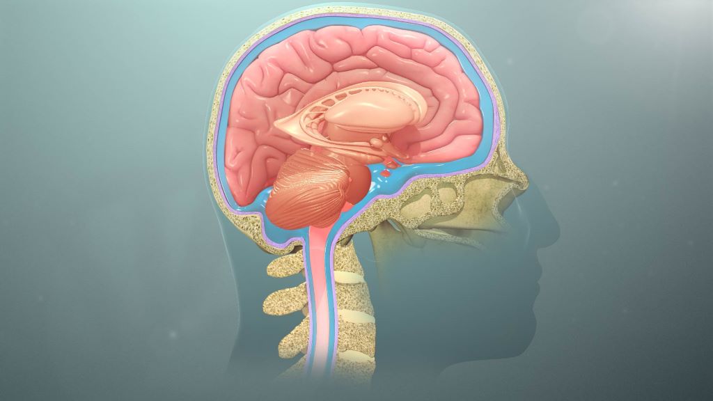
Deep inside your brain, a clear liquid is constantly on the move. Cerebrospinal fluid (CSF) cushions the brain, delivers nutrients, removes waste and keeps pressure stable. Think of it as an internal tide, circulating through cavities in the brain and around the spinal cord to keep this delicate organ in balance. When that flow is disrupted, the consequences can be serious.
For decades, doctors have relied mainly on static brain scans to guide treatment. But structure tells only part of the story. Researchers at the University of Pretoria (UP) are now focusing on something more dynamic: how fluid actually moves.
Two of the most common neurosurgical conditions worldwide – brain tumours and hydrocephalus (a dangerous build-up of fluid in the brain) – are closely tied to disturbed CSF circulation. Tumours can block or distort the pathways through which fluid moves. Hydrocephalus represents a more obvious breakdown, where fluid accumulates and pressure rises. In both cases, symptoms such as headaches, problems with vision and neurological decline are not simply caused by the presence of disease, but by changes in pressure and pulsating flow inside the skull.
At the Brain Tumour and Translational Neuroscience Centre (BTC@UP), scientists are investigating an unexpected window into this hidden system – without inserting monitors into the brain: the eye.
Professor Llewellyn Padayachy, Head of the Department of Neurosurgery at BTC@UP explains: “The optic nerve, which connects the eye to the brain, is surrounded by the same protective layers as the brain itself. CSF flows along this nerve, meaning changes in brain pressure can subtly affect structures at the back of the eye. By using advanced, non-invasive eye imaging, researchers can detect signs of altered fluid flow and pressure without inserting monitors into the brain.”
This matters enormously for children with hydrocephalus and patients with brain tumours who require long-term monitoring. It offers a safer, repeatable way to track disease progression and treatment response. In low- and middle-income countries, where hydrocephalus is common but access to advanced imaging and neurosurgical infrastructure may be limited, such non-invasive tools could reduce reliance on costly technology while still delivering meaningful clinical insight.
The research also helps refine innovation. Modern shunts and endoscopic procedures increasingly aim to restore more natural fluid circulation rather than simply drain excess fluid. Objective eye-based markers provide measurable ways to evaluate whether these technologies truly improve flow.
While the link between the eye and brain pressure has long been recognised, what is new is the integration of advanced imaging, physiological modelling and continuous monitoring. This approach treats CSF flow as a living system, and shifts care from reacting to late damage towards detecting subtle change earlier.
Why this research matters
This work reframes brain disease through a simple but powerful idea: health depends on flow. By learning to read the movements of brain fluid, even through the eye, researchers are paving the way for safer monitoring, smarter surgery and more equitable neurological care worldwide.
Fast fact
The most common surgical treatment for hydrocephalus is the surgical placement of a shunt, which has one of the highest failure rates of any medical device on the market.
Provided by the University of Pretoria.
