Day: March 17, 2026

‘Google Earth’ for Human Organs Made Available Online

A new open-access 3D portal that allows users to explore human organs in unprecedented detail, from the whole organ to individual cells, has been launched by an international team led by UCL scientists.

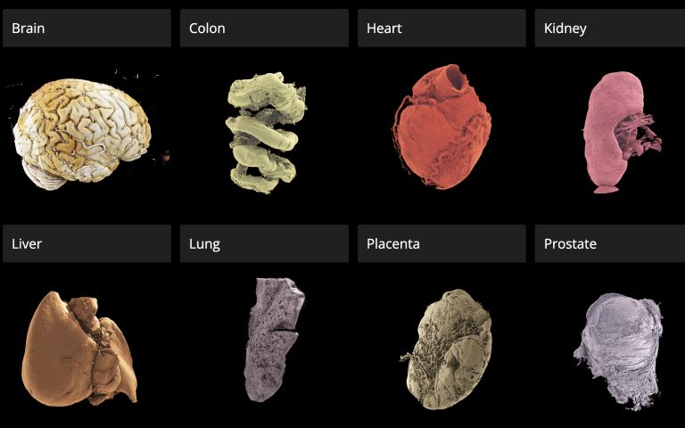
The Human Organ Atlas, described in a new paper in the journal Science Advances, brings together some of the most detailed images of 3D organs ever produced. It enables scientists, doctors, educators, students and the wider public to interactively “fly through” organs such as the brain, heart, lungs, kidney and liver, providing a new way of understanding human anatomy and human diseases.

The resource can be accessed directly through a standard web browser, without specialist software, at this link.

The Atlas is powered by an advanced X-ray imaging method called Hierarchical Phase-Contrast Tomography (HiP-CT), developed at the European Synchrotron (ESRF) in Grenoble, France. HiP-CT uses the ESRF’s Extremely Brilliant Source – a new generation of synchrotron source – which is up to 100 billion times brighter than conventional hospital CT scanners.

This allows researchers to scan entire intact ex vivo human organs (i.e., donated organs) non-destructively and then zoom in to near-cellular resolution (down to less than one micron, 50 times thinner than the size of a human hair).

The technique bridges a century-old gap in medicine between radiology and histology, and represents a major advance in biomedical imaging.

Professor Peter Lee (UCL Department of Mechanical Engineering), principal investigator of the Human Organ Atlas beamtime, said: “To create the Human Organ Atlas, we brought together scientists and medics from nine institutes worldwide. This grouping is continuing to expand, helping gain new insights into diseases from osteoarthritis to heart disease and changing how we learn about the human body.”

Dr Claire Walsh (UCL Department of Mechanical Engineering), Director of the Human Organ Atlas Hub, said: “The Human Organ Atlas shows what team science can achieve at its best – we went into this project wanting this data to be used by others and to help further the understanding of human physiology. The Human Organ Atlas is an incredible resource that will continue to grow. I am personally hugely excited to see how the AI community use the Human Organ Atlas in AI foundation models.”

From Covid-19 to cardiac and gynaecological disorders

Initially developed during the COVID-19 pandemic, the method has already led to high-impact publications and scientific advancements, revealing previously unseen microscopic vascular injury in the lungs of patients who died from Covid-19 or reshaping understanding of cardiac disorders. The technology has also been applied to other organs, providing new insights, for instance, into the way gynecological disorders develop.

Professor Judith Huirne, based at Amsterdam UMC, said: “The virtual 3D histological data derived from Human Organ Atlas hub provides us with valuable insights into the pathogenesis of gynecological disorders. This knowledge is crucial to bridging the current gaps in both understanding and gender disparities.”

This Human Organ Atlas portal is the result of more than five years of collaborative effort between many researchers, engineers, clinicians, and infrastructure specialists, united within the Human Organ Atlas Hub, a consortium involving nine institutes across Europe and the United States.

Since its inception, the team has been committed to open science. Dr Paul Tafforeau, ESRF scientist and pioneer of the imaging technique used to create the Human Organ Atlas, said: “From the beginning, we wanted these data to be accessible to everyone and build an open, shared scientific infrastructure at a global scale. This is a resource for researchers, doctors, educators – but also for anyone curious about how the human body is built.

A unique tool for AI, medicine and education

To the team’s knowledge, this is the highest-resolution open 3D dataset of intact human organs currently available. The Human Organ Atlas currently provides access to: (to be updated)

  • 62 organs, 319 full 3D datasets from 29 donors
  • 12 organ types, including brain, heart, lung, kidney, liver, colon, eye, spleen, placenta, uterus, prostate and testis
  • Multiscale scans, from whole-organ views down to near-cellular resolution (routinely down to 2 µm, as fine as 0.65 microns for some organs)

The portal has been designed to extend far beyond specialist research laboratories. Each dataset can reach hundreds of gigabytes or even over a terabyte in size. The largest one (a brain) is 14 Tb. To make the data usable worldwide, the portal provides:

  • Interactive browser-based visualisation (no special software required)
  • Downloadable datasets at multiple resolutions
  • Tutorials and software tools for analysis
  • Regular addition of new data

Beyond advancing anatomical and biomedical research, the atlas is expected to become a major resource for artificial intelligence. Large, high-quality 3D datasets are rare – limiting the development of advanced medical AI systems. The Human Organ Atlas provides a curated, hierarchical dataset ideally suited for training machine-learning models for segmentation, disease detection and super-resolution analysis.

At the same time, it offers powerful new opportunities for medical education and public engagement with science, allowing anyone to explore the human body out of curiosity.

Source: University College London

Timely Scan Could Save Lives of A&E Patients with Haematuria

Photo by Camilo Jimenez on Unsplash

One in ten emergency patients with visible blood in their urine die within three months of presenting at A&E, new research has found. The WASHOUT study, presented Monday 16 March at the European Association of Urology Congress (EAU26) in London, found that a scan within 48 hours could reduce this risk. 

Such a scan also ensured patients with cancer were diagnosed significantly faster. Around 1 in 4 people who presented at A&E with visible blood in their urine had an underlying cancer, with the most common being bladder cancer, the study found. 

Around 25 000 people visit UK A&E departments each year because they have blood in their urine. Currently, patients receive different care depending on which hospital they visit or even which doctor they see. This is because there are no guidelines built on real-world evidence for doctors to follow. Based on global figures, only around half (53%) of patients receive a scan and a third (35%) receive surgery, with others discharged home or admitted to the ward to watch how their symptoms progress, says the WASHOUT study. 

The WASHOUT study drew on global data to show that rapid action is critical for better patient outcomes. A CT scan or cystoscopy to look inside the bladder within 48 hours of arriving at A&E should determine the most appropriate next steps – such as whether the person should be treated for bladder cancer. Patients who didn’t receive investigative tests or appropriate treatment were 2.5% more likely to die within the next three months compared to those who did. They also spent more time in hospital and were more likely to be readmitted with the same problem within three months.

For patients with an underlying cancer, those who received investigative tests within the first 48 hours of admission were diagnosed within one day on average. In contrast, patients who were discharged without investigation faced a significantly longer wait, with diagnosis taking on average three weeks.

The research team is now taking steps to incorporate their findings into clinical guidelines, to help hospital staff provide the best treatment for these patients. 

The study looked at data from more than 8500 people across 380 hospitals around the world and followed their journey for 90 days after arriving at A&E with blood in their urine. It also considered other factors that might have affected results, including age, frailty and other underlying conditions. 

Nikita Bhatt, consultant urologist at St Vincent’s University Hospital, Dublin, led the research being presented at EAU26. She said: “This is the largest study exploring how we should treat people who present at A&E with blood in their urine. It’s a common problem affecting thousands of people around the world, and these patients are usually very unwell. But too often they fall through the gaps because it isn’t obviously tied to a specific disease. Our findings show how important it is that doctors take the necessary steps to identify the cause of the problem. For patients, the message is clear: if you have visible blood in your urine, don’t ignore it. See your doctor as soon as you can. If it doesn’t clear up, keep pushing until you find an answer. I hope our study gives patients the encouragement to do that.”

Jacqueline Emeks, a patient advocate on the WASHOUT study, who was diagnosed with a kidney infection and sepsis after arriving at A&E with visible blood in their urine, agrees: “These findings highlight that blood in the urine should trigger immediate action. It’s not something to watch and wait. For patients, this should mean quicker triage, earlier investigations and faster treatment, translating into safer care, fewer delays, and a better chance of avoiding severe illness or long-term harm. Patients know their bodies and deserve to be taken seriously. Blood in urine is a red flag until proven otherwise.”

Prof Dr Joost Boormans, a member of the EAU Scientific Congress Office and a urologist at the Erasmus University Medical Center, Rotterdam, said: “This is an important study highlighting the scale of the problem that emergency blood in the urine presents, both for patients and our already over-stretched healthcare systems. It’s difficult to draw strong conclusions about specific conditions because blood in the urine can be caused by many things, including cancer, and this group of patients is very diverse. But this study shows that timely investigative tests can accelerate diagnosis and reduce patients’ risk of readmission and long hospital stays, both being significantly high as shown in the WASHOUT study. As urologists in emergency care, we should be aware of these numbers and do more to get an immediate diagnosis for people with blood in urine, to reduce the burden on our healthcare systems and give our patients the best outcomes.”

Source: European Association of Urology

Prostate Cancer Screening on Par with Breast Cancer Screening

Credit: Darryl Leja National Human Genome Research Institute National Institutes Of Health

Prostate cancer screening compares favourably to screening for breast cancer in identifying significant cancers, reducing mortality and avoiding unnecessary harms, according to new research. The findings are presented on Sunday 15 March 2026 at the European Association of Urology Congress (EAU26) in London. The research is also accepted for publication in European Urology.

The researchers maintain that the similarities between the two forms of screening mean it is no longer rational to reject prostate cancer screening on one hand while endorsing screening for breast cancer on the other. Nevertheless, they recommend some caution given their research compares a trial with a population-based screening programme and across two different cancers. 

Although breast and prostate cancer are the most commonly diagnosed cancers in Europe amongst men and women respectively, screening for the diseases is vastly different. Organised breast cancer screening programmes have been established across Europe for more than three decades. Prostate cancer screening has lagged behind, primarily due to concerns around the effectiveness of the PSA blood test and the risks of overdiagnosis and overtreatment. Nevertheless, many men undergo variable, ‘opportunistic’ screening for the disease, mostly based on self-referral.

Several prostate cancer screening trials in Europe have now reported long-term outcomes, showing a reduced risk of death from prostate cancer [1]. This risk reduction is similar to that seen in breast screening programmes.

The new analysis compares the two types of cancer screening in terms of the effectiveness of the diagnostic tests and levels of overdiagnosis. The researchers, from the German Cancer Research Centre in Heidelberg, Germany, drew on data from the PROBASE prostate cancer screening trial in Germany and the country’s breast cancer screening programme.

They used data from 39,392 men who underwent an initial PSA blood test as part of the PROBASE trial at age 45 or 50. They compared this with data from just over 2.8 million women, aged 50–69, who had a mammography as part of Germany’s organised breast cancer screening programme. They found:

  • PSA blood testing followed by an MRI scan leads to a higher number of false positives than mammography (37-42% vs 10%).
  • A similar proportion of men and women were referred for biopsy (0.8-2.4% for men and 1.1% for women) as men in the PROBASE trial were triaged before referral using various factors to determine the likelihood of significant cancer (known as risk stratification)
  • Biopsies were far more likely to identify significant cancer in prostate screening than in breast screening (50-68% vs 10%), indicating that fewer men were referred for biopsy unnecessarily.
  • The percentages of invasive cancers identified were similar across both prostate and breast cancer screening (60-74% vs 73%).
  • Prostate cancer screening was more likely to identify non-aggressive cancers than breast cancer screening (26-31% vs. 22%). However, in prostate cancer the option of active surveillance is well-established, and the researchers maintain this would limit the risk of overtreatment. Active surveillance involves monitoring lower grade cancers and only starting treatment (radiotherapy or surgery) if they progress.

Dr Sigrid Carlsson, who leads Clinical Epidemiology of Early Cancer Detection at the German Cancer Research Centre (DKFZ) in Heidelberg, is lead author of the research. She said: “Until we have a population-based screening programme for prostate cancer, we can’t make an exact like-for-like comparison with breast cancer. But we can make some informed assumptions based on the data from our trial, which shows that if prostate cancer screening were extended to the wider population, then the outcomes are likely to be very similar to breast cancer. Although our study used German data, the findings are applicable to other countries. The final question we now need to answer is: what will this cost compared to what we are already paying for opportunistic screening? And that work is already underway.”

Tobias Nordström is a clinical urologist and Associate Professor at the Karolinska Institute, Sweden and a member of the EAU Scientific Congress Office. He said: “There is much that prostate cancer screening can learn from breast cancer screening and that is why this analysis is an important addition to our knowledge base. As these kinds of comparisons are very challenging, the results do need to be taken with a level of caution. That said, the clear overall similarities between the outcomes for breast and prostate cancer screening show that we are moving in the right direction, ensuring prostate cancer screening offers more benefits than harm.”

[1] See the 23-year follow-up from the European Randomised Study of Screening for Prostate Cancer (ERSPC) in the New England Journal of Medicine: European Study of Prostate Cancer Screening — 23-Year Follow-up | New England Journal of Medicine

Source: European Association of Urology

Rising CO₂ Levels are Reflected in Human Blood. Scientists Don’t Know What it Means

FabrikaCr / Getty Images

Alexander Larcombe, The Kids Research Institute Australia; Curtin University and Philip Bierwirth, Australian National University

Humans evolved in an atmosphere containing roughly 200–300 parts per million (ppm) of carbon dioxide (CO₂). Today, that figure sits above 420 ppm, higher than at any point in the history of our species.

We know this extra CO₂ is contributing to climate change, but could it also be changing the chemistry of our bodies?

In our recently published research we looked at two decades of information from one of the biggest health datasets in the world to start answering this question. We found some concerning trends.

What we found

We analysed blood chemistry data from the US National Health and Nutrition Examination Survey (NHANES), which collected samples from about 7000 Americans every two years between 1999 and 2020. We looked at three markers: CO₂, calcium and phosphorus.

CO₂ is mainly carried in blood in the form of bicarbonate (HCO₃⁻).

When CO₂ enters the blood, it is converted to bicarbonate. This process largely occurs inside red blood cells, and also produces hydrogen ions.

During short-term exposure to increased CO₂, this can make blood more acidic, and result in a modest increase in bicarbonate levels in the blood (to reduce acidity).

If the exposure continues for a long time the kidneys reduce the amount of bicarbonate lost in urine and also produce more bicarbonate. This has the net effect of higher bicarbonate levels in the blood, to counteract the persistent acidity.

Levels of calcium and phosphorus in the blood may also be affected, as they too play a role in regulating acidity in the blood. These processes are completely normal.

Over the 21 years from 1999 to 2020, we found that average blood bicarbonate levels rose by about 7%. Over the same period, atmospheric CO₂ concentrations rose by a similar proportion.

Atmospheric CO₂ has risen, along with increases in levels of carbonate in the blood and decreases in calcium and phosphorus. Larcombe & Bierwirth / Air Quality, Atmosphere & Health, CC BY

Meanwhile, blood calcium levels dropped by about 2% and phosphorus by around 7%.

If these trends continue, blood bicarbonate levels may exceed healthy levels in around 50 years. Calcium and phosphorus levels may fall below healthy levels, too, by the end of the century.

Our hypothesis is that rising CO₂ exposure could be contributing to these trends.

What’s causing the changes?

It’s important to be clear about what this study does and doesn’t show. It identifies population-level trends in blood chemistry that parallel rising atmospheric CO₂.

But correlation is not causation. The study does not adjust for factors such as diet, kidney function, diuretic use or obesity, which can influence the measurements and should be considered in future analyses.

There are other plausible contributors. One important consideration is indoor air.

Participants in the NHANES study likely spend most of their time indoors, where CO₂ concentrations often exceed 1000 ppm in poorly ventilated spaces. Other studies show time spent indoors has increased over the past two decades.

The NHANES dataset doesn’t capture this parameter, so we can’t directly assess this contribution. However, if more time indoors is contributing, it means total CO₂ exposure is rising even faster than atmospheric trends suggest. This arguably reinforces rather than alleviates the concern.

Other factors, such as shifting dietary patterns, changing rates of obesity, differences in physical activity and even variations in sample collection or processing across survey cycles, could also be important.

Can our bodies cope?

Some critics have argued that, based on what we know about how our bodies manage blood chemistry, we should have no trouble compensating for future increases in atmospheric CO₂, even under worst-case climate scenarios. For example, the lungs can increase ventilation and the kidneys can adjust to produce more bicarbonate.

For most healthy individuals, small long-term increases in outdoor CO₂ are not expected to meaningfully change the levels of bicarbonate, calcium or phosphorus in the blood.

This makes the population-level trends we observed puzzling. They could reflect a confounding rather than a direct CO₂ effect, but they do highlight how little data we have on long-term, real-world exposure.

A lack of long-term data

The argument that we can cope easily with higher CO₂ is based on short-term responses. Whether the same reasoning applies when CO₂ levels are higher across a person’s entire life remains largely untested.

There is, however, a growing body of evidence across many species which shows that even modest, environmentally relevant increases in CO₂ can produce subtle but measurable physiological effects.

In humans, short-term exposure at concentrations commonly found indoors (1000–2500 ppm) has been linked to reduced cognitive performance and changes in brain activity, though the mechanisms aren’t fully understood.

These new findings highlight a gap in evidence about long-term, real-world CO₂ exposure and human physiology. Unfortunately, there simply aren’t any studies assessing the physiological effects of breathing slightly elevated CO₂ over a lifetime.

This is particularly important for children, who will experience the longest cumulative exposure. And that’s why it’s vital to investigate this area further.

What this means

Our findings are not suggesting people will become suddenly unwell when atmospheric CO₂ reaches a certain level. What the data show is a signal that warrants attention.

If rising atmospheric CO₂ is contributing to gradual shifts in blood chemistry at a population level, then the composition of the atmosphere should be monitored alongside traditional climate indicators as a potential factor in long-term public health.

Reducing CO₂ emissions remains crucial for limiting global warming. Our findings suggest it may also be important for safeguarding aspects of human health that we’re only just beginning to understand.

Alexander Larcombe, Associate Professor and Head of Respiratory Environmental Health, The Kids Research Institute Australia; Curtin University and Philip Bierwirth, Emeritus Research Associate, Australian National University

This article is republished from The Conversation under a Creative Commons license. Read the original article.

University of Pretoria Study Exposes Harmful Impact of Medical Aid Forensic Audits on Physiotherapists

A University of Pretoria study has revealed troubling ethical and procedural gaps in the way medical schemes conduct forensic audits of physiotherapists, showing how practices intended to prevent billing irregularities are often leaving practitioners fearful, stigmatised and traumatised without proving guilt.

Audits of healthcare professionals are designed to detect billing irregularities and protect medical scheme funds. However, research conducted by Lesley Meyer, an extra-ordinary lecturer at University of Pretoria’s Department of Physiotherapy, explored the lived experiences of physiotherapists who have undergone such audits, and found that the audit practices conducted were experienced as punitive and harmful to practitioners rather than corrective.

Forensic audits in the healthcare sector are, in principle, supposed to protect medical scheme funds and by extension, patients’ contributions. In practice, however, Meyer’s research study, published in the latest edition of The South African Journal of Physiotherapy, found that these audits often extend beyond their legal scope and adversely affect the profession, while pocketing patient’s savings instead of returning these funds to the patients in accordance with the Medical Schemes Act.

Under Section 59(3) of the Medical Schemes Act of 1998, schemes may investigate inconsistencies in claims, but when alleged fraud, waste or abuse exceeds R100 000, the matter should be referred to the Health Professions Council of South Africa (HPCSA) or the South African Police Service. However, the study found that schemes mistrust these authorities, so they bypass that requirement by reclassifying potential fraud as administrative billing errors. This practice allows them to conduct internal investigations without external oversight, creating a loophole that has opened the door to misuse, coercive practices and a lack of accountability.“

Participants reported feeling unfairly targeted and singled out, describing the audit process as unfairly conducted. Many felt they were subjected to a witch hunt,” Meyer says, who is also a physiotherapist and runs a private outpatient practice with a special interest in chronic pain and trauma management. Participants described being treated as suspects rather than professionals and reported severe distress caused by a process that offers no transparency or recourse.

The study found that the problem is exacerbated by South Africa’s outdated billing system. The gazetted tariff codes, last updated by the Competition Commission in 2006, have remained unchanged. Due to the fact that medical aid schemes don’t accept new, unlisted codes, practitioners are forced to use outdated tariffs to describe modern, evidence-based treatments. In some instances, practitioners leave those treatments unbilled altogether.

The study found that practitioners were sometimes accused of overbilling or coding errors without being given access to the evidence used against them. Some described being pressured to sign an Admission of Debt (AOD) to avoid escalation, leading to payments that varied from R54 000 in a solo practice to R4,5 million for one group practice.

As reported in the study, the investigators’ tactics were perceived as coercive and participants were forced to either sign AODs or face continued blockages on payments, effectively turning them into cash practices. Physiotherapists operating as cash-based practices were blacklisted because scheme administrators could not use offset controls to manage claims.

Meyer explains: “For those who sign the AOD it means they’re admitting that they’re guilty, which is against the Health Professions Act, because if you are guilty, it means you’ve committed fraud, and you can lose your licence. But participants felt like they didn’t have a choice, because they weren’t getting any money from the schemes.”

The impact on clinical care

One of the key problems highlighted in the research is the lack of external oversight over medical schemes’ auditing procedures. While the Health Professions Council of South Africa regulates practitioner conduct, schemes are governed by the Council for Medical Schemes (CMS), however, a physiotherapist who participated in the study, who complained to the CMS received no response. This gap leaves practitioners vulnerable to arbitrary decisions and offers no appeal mechanism when they believe they have been treated unfairly.

Meyer says the distress caused by the audits has clear hallmarks of trauma with participants describing the trauma they experience being akin to post-traumatic stress disorder, triggering physical reactions such as going into a cold sweat when encountering reminders of the medical fund and enduring stigma from being blacklisted.

One participant said: “Seven months of watching my father die was easier than this experience.

”Meyer’s research shows that these experiences aren’t isolated incidents but systemic. Interviews revealed a pattern of practitioners who felt coerced into compliance due to their fear of professional ruin.

The study revealed that physiotherapists perceived the audit process as vindictive rather than beneficial. The physiotherapists also felt that the forensic investigators perceived them as being guilty from the start, without considering alternative reasons for irregular billing patterns.

These hostile auditing processes contradict the principle of procedural fairness, Meyer says, which requires fair treatment, transparency, impartiality and an opportunity to be heard.The way forwardThe study recommends teaching undergraduate and registered physiotherapists about forensic literacy. Therefore, Meyer created five lectures based on her findings’ which have been implemented with the fourth-year physiotherapy students as part of the IHL module at the University of Pretoria, to empower students and increase their resilience when faced with forensic audits in private practice.

Moreover, the study recommends a framework that allows practitioners to be heard and protected while ensuring that accountability remains central. Such a framework includes establishing an independent oversight body, standardising investigative procedures and ensuring audited practitioners can access evidence, respond to allegations and appeal decisions. Meyer will continue with this framework through a PhD.

A significant development since Meyer’s study was completed, is the release of the final report by an independent legal panel that reviewed how Section 59(3) of the Medical Schemes Act is applied in forensic audits of healthcare professionals. Meyer says the report confirmed many of the issues raised in her research, including retrospective audits, a lack of transparency and potential misuse of power by medical schemes.“

The release of this report is an important step toward institutional accountability and reform,” Meyer says. “However, the full implementation of its recommendations remains critical to ensure fair audit practices and to restore trust among healthcare providers.”

“The people I interviewed were not trying to avoid accountability. They wanted fairness. They wanted to be heard. If we don’t address the lack of oversight, we risk losing good practitioners and damaging trust in the healthcare system itself.”

Read the full study here