Investigators have developed an artificial intelligence-assisted diagnostic system that can estimate bone mineral density in both the lumbar spine and the femur of the upper leg, based on X-ray images. The advance is described in a study published in the Journal of Orthopaedic Research.
A total of 1454 X-ray images were analysed using the scientists’ system. Performance rates for the lumbar and femur of patients with bone density loss, or osteopenia, were 86.4% and 84.1%, respectively, in terms of sensitivity. The respective specificities were 80.4% and 76.3%. (Sensitivity reflected the ability of the test to correctly identify people with osteopenia, whereas specificity reflected its ability to correctly identify those without osteopenia). The test also had high sensitivity and specificity for categorising patients with and without osteoporosis.
“Bone mineral density measurement is essential for screening and diagnosing osteoporosis, but limited access to diagnostic equipment means that millions of people worldwide may remain undiagnosed,” said corresponding author Toru Moro, MD, PhD, of the University of Tokyo. “This AI system has the potential to transform routine clinical X-rays into a powerful tool for opportunistic screening, enabling earlier, broader, and more efficient detection of osteoporosis.”
Dementia poses a major health challenge with no safe, affordable treatments to slow its progression. Researchers at Lawson Research Institute (Lawson), the research arm of St. Joseph’s Health Care London, are investigating whether Ambroxol – a cough medicine used safely for decades in Europe – can slow dementia in people with Parkinson’s disease.
Published in JAMA Neurology, this 12-month clinical trial involving 55 participants with Parkinson’s disease dementia (PDD) monitored memory, psychiatric symptoms and GFAP, a blood marker linked to brain damage.
Parkinson’s disease dementia causes memory loss, confusion, hallucinations and mood changes. About half of those diagnosed with Parkinson’s develop dementia within 10 years, profoundly affecting patients, families and the health care system.
Led by Cognitive Neurologist Dr Stephen Pasternak, the study gave one group daily Ambroxol while the other group received a placebo.
“Our goal was to change the course of Parkinson’s dementia,” says Pasternak. “This early trial offers hope and provides a strong foundation for larger studies.”
Key findings from the clinical trial include:
Ambroxol was safe, well-tolerated and reached therapeutic levels in the brain.
Psychiatric symptoms worsened in the placebo group but remained stable in those taking Ambroxol.
Participants with high-risk GBA1 gene variants showed improved cognitive performance on Ambroxol.
A marker of brain cell damage (GFAP) increased in the placebo group but stayed stable with Ambroxol, suggesting potential brain protection.
Although Ambroxol is approved in Europe for treating respiratory conditions and has a long-standing safety record – including use at high doses and during pregnancy – it is not approved for any use in Canada or the U.S.
“Current therapies for Parkinson’s disease and dementia address symptoms but do not stop the underlying disease,” explains Pasternak. “These findings suggest Ambroxol may protect brain function, especially in those genetically at risk. It offers a promising new treatment avenue where few currently exist.”
An old drug with new possibilities
Ambroxol supports a key enzyme called glucocerebrosidase (GCase), which is produced by the GBA1 gene. In people with Parkinson’s disease, GCase levels are often low. When this enzyme doesn’t work properly, waste builds up in brain cells, leading to damage.
Pasternak learned about Ambroxol during a fellowship at The Hospital for Sick Children (SickKids) in Toronto, where it was identified as a treatment for Gaucher disease – a rare genetic disorder in children caused by a deficiency of GCase. He is now applying that research to explore whether boosting GCase with Ambroxol could help protect the brain in Parkinson’s related diseases.
“This research is vital because Parkinson’s dementia profoundly affects patients and families,” says Pasternak. “If a drug like Ambroxol can help, it could offer real hope and improve lives.”
Funded by the Weston Family Foundation, this study is an important step toward developing new treatments for Parkinson’s disease and other cognitive disorders, including dementia with Lewy bodies. Pasternak and his team plan to start a follow-up clinical trial focused specifically on cognition later this year.
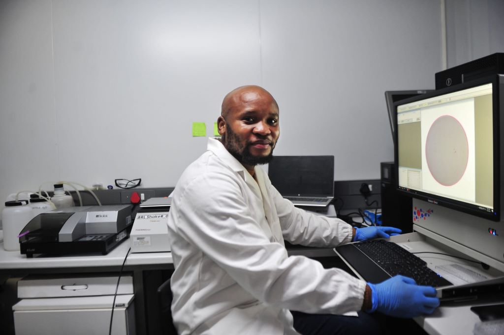
During South Africa’s COVID-19 hard lockdown, Dr Sandile Cele became the first to successfully grow the beta variant of SARS-CoV-2 in the lab. PHOTO: Rosetta Msimango/Spotlight
In a Durban laboratory in 2020, there was dancing and scientists jumping with joy when Dr Sandile Cele realised they had finally successfully “grown” the SARS-CoV-2 Beta variant. It was the holiday season and Cele and a few colleagues had sacrificed their Christmas to continue research at an otherwise deserted laboratory.
The Beta variant (501Y.V2) was first detected in the Eastern Cape in October 2020 and was announced to the public on 18 December that year.
“It was December 2020 and Tulio [Professor Tulio de Oliveira] had just flagged the beta variant and we had been struggling trying to grow it, really struggling for about two weeks,” says Cele. “But then as a scientist, you have to think outside the box and eventually it [the virus] did catch on. I was with Professor Alex Sigal that day in the laboratory. We were so excited. There was a lot of dancing in the lab, jumping up and down…”
The 35-year-old’s work on the Beta and Omicron variants helped propel South Africa to the forefront of COVID-19 research. Cele is the scientist credited with growing both Beta and Omicron in record time as the world reeled under lockdown pressure. Last year, he was awarded a special ministerial Batho Pele excellence award for his contribution to COVID-19 research in South Africa.
The moment of greatest fulfilment
Speaking to Spotlight, Cele says growing the beta variant was the moment of greatest fulfilment in his career so far.
“It was just a crazy, crazy moment. Like, you know when you are with your superior, usually you meet on a basis of respect. I mean, you talk seriously. They ask a question, you answer, and so on. But [at] that moment, all that got thrown out the window. We were celebrating. So yes, it was really special.”
At the time, they were leaping with joy inside PPE (personal protective equipment), including specialised masks, double gloves, plastic sleeves, and boots. Cele points out that due to all the safety measures in place, infection risk was smaller in their lab than at an average mall.
He was working inside a state-of-the-art biosafety level 3 (BSL-3) laboratory at the Africa Health Research Institute (AHRI). The laboratory is on the third floor of the University of KwaZulu-Natal’s medicine building. In the same eight-storey glass and face brick building, on the first floor, de Oliveira had been studying virus samples for genetic clues at KRISP, the KwaZulu-Natal Research and Innovation Sequencing Platform, from where the discovery of Beta and Omicron was first announced.
How he did it – growing the beta variant
Cele explains that viruses are isolated or “outgrown” by infecting cells in the laboratory, using swab samples from infected individuals.
“Growing a virus simply means isolating it from an infected host (humans) and making more of it in the lab for research purposes,” Cele explains. “You cannot study a virus within an infected person, especially a new virus. You need to have it in the lab for identification and clarification. Usually, you get small quantities from an infected person, thus you have to expand or grow – or make more of it – for research.”
Photo by Shvets Production on Pexels
However, the beta variant had not responded like previous SARS-CoV-2 variants. At the time, Cele found a creative solution using both human and monkey cell lines. First, he infected human cell lines with the beta variant, incubating the assay for four days. Then he used the infected human cell lines to infect monkey cell lines, which successfully lead to production of the virus.
Their moment of triumph arrived when they noticed the monkey cell lines starting to die, meaning that the virus was growing. The isolated virus could then be used in the laboratory to run experiments, like testing vaccine efficacy.
“Looking at the cells under the microscope, you can see them starting to die,” he says. “That they’re not happy. That they have been infected, which then obviously needed to be confirmed.”
While Cele’s Durban mentors – de Oliveira and Sigal – kept the public abreast of research developments, the young scientist kept his head down, pouring over his microscopes. “The world was going crazy, everything was crazy, but I had work to do,” he says.
‘a rising star’
During the interview, Cele readily shares anecdotes and laughs often.
From Ndwedwe, a rural area forty kilometres north of Durban, Cele joined Sigal’s laboratory team at the AHRI in 2014, where he studied HIV drug resistance and later COVID-19. His PhD obtained from UKZN in 2021, focused specifically on understanding the beta variant and its escape from antibodies.
“Actually, Professor Alex Sigal really took a chance on me,” he says. “Because on that post for a laboratory technologist, they stipulated that they wanted someone with three years experience. And I had only been doing my internship [at the Technology Innovation Agency] for eight months.”
But Sigal’s faith paid off, and he subsequently praised Cele in national press interviews on COVID-19. “Sandile is a rising star who spent all his holidays in a laboratory,” Sigal told journalists in January 2021.
Last year, the Bill and Melinda Gates Foundation invited Cele to present his findings at the Grand Challenges Annual Meeting in Brussels. This was his first time abroad. “It was my first time traveling outside South Africa and my first time talking in front of so many people. I presented my go-to talk – based on a paper I did on COVID-infection and HIV – and it went well,” he says.
Earlier this year, Cele was named one of Mail & Guardian’s 200 trailblazing young South Africans in the technology and innovation category. At the time, he could not attend the gala event as he was at the University of Nairobi in Kenya for training relating to a project involving HIV research for the Aurum Institute. Cele started a new job at the Aurum Institute in Johannesburg in March.
Over Zoom, Cele is speaking from his new home in Johannesburg. He is wearing a fluffy blue robe over his clothes, laughing as he says how cold Johannesburg is coming from Durban.
A sudden death
In Ndwedwe, Cele was one of ten boys born to his father, who was away from home often for work. Describing his mother as “a busy lady”, Cele says she was the one who shaped his young everyday life. Growing up in a mud hut without electricity and running water, he recalls how his mother would get up early every morning to prepare vetkoek, which she sold at a local school, and to boil water so her children could have a bath before leaving for school.
In the afternoons, he would look after his father’s goats and play soccer. He says that as a child he preferred herding goats to cows, as goats grazed for only about five hours, whereas cows took all day to eat their fill. From Grade 9 on, he attended school in Durban, at Overport Secondary School.
A childhood memory that inspired him? “Before my mother died, she sat us down and said one day I will be gone and I want you to know there are no shortcuts in life. Work hard and look after one another and you will be okay.”
His mother’s death was sudden, following complications from minor surgery.
“Like, I came back from school on a Friday only to find my father wasn’t around and had left a note… On the Saturday morning, I found out my mother had passed. And I think she went for, I don’t know, an operation or something. But as a kid, I guess they didn’t tell us because they thought it was something minor; that she would get operated [on], then go back home. I’m not really sure what happened. So, yes, it was a sudden death.”
The year after his mother died, Cele’s matric marks suffered. He says his final grade 12 marks had been 48% for maths, 53% for physics, and 66% for biology.
“I wasn’t really studying, I couldn’t really concentrate,” he says. “There was a lot going on when I was doing my matric. My mother passing away… and also the move from a rural school to the city where we were taught in English, everything in English.”
Cele came to study biology quite at random. He applied to study at UKZN only in October of his matric year – with admissions to most of the university’s courses having closed the previous month. He picked one of the last remaining options, which had been biology.
Soon, the young student started excelling. Cele obtained his BSc Biomedical Sciences degree with a Dean’s commendation and his Honours in Medical Microbiology, summa cum laude. He completed his Masters in Biochemistry with an upper-class pass.
To the Mail and Guardian, he shared advice he would give to his younger self: “Do not be afraid, you are a force to be reckoned with.”
Cele’s driving passion is to advance public healthcare, which he will continue to do at the Aurum Institute – an organisation that amongst others does research into Africa’s tuberculosis and HIV response. Cele has a ten-year-old son who lives in Durban.
Note:The Bill and Melinda Gates Foundation is mentioned in this article. Spotlight receives funding from the foundation, but is editorially independent – an independence that the editors guard jealously. Spotlight is a member of the South African Press Council.
Detail from Small’s reprocessed cryo-EM data zooming in on an unoccupied area of the SARS-CoV-2 NiRAN domain. (Courtesy of Campbell lab)
The COVID pandemic illustrated how urgently we need antiviral medications capable of treating coronavirus infections. To aid this effort, researchers quickly homed in on part of SARS-Cov-2’s molecular structure known as the NiRAN domain – an enzyme region essential to viral replication that’s common to many coronaviruses. A drug targeting the NiRAN domain would likely work broadly to shut down a range of these pathogens, potentially treating known diseases like COVID as well as helping to head off future pandemics caused by related viruses.
In 2022, scientists (Yan et. al.) published a structural model describing exactly how this domain works. It should have been a tremendous boon for drug developers.
But the model was wrong.
“Their work contains critical errors,” says Gabriel Small, a graduate fellow in the laboratories of Seth A. Darst and Elizabeth Campbell at Rockefeller. “The data does not support their conclusions.”
Now, in a new study published in Cell, Small and colleagues demonstrate exactly why scientists still don’t know how the NiRAN domain works. The findings could have sweeping implications for drug developers already working to design antivirals based on flawed assumptions, and underscore the importance of rigorous validation.
“It is absolutely important that structures be accurate for medicinal chemistry, especially when we’re talking about a critical target for antivirals that is the subject of such intense interest in industry,” says Campbell, head of the Laboratory of Molecular Pathogenesis. “We hope that our work will prevent developers from futilely trying to optimise a drug around an incorrect structure.”
A promising lead
By the time the original paper was published in Cell, the Campbell and Darst labs were already quite familiar with the NiRAN domain and its importance as a therapeutic target. Both laboratories study gene expression in pathogens, and their work on SARS-CoV-2 focuses in part on characterizing the molecular interactions that coordinate viral replication.
The NiRAN domain is essential for helping SARS-CoV-2 and other coronaviruses cap their RNA, a step that allows these viruses to replicate and survive. In one version of this process, the NiRAN domain uses a molecule called GDP to attach a protective cap to the beginning of the virus’s RNA. Small previously described that process in detail, and its structure is considered solved. But the NiRAN domain can also use a related molecule, GTP, to form a protective cap. Determined to develop antivirals that comprehensively shut down the NiRAN domain, scientists were keen to discover the particulars of the latter GTP-related mechanism.
In the 2022 paper, researchers described a chain of chemical steps, beginning with a water molecule breaking a bond to release the RNA’s 5′ phosphate end. That end then attaches to the beta-phosphate end of the GTP molecule, which removes another phosphate and, with the help of a magnesium ion, transfers the remaining portion of the GTP molecule to the RNA, forming a protective cap that allows the virus to replicate and thrive.
The team’s evidence? A cryo-electron microscopy image that showed the process caught in action. To freeze this catalytic intermediate, the team used a GTP mimic called GMPPNP.
Small read the paper with interest. “As soon as they published, I went to download their data,” he says. It wasn’t there. This raised a red flag—data is generally available upon release of a structural biology paper. Months later, however, when Small was finally able to access the data, he began to uncover significant flaws. “I tried to make a figure using their data, and realized that there were serious issues,” he says. Small brought his concerns to Campbell and Darst.
They agreed. “Something was clearly wrong,” Campbell says. “But we decided to give the other team the benefit of the doubt, and reprocess all of their data ourselves.”
An uphill battle
It was painstaking work, with Small leading the charge. Working frame by frame, he compared the published atomic model to the actual cryo-EM map and found something striking: the key molecules that Yan and colleagues claimed to have seen, specifically, the GTP mimic GMPPNP and a magnesium ion in the NiRAN domain’s active site, simply were not there.
Not only was there no supporting image data, but the placement of these molecules in the original model also violated basic rules of chemistry, causing severe atomic clashes and unrealistic charge interactions. Small ran additional tests, but even advanced methods designed to pick out rare particles turned up empty. He could find no evidence to support the model previously produced by Yan and colleagues.
Once the Rockefeller researchers validated their results, they submitted their findings to Cell. “It was very important that we publish our corrective manuscript in the same journal that published the original model,” Campbell says, noting that corrections to high-profile papers are often overlooked when published in lower tier journals.
Otherwise, this confusion in the field could cause problems that reach far beyond the lab bench, Campbell adds – a costly reminder that rigorous basic biomedical research is not just academic, but essential to real-world progress. “Companies keep their cards close to their chests, but we know that several industry groups are studying this,” she says. “Efforts based on a flawed structural model could result in years of wasted time and resources.”
A blood-test analysis developed at Stanford Medicine can determine the “biological ages” of 11 separate organ systems in individuals’ bodies and predict the health consequences.
Beside our chronological age, research has shown that we also have what’s called a “biological age,” a cryptic but more accurate measure of our physiological condition and likelihood of developing aging-associated disorders from heart trouble to Alzheimer’s disease.
How old someone’s internal organs are is a challenge to determine compared to looking at wrinkles and greying hair. Internal organs are ageing at different speeds, too, according to a new study by Stanford Medicine investigators.
“We’ve developed a blood-based indicator of the age of your organs,” said Tony Wyss-Coray, PhD, professor of neurology and neurological sciences and director of the Knight Initiative for Brain Resilience at the Wu Tsai Neurosciences Institute. “With this indicator, we can assess the age of an organ today and predict the odds of your getting a disease associated with that organ 10 years later.”
They can even predict who is most likely to die from medical conditions associated with one or more of the 11 separate organ systems the researchers looked at: brain, muscle, heart, lung, arteries, liver, kidneys, pancreas, immune system, intestine and fat.
The brain is the gatekeeper of longevity. If you’ve got an old brain, you have an increased likelihood of mortality. If you’ve got a young brain, you’re probably going to live longer.”
The biological age of one organ, the brain, plays an outsized role in determining how long you have left to live, Wyss-Coray said.
“The brain is the gatekeeper of longevity,” he said. “If you’ve got an old brain, you have an increased likelihood of mortality. If you’ve got a young brain, you’re probably going to live longer.”
Wyss-Coray is the senior author of the study, published online July 9 in Nature Medicine. The lead author is Hamilton Oh, PhD, a former graduate student in Wyss-Coray’s group.
Eleven organ systems, 3000 proteins, 45 000 people
The scientists used 44 498 randomly selected participants, ages 40 to 70, who were drawn from the UK Biobank. This ongoing effort has collected multiple blood samples and updated medical reports from some 600 000 individuals over several years. These participants were monitored for up to 17 years for changes in their health status.
Wyss-Coray’s team made use of an advanced commercially available laboratory technology that counted the amounts of nearly 3000 proteins in each participant’s blood. Some 15% of these proteins can be traced to single-organ origins, and many of the others to multiple-organ generation.
The researchers fed everybody’s blood-borne protein levels into a computer and determined the average levels of each of those organ-specific proteins in the blood of those people’s bodies, adjusted for age. From this, the scientists generated an algorithm that found how much the composite protein “signature” for each organ being assessed differed from the overall average for people of that age.
Based on the differences between individuals’ and age-adjusted average organ-assigned protein levels, the algorithm assigned a biological age to each of the 11 distinct organs or organ systems assessed for each subject. And it measured how far each organ’s multiprotein signature in any given individual deviated in either direction from the average for people of the same chronological age. These protein signatures served as proxies for individual organs’ relative biological condition. A greater than 1.5 standard deviation from the average put a person’s organ in the “extremely aged” or “extremely youthful” category.
One-third of the individuals in the study had at least one organ with a 1.5-or-greater standard deviation from the average, with the investigators designating any such organ as “extremely aged” or “extremely youthful.” One in four participants had multiple extremely aged or youthful organs.
For the brain, “extremely aged” translated to being among the 6% to 7% of study participants’ brains whose protein signatures fell at one end of the biological-age distribution. “Extremely youthful” brains fell into the 6% to 7% at the opposite end.
Health outcomes foretold
The algorithm also predicted people’s future health, organ by organ, based on their current organs’ biological age. Wyss-Coray and his colleagues checked for associations between extremely aged organs and any of 15 different disorders including Alzheimer’s and Parkinson’s diseases, chronic liver or kidney disease, Type 2 diabetes, two different heart conditions and two different lung diseases, rheumatoid arthritis and osteoarthritis, and more.
Risks for several of those diseases were affected by numerous different organs’ biological age. But the strongest associations were between an individual’s biologically aged organ and the chance that this individual would develop a disease associated with that organ. For example, having an extremely aged heart predicted higher risk of atrial fibrillation or heart failure, having aged lungs predicted heightened chronic obstructive pulmonary disease (COPD) risk, and having an old brain predicted higher risk for Alzheimer’s disease.
The association between having an extremely aged brain and developing Alzheimer’s disease was particularly powerful: 3.1 times that of a person with a normally aging brain. Meanwhile, having an extremely youthful brain was especially protective against Alzheimer’s – barely one-fourth that of a person with a normally aged brain.
In addition, Wyss-Coray said, brain age was the best single predictor of overall mortality. Having an extremely aged brain increased subjects’ risk of dying by 182% over a roughly 15-year period, while individuals with extremely youthful brains had an overall 40% reduction in their risk of dying over the same duration.
Predicting the disease, then preventing it
“This approach could lead to human experiments testing new longevity interventions for their effects on the biological ages of individual organs in individual people,” Wyss-Coray said.
Medical researchers may, for example, be able to use extreme brain age as a proxy for impending Alzheimer’s disease and intervene before the onset of outward symptoms, when there’s still time to arrest it, he said.
Careful collection of lifestyle, diet and prescribed- or supplemental-substance intake in clinical trials, combined with organ-age assessments, could throw light on the medical value of those factors’ contributions to the aging of various organs, as well as on whether existing, approved drugs can restore organ youth before people develop a disease for which an organ’s advanced biological age puts them at high risk, Wyss-Coray added.
If commercialised, the test could be available in the next two to three years, Wyss-Coray said. “The cost will come down as we focus on fewer key organs, such as the brain, heart and immune system, to get more resolution and stronger links to specific diseases.”