Tag: tooth loss

Zinc-based Compound Could be a New Cavity Fighter

Scientists at NYU are developing a zinc-based treatment for tooth decay that combats bacteria, blocks pain, and avoids staining teeth – all without drilling

Photo by Caroline Lm on Unsplash

Tooth decay is the most common health condition worldwide. While it is preventable and treatable, billions of people are living with cavities and the pain that accompanies them.

Given the massive scale of the problem, there’s a growing movement in dentistry to treat cavities without drilling and filling them. One such approach is applying a clear liquid called silver diamine fluoride to the surface of teeth. Silver diamine fluoride is already FDA-approved to treat tooth sensitivity, and recent NYU research shows that the compound’s antimicrobial properties also make it effective at preventing cavities and stopping small cavities from progressing into larger ones. Because it’s inexpensive and easy to administer, it can be given in schools, in rural areas lacking dentists, or to patients who may have difficulty with dental care, including those with disabilities. 

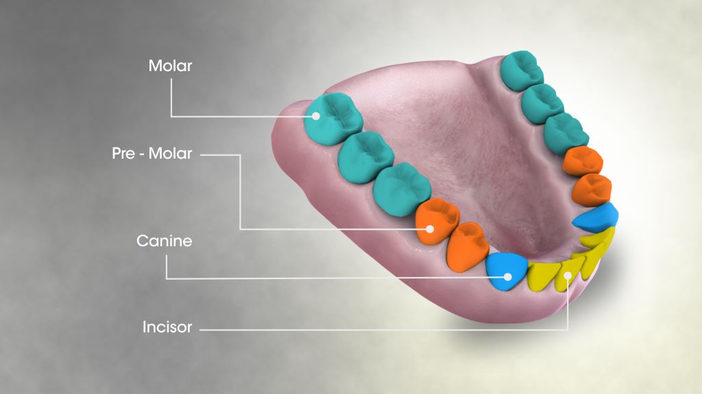
But treatment with silver diamine fluoride comes with one notable drawback: when the silver in it interacts with tooth decay, it turns the treated surface black. While this is not a significant issue for molars at the back of the mouth or baby teeth that fall out, it’s not a great option for teeth seen in a smile.

“Once your teeth are treated with silver diamine fluoride, that stain is permanent, which is a barrier for many people wanting to use the product,” explains Marc Walters, professor of chemistry at NYU. 

Walters has long studied silver and other elements used in medicine to carry drugs and imaging contrast agents. Several years ago, he was approached by researchers at NYU College of Dentistry seeking to better understand how silver stains teeth in order to avoid that outcome.

From silver to zinc

Walters had an idea. What if another mineral could be used that was also colourless and antimicrobial but didn’t turn teeth black? This question led him to zinc, an important nutrient found in foods like oysters and beef, as well as in over-the-counter products intended to shorten the duration of colds. Zinc is also used in dentistry, including in toothpaste and mouthwash to fight bacteria and bad breath, as well as in some denture adhesives and cementing agents to affix crowns or temporary fillings.

Walters began studying a zinc phosphate compound to see how it interacts with cavities, and crucially, to determine whether it can permeate deep into teeth. In order to address pain and hypersensitivity, the compound would need to reach the tooth’s dentin, the porous material sandwiched between the hard enamel outer layer and the nerves within. Dentin contains an abundance of microscopic, hollow channels – in fact, 40 000 of these tubules are packed into each square millimetre of dentin. 

“We had to develop a solution to give dentists that will be taken up in these very small openings and go deep enough in the tubules so that the material will be retained,” Walters explains.

Walters applied phosphate followed by zinc to slices of a human tooth. Under the microscope, he saw deposits of the compound deep inside the dentin tubules. But while the zinc phosphate successfully permeated the teeth, he knew that a simpler approach that didn’t require applying two treatments would be easier for dentists. “Two steps is one too many,” says Walters.

Drawing inspiration from silver diamine fluoride, Walters developed another zinc-based molecule called zinc tetramine difluoride, which forms a colourless zinc oxide deep inside dentin tubules. The agent starts out as a liquid that is sensitive to concentration and pH. When painted onto a tooth and absorbed, the conditions within dentin tubules prompt a chemical change that quickly turns it into a solid, blocking the tubules and slowly releasing the antimicrobial zinc into the tooth.

His team is continuing to develop several related compounds for the treatment of cavities and has applied for patents of these zinc-based materials in several countries.

Fast and slow

Having both fact-acting and long-lasting properties would offer an ideal combination for fighting cavities and tooth sensitivity, given that many current treatments for sensitive teeth require multiple applications and can take days or weeks to work. 

“In one of our studies, two minutes after treatment with our agent, we can see using the electron microscope that the zinc forms long cylinders of mineral that occupy the tubules,” says Walters. “Blocking the dentin tubules cuts off access to the nerves that are much deeper in dentin. It’s like putting a cork in place that shuts off the lower portion of the tubule from the outside environment – and this happens within a minute or two.”

Walters shows an image of a tubule under the microscope that was filled with the zinc compound.

In additional tests, Walters found that zinc oxide persisted in tooth samples for at least one to two months. The goal is to develop a product that lasts for months or even years inside of teeth, stopping hypersensitivity and fighting bacteria on an ongoing basis. 

“Not only do you have the analgesic result of having tubules blocked, but you also have a very low solubility agent that can slowly release the zinc into the tubule to prevent the growth of Streptococcus mutans and other bacteria,” Walters adds.

The journey from lab to shelves

With a promising zinc nanocrystal agent in hand, Walters sought out other experts at NYU and beyond. His work caught the attention of Southern Dental Industries (SDI), an Australian company that makes restorative dental materials, including silver diamine fluoride. The company purchased the license for the zinc technology and NYU is working with them to develop it.

Closer to home, Walters began collaborating with Deepak Saxena, professor of molecular pathobiology and director of research innovation and entrepreneurship at NYU College of Dentistry.

Saxena and Walters are collaborating on a new NIH grant to further develop the zinc-based treatment.

As a result of bringing together this diverse expertise Saxena and Walters received a award from NYU, and last month, secured a grant from the NIH

The NIH grant will fund feasibility studies for Walters’s team to further develop the formulation and confirm its ability to block tubules in a range of dentin samples. It will also fund research through Periomics Care in which Saxena’s team will study the agent’s antimicrobial properties. Specifically, they will look to see if the zinc creates a “zone of inhibition” – preventing the growth of decay-causing bacteria in the vicinity of it or even killing bacteria that comes in contact with it.  

“The mouth is full of bacteria. A compound needs to have good antimicrobial activity, which can occur from ionic imbalance, the properties of the zinc, or by the fluoride,” Saxena says. “If a compound does not stain, has good antimicrobial activity, plus it blocks the tubules, then it should be successful in stopping tooth decay and be aesthetically accepted.” 

Saxena and Walters are already planning for the next phase of their research, which will include additional studies on the compound’s formulation, effectiveness, toxicity, and shelf life. Ultimately, if these studies go well, the researchers and SDI will approach the FDA for permission to do a clinic trial.

One factor working in their favour: because zinc phosphate has long been used as a dental adhesive, it’s known to be safe and the FDA has already approved it in other forms. These existing products may pave the way for faster research and development of a cavity treatment compared to untested elements, which can take many years to develop.

The future of dentistry

A new non-invasive treatment for cavities could be a game-changer in oral health. “We know that there’s a need – and a market – for a product that stops tooth decay that is effective, cheap, easy to use, and non-staining, given the rise in global numbers of untreated cavities,” Saxena says. 

Dentists could use it to treat cavities without needing to scrape or drill out the cavity in preparation. Squirmy kids would need less time in the dentist’s chair. Older adults who get cavities near the roots of their teeth as their gums recede could have a new option for stopping sensitivity and decay in difficult-to-treat areas. If safe and effective, perhaps small quantities could even be available on drugstore shelves and sold directly to consumers.

For Walters and Saxena, their goal is a future with less tooth decay and pain – and if their studies of zinc confirm its potential, silver-stained teeth may be a thing of the past.

Source: New York University

Tooth Pain Nerves Serve Another Purpose: Tooth Protectors

Types of teeth. Credit: Scientific Animation CC4.0

Until now the sensory neurons inside the tooth were primarily thought to send pain signals to the brain, but a new study shows those neurons are multitaskers that also trigger a jaw-opening reflex that almost instantaneously prevents damage and further injury to teeth.

The reflex that pops open the lower jaw was a widely known craniofacial reflex, but until this study the cellular origins of this phenomenon were not known.

University of Michigan researchers in sensory neuroscience, dentistry and mechanical engineering found the origin using special live imaging, behaviour-tracking tools and mice molars to uncover the neurons’ additional role of monitoring the inner tooth and outer enamel.

The discovery and understanding of this additional role shows how important healthy, active nerves are for preserving teeth.

“We suspected there was a more fundamental role for tooth nerves,” said Joshua Emrick, senior author of the study and assistant professor at the U-M School of Dentistry. “When we consider regenerating a tooth pulp, we need to bring back the nerves.”

Emrick’s research team looked at how nerve cells reacted to stimulation of the molar teeth of mice in real time. Their experiments revealed a newly defined, protective role for intradental High-Threshhold Mechanoreceptors, highly specialized sensory neurons that respond to tooth damage. These HTMRs detect dangerous threats and send the message rapidly to the brain for instantaneous action.

“Our study challenges the prior assumption that nerves inside the tooth primarily function to elicit pain and force us straight to the dentist for help,” Emrick said. “If you’ve ever accidentally bitten down on your fork, you’ve probably experienced a startling jolt, but also stopped short of fracturing your teeth. You may thank these intradental HTMRs for that.”

The reflex is really about self-preservation.

“We think protection of the teeth through this jaw-opening reflex is highly conserved among mammals that haven’t developed the ability to replace teeth – like humans or in the molar teeth of mice,” Emrick said. “Our work reports an ability to use these neurons to also elicit pain which will open up possibilities for developing new methods for relieving toothache at the dentist’s office.”

To break it down further, the study, published in Cell Reports, showed that when enamel or dentin is damaged, the neurons fire a response. Follow-up experiments determined what happened after the HTMRs were activated. As previously known, the group identified that they trigger acute pain, but more surprisingly they also witnessed a rapid jaw-opening reflex within 5 to 15 milliseconds of the activation.

While the authors focused their work on understanding how the HTMRs function within the tooth, this important subclass of sensory neurons may protect other oral and body structures from damage.
Elizabeth Ronan, postdoctoral fellow at the School of Dentistry and lead author of the work, said the findings are the start of a deeper understanding.

“While we typically think of sensation as giving rise to our perceived external experience of the world, sensory neurons are equally essential in protecting and maintaining our tissues throughout life,” she said. “Much remains to be discovered regarding how sensory neurons function within individual tissues, especially internal ones such as the teeth.”

Source: University of Michigan

Study Finds that Titanium Particles are Common Around Dental Implants

Photo by Tima Miroshnichenko on Pexels

Titanium micro-particles in the oral mucosa around dental implants are common. This is shown in a new study from the University of Gothenburg, which also identified 14 genes that may be affected by these particles.

According to the researchers, there is no reason for concern, but more knowledge is needed.

“Titanium is a well-studied material that has been used for decades. It is biocompatible and safe, but our findings show that we need to better understand what happens to the micro-particles over time. Do they remain in the tissue or spread elsewhere in the body?” says Tord Berglundh, senior professor of periodontology at Sahlgrenska Academy, University of Gothenburg.

Found at all implants

Previous research has shown that titanium particles may occur in inflamed tissues around dental implants. The new study, published in Communications Medicine, showed that titanium micro-particles were consistently found at all examined implants—even those without signs of inflammation.

The researchers analysed tissue samples from 21 patients with multiple adjacent implants. Samples were taken both at healthy implants and at implants affected by peri-implantitis, an inflammatory disease in the tissue around the implant. Each patient thus served as their own control. The density of particles varied between patients, but not between sites with and without peri-implantitis within the same patient. The analyses were conducted in collaboration with Uppsala University, where researchers used an advanced method called µ-PIXE to map the distribution of titanium particles in the tissue samples.

Affected genes

Peri-implantitis is a microbial biofilm-associated inflammatory disease around dental implants, with features similar to those of periodontitis around teeth. The inflammatory process is complex and the resulting destruction of supporting bone in peri-implantitis may lead to loss of the implant. 

“We observed that tissue samples with higher concentrations of titanium particles had an altered gene expression, especially genes related to inflammation and wound healing. We identified 14 such genes, but it is unclear whether the particles influence the local immune response or if the difference in gene expression reflects inter-individual variability in inflammatory conditions,” says Carlotta Dionigi, specialist in periodontology and researcher at the Department of Periodontology, Sahlgrenska Academy, University of Gothenburg.

The researchers suspect that titanium particles are released during the surgical installation procedure, when the screw-shaped implant is inserted into the prepared canal in the alveolar bone. In this context, the observation on differences in micro-particle densities between various implant systems deserves attention, since the surface structure of the implant may influence the deposition of micro-particles. This is now an important topic for continued research.

Source: University of Gothenburg

Diabetes may Weaken Teeth, Promoting Tooth Decay

Source: Unsplash CC0

People with both Type 1 and Type 2 diabetes are prone to tooth decay, and a new study published in Archives of Oral Biology may explain why: reduced strength and durability of enamel and dentin, the hard substance under enamel that gives structure to teeth.

Researchers induced Type 1 diabetes in 35 mice and used a Vickers microhardness tester to compare their teeth with those of 35 healthy controls over 28 weeks. Although the two groups started with comparable teeth, enamel grew significantly softer in the diabetic mice after 12 weeks, and the gap continued to widen throughout the study. Significant differences in dentin microhardness arose by week 28.

“We’ve long seen elevated rates of cavity formation and tooth loss in patients with diabetes, and we’ve long known that treatments such as fillings do not last as long in such patients, but we did not know exactly why,” said Mohammad Ali Saghiri, an assistant professor of restorative dentistry at the Rutgers School of Dental Medicine.

The study advances a multiyear effort by Assistant Prof Saghiri and other researchers to understand how diabetes affects dental health and to develop treatments that counter its negative impact. Previous studies have established that people with both types of diabetes have significantly elevated rates of most oral health issues, both in the teeth and the soft tissues that surround them. Assistant Prof Saghiri and other researchers also have demonstrated that diabetes can interfere with the ongoing process of adding minerals to teeth as they wear away from normal usage.

“This is a particular focus of mine because the population of people with diabetes continues to grow rapidly,” Assistant Prof Saghiri said. “There is a great need for treatments that will allow patients to keep their teeth healthy, but it has not been a major area for research.”

Source: EurekAlert!

New Machine Learning Tools Could Save Teeth

Photo by Kevin Bation on Unsplash

Machine learning tools could help identify those at greatest risk for tooth loss and refer them for further dental assessment for early interventions to avert or delay the conditions.

The study by researchers at the Harvard School of Dental Medicine compared five different algorithms using various combinations of variables to screen for risk. The results showed those that factored medical characteristics and socioeconomic variables, including race, education, arthritis, and diabetes, outperformed algorithms that relied on dental clinical indicators alone.

“Our analysis showed that while all machine-learning models can be useful predictors of risk, those that incorporate socioeconomic variables can be especially powerful screening tools to identify those at heightened risk for tooth loss,” said study lead investigator Hawazin Elani, assistant professor of oral health policy and epidemiology at HSDM.

The approach could be used to screen people globally and in a variety of health care settings even by non-dental professionals, she added. This approach could be applied around the world, even allowing non-dental professionals to screen patients.

Tooth loss can affect quality of life, well-being, nutrition, and social interactions. It is also associated with dementia. If the earliest signs of dental disease are identified, then the process can be delayed or averted with prompt treatment. However, many people with dental disease may not see a dentist until the process is too far gone. This is where screening tools could help identify those at highest risk and refer them for further assessment, the team said.

For the study, the researchers used data on nearly 12 000 adults from the National Health and Nutrition Examination Survey to design and test five machine-learning algorithms and assess their predictions for both complete and incremental tooth loss among adults based on socioeconomic, health, and medical characteristics.

A key point is that algorithms were designed to assess risk without a dental exam, though anyone at risk for tooth loss would still need one. The study’s findings point to the importance of socioeconomic factors.

“Our findings suggest that the machine-learning algorithm models incorporating socioeconomic characteristics were better at predicting tooth loss than those relying on routine clinical dental indicators alone,” Elani said. “This work highlights the importance of social determinants of health. Knowing the patient’s education level, employment status, and income is just as relevant for predicting tooth loss as assessing their clinical dental status.”

Low socioeconomic status populations have long been known to have greater rates of tooth loss, likely due to lack of regular access to dental care, among other reasons, the team said.

“As oral health professionals, we know how critical early identification and prompt care are in preventing tooth loss, and these new findings point to an important new tool in achieving that,” said Jane Barrow, associate dean for global and community health and executive director of the Initiative to Integrate Oral Health and Medicine at HSDM. “Dr. Elani and her research team shed new light on how we can most effectively target our prevention efforts and improve quality of life for our patients.”

Source: Harvard Medical School

Journal information: Hawazin W. Elani et al, Predictors of tooth loss: A machine learning approach, PLOS ONE (2021). DOI: 10.1371/journal.pone.0252873