Tag: 13/10/25

Amazing Images Show How Antibiotics Shred Bacterial Armour

Bacteria response to antibiotics

UK researchers have shown for the first time in stunning detail how life-saving antibiotics act against harmful bacteria. The team, led by UCL and Imperial College London, has shown for the first time how a class of antibiotics called polymyxins are able to pierce the armour of E. coli and kill the microbes. 

The findings, published in the journal Nature Microbiology, could lead to new treatments for bacterial infections – especially urgent since drug-resistant infections already kill more than a million people a year.

Polymyxins were discovered more than 80 years ago and are used as a last-resort treatment for infections caused by “Gram negative” bacteria.

These bacteria have an outer surface layer that acts like armour and prevents certain antibiotics from penetrating the cell. Polymyxins are known to target this outer layer, but how they disrupt it and then kill bacteria is still not understood.

Through capturing these incredible images of single cells, we’ve been able to show that this class of antibiotics only work with help from the bacterium, and if the cells go into a hibernation-like state, the drugs no longer work – which is very surprising

Dr Andrew Edwards Department of Infectious Disease

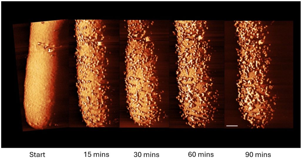
In the new study, the research team revealed in high-resolution images and biochemical experiments how the antibiotic Polymyxin B rapidly caused bumps and bulges to break out on the surface of an E. coli bacterial cell.

These protrusions, which appeared within minutes, were followed by the bacterium rapidly shedding its outer armour.

The antibiotic, the researchers concluded, had triggered the cell to produce and shed its armour.

The more the cell tried to make new amour, the more it lost the amour it was making, at such a rate that it left gaps in its defences, allowing the antibiotic to enter the cell and kill it.

However, the team found that this process – protrusions, fast production and shedding of armour, and cell death – only occurred when the cell was active. In dormant (sleeping) bacteria, armour production is switched off, making the antibiotic ineffective.

Co-senior author Dr Andrew Edwards, from the Department of Infectious Disease at Imperial College London, said: “For decades we’ve assumed that antibiotics that target bacterial armour were able to kill the microbes in any state, whether they’re actively replicating or they were dormant. But this isn’t the case.

“Through capturing these incredible images of single cells, we’ve been able to show that this class of antibiotics only work with help from the bacterium, and if the cells go into a hibernation-like state, the drugs no longer work – which is very surprising.”

Becoming dormant allows bacteria to survive unfavourable conditions such as a lack of food. They can stay dormant for many years and “wake up” when conditions become more favourable. This can allow bacteria to survive against antibiotics, for instance, and reawaken to cause recurrent infections in the body.

Co-senior author Professor Bart Hoogenboom, based at the London Centre for Nanotechnology at UCL, said: “Polymyxins are an important line of defence against Gram-negative bacteria, which cause many deadly drug-resistant infections. It is important we understand how they work.

“Our next challenge is to use these findings to make the antibiotics more effective. One strategy might be to combine polymyxin treatment – counterintuitively – with treatments that promote armour production and/or wake up ‘sleeping’ bacteria so these cells can be eliminated too.

“Our work also shows we need to take into account what state bacteria are in when we are assessing the effectiveness of antibiotics.”

The E. coli cells were imaged at the London Centre for Nanotechnology at UCL. A tiny needle, only a few nanometres wide, was run over the bacterial cell, “feeling” the shape to create an image (a technique called atomic force microscopy) at much higher resolution than would be possible using light.

Co-author Carolina Borrelli, a PhD student at the London Centre for Nanotechnology at UCL, said: “It was incredible seeing the effect of the antibiotic at the bacterial surface in real-time. Our images of the bacteria directly show how much polymyxins can compromise the bacterial armour. It is as if the cell is forced to produce ‘bricks’ for its outer wall at such a rate that this wall becomes disrupted, allowing the antibiotic to infiltrate.”

The team compared how active (growing) and inactive E. coli cells responded to polymyxin B in the lab, finding that the antibiotic efficiently eliminated active cells but did not kill dormant cells.

They also tested the E. coli cells’ response with and without access to sugar (a food source that wakes up dormant cells). When sugar was present, the antibiotic killed previously dormant cells, but only after a delay of 15 minutes – the time needed for the bacteria to consume the sugar and resume production of its outer armour.

In conditions where the antibiotic was effective, the researchers detected more armour being released from the bacteria. They also observed the bulges occurring across the surface of the cell.

In conditions where it was ineffective, the antibiotic bound itself to the outer membrane but caused little damage.

Co-author Dr Ed Douglas, from Imperial, said: “We observed that disruption of the outermost armour of the bacteria only occurred when the bacteria were consuming sugar. Once we knew that, we could quickly figure out what was happening.”

Co-author Professor Boyan Bonev, of the University of Nottingham, said: “Working together has given us unique insights into bacterial physiology and morphology under stress that have remained hidden for decades. Now we understand better the weak points of bacteria.”

Source: Imperial College London

Could Slime Mould Microbes Be a Source of Potent Antimicrobials?

Photo by National Cancer Institute on Unsplash

The cellular slime mold Dictyostelium discoideum is a soil microbe that produces diverse natural products with potential antibiotic activity. Previously, three chlorinated compounds had been detected in Dictyostelium, but only the most abundant compound (CDF-1) was identified and shown to be almost as effective an antimicrobial as ampicillin.  In research published in FEBS Open Bio, investigators optimised lab culture conditions of Dictyostelium cells to boost the levels of low-abundance chlorinated compounds and to characterise their antimicrobial properties.

The optimized culture conditions took advantage of propionic acid and zinc supplementation to increase the yield of the chlorinated compounds, leading to the identification of CDF-2 and CDF-3 in addition to CDF-1. The molecular structure of CDF-2 and CDF-3 was similar to that of CDF-1, aside from the length of a molecular structure called an acyl side chain. When their antibacterial activity was tested, similarly to CDF-1, CDF-2 and CDF-3 exhibited stronger activity against Gram-positive bacteria than ampicillin but limited activity against Gram-negative bacteria.

Because these compounds are conserved across distantly related Dictyostelium species, CDFs may fulfill a critical role in protecting against harmful bacteria.

“Soil presents both opportunities and dangers for the Dictyostelium amoeba, and we believe this amoeba responds by producing specialised chemicals to attract, repel, or eliminate friends, prey, and predators. We are just starting to discover these chemicals, including this new, potent antibiotic,” said corresponding author Tamao Saito, PhD, of Sophia University, in Japan.

Source: Wiley

New One-hour, Low-cost HPV Test Could Transform Cervical Cancer Screening

Materials used to run the HPV LAMP assay. A cytology brush is used to collect a cervicovaginal swab sample into ThinPrep buffer. Samples are lysed in screw-on tubes and lysate is added to LAMP reagents in PCR tubes. The assay is run on the Axxin T8-ISO heater/fluorimeter.

A team of researchers led by Rice University, in collaboration with colleagues in Mozambique and the US, has developed a simple, affordable human papillomavirus (HPV) test that delivers results in less than an hour with no specialised laboratory required. The breakthrough could provide an option for women in low-resource settings to be screened and treated for cervical cancer in a single clinic visit, a step that global health experts say could save countless lives. The research was recently published in Nature Communications.

Cervical cancer is considered easily preventable, yet it remains one of the deadliest cancers for women worldwide. According to the World Health Organization (WHO), each year more than 350 000 women die from the disease, and nearly 90% of those deaths occur in low- and middle-income countries where access to regular cervical cancer screening is limited. Persistent infection with high-risk types of HPV causes nearly all cases of cervical cancer. While vaccines are helping reduce HPV infections globally, most women at risk today are adults who did not get the vaccine in childhood. For them, regular and reliable screening is the only path to early detection and lifesaving treatment.

“Cervical cancer is almost entirely preventable, yet it still claims hundreds of thousands of lives each year,” said first author Maria Barra, a bioengineering graduate student at Rice. “Our goal was to build a test accurate enough to guide treatment, fast enough to use during a clinic visit and inexpensive enough to scale. This assay meets all three goals.”

The WHO recommends HPV DNA testing as the gold standard for cervical cancer screening, but existing HPV DNA tests often require expensive lab equipment and trained laboratory technicians – barriers that make widespread use in low-resource settings unattainable. As a result, many women are not screened for cervical cancer. Even where screening programs exist, results may take days or weeks to return. Patients leave to await results. However, where care facilities are remote, few in number and difficult to access, patients are often unable to return for treatment, leaving precancerous lesions to progress unchecked. A faster test without reliance on a lab could provide results and prompt treatment during the same patient visit.

“This is the kind of pragmatic innovation we focus on when engineering for global health – fewer steps, lower cost, higher impact,” said Rebecca Richards-Kortum, Professor of Bioengineering and co-director of the Rice360 Institute for Global Health Technologies at Rice. “Our data show you can bring lab-grade molecular screening to almost any setting without sacrificing reliability. Providing accurate results quickly enables clinicians to start treatment without delay.”

The new test uses a method called loop-mediated isothermal amplification (LAMP), which simplifies DNA detection by running at a single temperature. Instead of requiring DNA extraction – a complicated step in many existing tests – this process is extraction-free. A swab sample is chemically lysed, added directly to the LAMP reagents and incubated for about 45 minutes in a portable heater then read by fluorescence.

The test detects three of the most dangerous HPV types (HPV16, HPV18 and HPV45), which together cause about 75% of all cervical cancers. It also includes a built-in cellular control to ensure that the sample was collected properly.

In clinical studies, the test showed 100% agreement with the reference standard in 38 samples from Houston and 93% agreement in 191 samples from Maputo, Mozambique. The cost of the test is projected to be less than $8 each, and the portable device it runs on is battery-operated, making it ideal for clinics without consistent electricity.

“High mortality rates from cancer are closely associated with delays in diagnoses and limited access to early treatment,” said Cesaltina Lorenzoni, head of the National Cancer Control Program at the Mozambican Ministry of Health, director of science and teaching at Maputo Central Hospital and professor of pathology at the Eduardo Mondlane University Faculty of Medicine. “Point-of-care technologies that can aid clinicians in identifying cancer and guide treatment options in a single patient visit could be lifesaving in clinical settings in Maputo. This assay performed very well in our clinical setting and holds promise of delivering the kind of rapid, specific, cost-effective cancer detection that would meaningfully improve outcomes for women in our country.”

The WHO has set ambitious targets to screen 70% of women worldwide by 2030 as part of its public health campaign to eliminate cervical cancer. Meeting that goal will require screening millions of women in various global settings that lack advanced lab equipment or resources.

By cutting out expensive instruments, minimising sample handling and delivering rapid, accurate results, the LAMP assay represents a significant step toward realistically achieving the WHO goal. Critically, it opens the door to “screen-and-treat” strategies, where if a positive result is found, the patient can be treated on the same medical visit, reducing treatment delays and loss to follow-ups.

The team is currently working to expand the test to cover additional high-risk HPV types and is also working on lyophilised (freeze-dried) reagents that don’t require refrigeration, further increasing the test’s usability in rural or resource-limited areas. The team also plans to conduct usability studies with frontline health workers to refine the design before larger clinical rollouts.

“Our goal is a complete, field-ready kit that community clinics can use anywhere,” Richards-Kortum said. “If we can help health systems move to same-day screen-and-treat, we can move towards a future where cervical cancer can be eliminated globally.”

Source: Rice University

Certain Fatty Acids can ‘Supercharge’ T-Cells’ Antitumour Immunity

A research team at the LKS Faculty of Medicine of the University of Hong Kong (HKUMed) discovered that certain dietary fatty acids can supercharge the human immune system’s ability to fight cancer. The team found that a healthy fatty acid found in olive oil and nuts, called oleic acid (OA), enhances the power of immune γδ-T cells, specialised cells known for their cancer-fighting properties.

Conversely, they found that another fatty acid, called palmitic acid (PA), commonly found in palm oil and fatty meats, diminishes the ability of these immune cells to attack tumours. This groundbreaking study, published in the academic journal Signal Transduction and Targeted Therapy, offers an innovative approach using dietary OA supplementation to strengthen the antitumour immunity of γδ-T cells.

Dietary fatty acids and cancer immunotherapy

Dietary fatty acids are essential for health, helping with growth and body functions. They may also play a role in cancer prevention and treatment, but understanding how they affect cancer is challenging because of the complexity of people’s diets and the lack of detailed studies. Recently, scientists have learned that fatty acids can influence the immune system, especially in how it fights cancer. Specialised immune cells, called γδ-T cells, are particularly good at attacking tumours. These cells, once activated, have helped some lung and liver cancer patients live longer. However, this therapy is not effective for all patients, partly because the variation of the metabolic status, such as fatty acid metabolism, can influence its efficacy in the patients.

Oleic acid may improve cancer treatment outcomes

The research team identified a correlation between PA and OA levels and the efficacy of cancer therapies. ‘Our research suggests that dietary fatty acid supplementation, particularly with foods rich in OA, such as olive oil and avocados, could enhance γδ-T cell immunosurveillance, leading to more effective cancer treatments,’ said Professor Tu Wenwei from the Department of Paediatrics and Adolescent Medicine, School of Clinical Medicine, HKUMed, who led the study.

The team also discovered that another fatty acid, called PA, can weaken these immune cells and how OA can counteract this. ‘The results indicate that cancer patients should avoid PA and consider OA supplementation in their diets to improve clinical outcomes of γδ-T cell-based cancer therapies,’ added Professor Tu.

Significant impact from simple dietary changes

Professor Tu said, ‘This study is the first to show that the fatty acids we eat can directly affect how well our immune cells fight cancer.’ It reveals how PA can harm these cells and how OA helps them through a specific process involving a protein called IFNγ. By analysing blood samples, the researchers confirmed that the levels of these fatty acids are linked to the outcome of cancer immunotherapy.

‘For cancer patients, this discovery suggests simple changes, like eating more foods rich in OA (such as olive oil, avocados and nuts) and cutting back on PA (found in processed foods, palm oil and fatty meats), could improve the effectiveness of cancer treatments. The study also points to novel strategies, like combining dietary changes with specific drugs to further boost the immune system,’ added Professor Tu.

This study demonstrates that personalised nutrition may serve as an effective strategy to enhance immune function and support cancer treatment. It also suggests that new drugs targeting the processes affected by these fatty acids could enhance the power of γδ-T cell therapies. By integrating nutritional interventions with immunotherapy, this discovery could help more cancer patients achieve better outcomes.

Source: University of Hong Kong

Aplastic Anaemia: A Stem Cell Donor Is the Difference Between a Future and a Fight for Life 

Both Leathan (L) and Godfrey (R) have aplastic anaemia, which can treated with a stem cell donation. Leathan received stem cells from his twin sister, who is a perfect match. But Godfrey must travels from KwaMhlanga to Pretoria for life-sustaining blood transfusions.

When aplastic anaemia struck two young South Africans, their fates diverged dramatically. While one received a life-saving stem cell transplant, the other continues to fight every day. The rare blood disease affects fewer than six people per million, but for Leathan and Godfrey, the statistics became deeply personal. 

Understanding Aplastic Anaemia: When Hope Meets Science 

Aplastic anaemia is a devastating condition where the bone marrow fails to produce sufficient blood cells, leaving patients vulnerable to infections, bleeding, and severe anaemia. Given this rare disease’s high mortality rates, prompt recognition and immediate action are critical for survival. “The challenge with aplastic anaemia is that early symptoms can be subtle,” explains Dr Gugulethu Jali, a Clinical Haematologist and Haematopathologist at the Department of Health Kwa-Zulu Natal. “However, advances in treatment, particularly hematopoietic stem cell transplantation (HSCT), have transformed the prognosis, with survival rates now exceeding 80% when matched donors are found.” 

Leathan’s Journey: From Crisis to Recovery 

Seventeen-year-old Leathan had his whole life mapped out. The passionate soccer player dreamed of becoming a criminal lawyer, balancing his love for the game with serious academic ambitions. But subtle symptoms began to appear, including weight loss and nosebleeds that seemed minor at first. 

When he suddenly collapsed at home, his family rushed him to hospital where doctors discovered his blood levels were critically low. Tests revealed that his bone marrow had completely stopped producing blood cells. Without immediate intervention, he would need blood transfusions and platelets for the rest of his life. 

But Leathan had something that changes everything in aplastic anaemia cases: a perfect genetic match. His twin sister, without hesitation, donated her stem cells , giving her brother the ultimate gift of life. 

Today, Leathan represents the success story that medical advances have made possible. Since the transplant, he has not needed further transfusions, and his blood counts are steadily stabilising. However, he may still need additional stem cell support to fully restore his health.  

Currently, he’s on the path back to his soccer dreams and law school aspirations, a living example of what’s achievable when the right match is found. 

Godfrey’s Battle: The Same Disease, Different Circumstances 

While Leathan’s recovery shows what’s possible, eleven-year-old Godfrey from KwaMhlanga, Mpumalanga, is still living with the daily reality of aplastic anaemia. Like Leathan, Godfrey was once full of energy and loved soccer. 

Then the familiar pattern began to emerge: Godfrey started moving more slowly, struggling with everyday tasks that once came easily. When uncontrollable bleeding began, his family knew something was seriously wrong. After a long diagnostic journey that began in 2019, Godfrey received the same diagnosis Leathan had faced: aplastic anaemia. 

Unlike Leathan, Godfrey doesn’t have a twin sister who’s a perfect match. Instead, every month, he travels from KwaMhlanga to Pretoria for life-sustaining blood transfusions. The physical and emotional toll has been devastating. He was unable to pass Grade 5 last year, not because he lacks ability, but because fighting for your life leaves little energy for schoolwork. 

Your Role in Changing Godfrey’s Story 

For Godfrey to follow the same path as Leathan, he needs his genetic match. That person could be you. 

Compatible donors are often found within similar ethnic backgrounds, making diversity in donor registries crucial for patients like Godfrey. If you’re between 17 and 55 and in good health, registering as a stem cell donor takes minutes and costs nothing. Register today at https://www.dkms-africa.org/save-lives.  

Hospital Association of South Africa Announces New Board of Directors

Photo by Kindel Media

The Hospital Association of South Africa (HASA) has announced the appointment of a new Board of Directors following its Annual General Meeting held on Monday, 6 October 2025, in Sandton.

Gale Shabangu from Mediclinic Southern Africa has been elected Chairperson, succeeding Melanie Da Costa from Netcare. Mark Bishop from Lenmed will serve as Deputy Chairperson.

Shabangu is widely recognised for her leadership in advancing inclusive, values-driven corporate cultures across South Africa’s private sector.

The newly elected Board represents a broad cross-section of the private hospital industry, from day hospitals, large hospital groups and smaller hospital operators, bringing together strategic insight, operational experience to strengthen HASA’s role in advancing the country’s healthcare priorities.

HASA Chief Executive Officer, Dr Dumisani Bomela, welcomed the new Board and extended appreciation to the outgoing Chairperson and Board members, and said: “I am pleased to share that HASA has elected a new Board of Directors for 2025/2026 to help steer the Association through the next phase of its journey. We also wish to extend our sincere gratitude to Melanie Da Costa, our outgoing Chairperson, for her dedicated leadership over many years, and for her invaluable insights and contributions, in particular on health policy matters, during her tenure.”

This new Board marks a moment of renewal for HASA, with several young professionals taking their place at the Board table, ensuring the Association plays an even more constructive role in advancing South Africa’s healthcare reform agenda. The collective expertise and insight of our members will ensure that the private hospital sector continues to be a strong partner in building an inclusive, resilient and high-performing health system.”

The HASA Board for 2025/2026 is as follows:

  • Amrita Raniga
  • Andre Joseph
  • Bert Von Wielligh
  • Biancha Mentoor
  • Charles Vikisi
  • Gale Shabangu (Chairperson)
  • Hendrica Ngoepe
  • Mark Bishop (Deputy Chairperson)
  • Milton Streak
  • Pranthna Sookoo
  • Vishnu Rampartab

Alternate Directors:

  • Ashley Chengadoo
  • Mary-Ann Nabbie
  • Melanie Stander