Scientists have produced the first detailed characterisation of the changes that weight loss causes in human fat tissue by analysing hundreds of thousands of cells. They found a range of positive effects, including clearing out of damaged, ageing cells and increased metabolism of harmful fats.
The researchers say the findings help to better understand how weight loss leads to health improvements at a molecular level. In the future this could help to inform the development of therapies for diseases such as type 2 diabetes.
The study
The study, published in Nature, compared samples of fat tissue from healthy weight individuals with samples from people with severe obesity, meaning a BMI over 35, undergoing bariatric weight loss surgery.
The weight loss group had fat samples taken during surgery and more than five months after surgery, at which point they had lost an average of 25kg.
Lipid recycling
The researchers, who were from the Medical Research Council (MRC) Laboratory of Medical Sciences and Imperial College London, analysed gene expression in more than 170,000 cells that made up the fat tissue samples, from 70 people.
They unexpectedly found that weight loss triggers the breakdown and recycling of fats called lipids.
This recycling process could be responsible for burning energy and reversing the harmful build-up of lipids in other organs like the liver and pancreas.
The researchers say that further study will be needed to establish if lipid recycling is linked to the positive effects of weight loss on health, such as remission of type 2 diabetes.
Senescent cells
They also found that the weight loss cleared out senescent cells, which are ageing and damaged cells that accumulate in all tissues.
The senescent cells cause harm because they no longer function properly and release signals that lead to tissue inflammation and scarring.
Immune system
In contrast, the researchers found that weight loss did not improve the effects of obesity on certain aspects of the immune system.
They found that inflammatory immune cells, which infiltrated the fat of people with obesity, did not fully recover even after weight loss.
This type of inflammatory cell memory could be harmful in the long term if people regain weight.
Detailed map of what drives health benefits
Dr William Scott, from the MRC Laboratory of Medical Sciences and from Imperial College London, who led the study, said:
We’ve known for a long time that weight loss is one of the best ways to treat the complications of obesity, such as diabetes, but we haven’t fully understood why. This study provides a detailed map of what may actually be driving some of these health benefits at a tissue and cellular level.
Fat tissues have many underappreciated health impacts, including on blood sugar levels, body temperature, hormones that control appetite, and even reproductive health.
We hope that new information from studies like ours will start to pave the way for developing better treatments for diabetes and other health problems caused by excess body fat.
The largest review of ‘gold standard’ antidepressant withdrawal studies to date has identified the type and incidence of symptoms experienced by people discontinuing antidepressants, finding most people do not experience severe withdrawal.
In a systematic review and meta-analysis of previous randomised controlled trials relating to antidepressant withdrawal, a team of researchers led by Imperial College London and King’s College London concluded that, while participants who stopped antidepressants did experience an average of one more symptom than those who continued or were taking placebos, this was not enough to be judged as significant. The results are out now in JAMA Psychiatry.
The most common symptoms were dizziness, nausea, vertigo and nervousness. Importantly, depression was not a symptom of withdrawal from antidepressants, and was more likely to reflect illness recurrence.
Researchers at Imperial College London, King’s College London, UCL and UK collaborators say their study provides much needed, clearer guidance for clinicians, patients and policymakers.
Dr Sameer Jauhar, lead author, at Imperial College London, said: “Our work should reassure the public because we replicated other findings, from high-quality studies, and have highlighted the clinical symptoms to look out for. Despite previous concern about stopping antidepressants, our work finds that most people do not experience severe withdrawal, in terms of additional symptoms. Importantly, depression relapse was not linked to antidepressant withdrawal in these studies, suggesting that if this does occur, people will need to see their health professional to rule out a recurrence of their depressive illness.”
Clinical academics from around the UK worked collaboratively to conduct the largest and most rigorous analysis of randomised controlled trials in antidepressant withdrawal, examining data from 50 trials across multiple conditions. The data involved a total of 17,828 participants, with an average age of 44 years, of whom 70% were female. Two meta-analyses were conducted, one of the trials that used a standardised measure known as the Discontinuation Emergent Signs and Symptoms scale (DESS), and the other of the trials that used various other scales.
Across antidepressants, irrespective of type taken, the number of extra symptoms generally equated to one more symptom on the 43-symptom item scale. In placebo-controlled randomised controlled trials, the most common symptoms across antidepressants were dizziness (7.5% vs 1.8%), nausea (4.1% vs 1.5%), vertigo (2.7% vs 0.4%) and nervousness (3% vs 0.8%).
Experiencing just one symptom is below the 4 or more cutoff for clinically important discontinuation syndrome.
The nature and rates of different symptoms varied between antidepressants, and some symptoms were also seen with placebo. This helped to clarify which symptoms were likely to be illness recurring, such as the participant relapsing into depression.
The data involved different types of antidepressants, including the serotonin-norepinephrine reuptake inhibitors (SNRIs) venlafaxine and duloxetine; the selective serotonin reuptake inhibitors escitalopram, sertraline and paroxetine; agomelatine, which is a melatonin receptor agonist and selective serotonin receptor antagonist; and vortioxetine, which inhibits the reuptake of serotonin as well as partial agonist and antagonist effects on various serotonin receptors.
The most symptoms were seen with discontinuance of venlafaxine, where approximately 20% of people suffered from dizziness, compared to 1.8% taking placebo. With vortioxetine, fewer than one extra symptom was seen on the standardised discontinuation scale. No extra symptoms were seen with agomelatine.
Relapse of depression was not seen in those withdrawing from antidepressants, even in people with existing depression.
The review included studies with different discontinuation regimes, but in the majority of studies (44), people either discontinued abruptly or tapered over 1 week.
Michail Kalfas, of the Institute of Psychiatry, Psychology & Neuroscience at King’s College London, said: “While uncommon, our study highlights that there could be a sub-group of people who develop more severe withdrawal symptoms than the wider population of antidepressant users. Our focus must now turn to look at the pharmacological basis for this reaction, and ask whether it relates to the way they metabolise these drugs.”
In terms of study limitations, 38 of the trials followed people up for up to two weeks post-discontinuation (the time period one would expect most discontinuation symptoms to occur), so researchers say this limits long-term conclusions. However, they note that findings from the 2021 UCL-led ANTLER trial involving long-term antidepressant users – which was included in this review – suggested severe withdrawal is infrequent, even after prolonged use.
The study follows recent concerns about the effects of stopping antidepressants, as well as various guidance changes on their prescribing. This current meta-analysis helps resolve the debate by showing that withdrawal is a real and drug-specific phenomenon, though not an inevitable outcome.
Although postoperative complications, such as infections, can pose significant health risks to children after undergoing surgical procedures, timely detection following hospital discharge can prove challenging.
A new study from Northwestern University, along with other institutions, is the first to use consumer wearables to quickly and precisely predict postoperative complications in children and shows potential for facilitating faster treatment and care. The study appears in Science Advances.
“Today, consumer wearables are ubiquitous, with many of us relying on them to count our steps, measure our sleep and more,” said senior author Arun Jayaraman, professor at Northwestern University Feinberg School of Medicine and a scientist at Shirley Ryan AbilityLab. “Our study is the first to take this widely available technology and train the algorithm using new metrics that are more sensitive in detecting complications. Our results suggest great promise for better patient outcomes and have broad implications for paediatric health monitoring across various care settings.”
How the study worked
As part of the study, commercially available Fitbit devices were given to 103 children for 21 days immediately after appendectomy, the most common surgery in children, which results in complications up to 38% of the time. Rather than just using the metrics automatically captured by the Fitbit to identify signs of complications (eg, low activity, high heart rate, etc.), Shirley Ryan AbilityLab scientists trained the algorithm using new metrics related to the circadian rhythms of a child’s activity and heart rate patterns.
In the process, they found such metrics were more sensitive to picking up complications than the traditional metrics. In fact, in analysing the data, scientists were able to retrospectively predict postoperative complications up to three days before formal diagnosis with 91% sensitivity and 74% specificity.
“Historically, we have been reliant upon subjective reporting from children – who often have greater difficulty articulating their symptoms – and their caregivers following hospital discharge. As a result, complications are not always caught right away,” said study author Dr Fizan Abdullah, who at the time of the study was an attending physician of paediatric surgery at Ann & Robert H. Lurie Children’s Hospital of Chicago and a professor at Feinberg. “By using widely available wearables, coupled with this novel algorithm, we have an opportunity to change the paradigm of postoperative monitoring and care – and improve outcomes for kids in the process.”
What’s next?
This research is part of a four-year National Institutes of Health-funded project. As a next step, scientists plan to transition this approach into a real-time (vs retrospective) system that analyses data automatically and sends alerts to children’s clinical teams.
“This study reinforces wearables’ potential to complement clinical care for better patient recoveries,” said Hassan M.K. Ghomrawi, vice chair of research and innovation in the department of orthopaedic surgery at University of Alabama at Birmingham. “Our team is eager to enter the next phase of research exploration.”
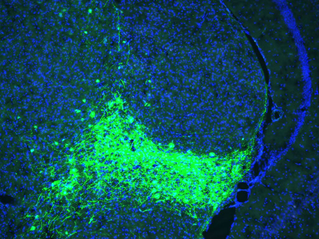
Salk scientists uncover a key neural circuit in mice that gives pain its emotional punch, opening new doors for treating fibromyalgia, migraine, and PTSD
CGRP-expressing neurons (green) in the parvocellular subparafascicular nucleus (SPFp) of the thalamus. Credit: Salk Institute
More than just a physical sensation, pain also carries emotional weight. That distress, anguish, and anxiety can turn a fleeting injury into long-term suffering.
Salk Institute researchers have now identified a brain circuit that gives physical pain its emotional tone, revealing a new potential target for treating chronic and affective pain conditions such as fibromyalgia, migraine, and post-traumatic stress disorder (PTSD).
Published in PNAS, the study identifies a group of neurons in a central brain area called the thalamus that appears to mediate the emotional or affective side of pain in mice. This new pathway challenges the textbook understanding of how pain is processed in the brain and body.
“For decades, the prevailing view was that the brain processes sensory and emotional aspects of pain through separate pathways,” says senior author Sung Han, associate professor and holder of the Pioneer Fund Developmental Chair at Salk. “But there’s been debate about whether the sensory pain pathway might also contribute to the emotional side of pain. Our study provides strong evidence that a branch of the sensory pain pathway directly mediates the affective experience of pain.”
The physical sensation of pain allows immediate detection, assessment of its intensity, and identification of its source. The affective part of pain is what makes it so unpleasant – the emotional discomfort motivates avoidance.
This is a critical distinction. Most people start to perceive pain at the same stimulus intensities, meaning the sensory side of pain is processed similarly. But the ability to tolerate pain varies greatly. The degree of suffering or feeling threatened by pain is determined by affective processing, and if that becomes too sensitive or lasts too long, it can result in a pain disorder. This makes it important to understand which parts of the brain control these different dimensions of pain.
Sensory pain was thought to be mediated by the spinothalamic tract, a pathway that sends pain signals from the spinal cord to the thalamus, which then relays them to sensory processing areas across the brain.
Affective pain was generally thought to be mediated by a second pathway called the spinoparabrachial tract, which sends pain information from the spinal cord into the brainstem.
However, previous studies using older research methods have suggested the circuitry of pain may be more complex. This long-standing debate inspired Han and his team to revisit the question with modern research tools.
Using advanced techniques to manipulate the activity of specific brain cells, the researchers discovered a new spinothalamic pathway in mice. In this circuit, pain signals are sent from the spinal cord into a different part of the thalamus, which has connections to the amygdala, the brain’s emotional processing center. This particular group of neurons in the thalamus can be identified by their expression of CGRP (calcitonin gene-related peptide), a neuropeptide originally discovered in Professor Ronald Evans’ lab at Salk.
When the researchers “turned off” (genetically silenced) these CGRP neurons, the mice still reacted to mild pain stimuli, such as heat or pressure, indicating their sensory processing was intact. However, they didn’t seem to associate lasting negative feelings with these situations, failing to show any learned fear or avoidance behaviors in future trials. On the other hand, when these same neurons were “turned on” (optogenetically activated), the mice showed clear signs of distress and learned to avoid that area, even when no pain stimuli had been used.
“Pain processing is not just about nerves detecting pain; it’s about the brain deciding how much that pain matters,” says first author Sukjae Kang, a senior research associate in Han’s lab. “Understanding the biology behind these two distinct processes will help us find treatments for the kinds of pain that don’t respond to traditional drugs.”
Many chronic pain conditions—such as fibromyalgia and migraine—involve long, intense, unpleasant experiences of pain, often without a clear physical source or injury. Some patients also report extreme sensitivity to ordinary stimuli like light, sound, or touch, which others would not perceive as painful.
Han says overactivation of the CGRP spinothalamic pathway may contribute to these conditions by making the brain misinterpret or overreact to sensory inputs. In fact, transcriptomic analysis of the CGRP neurons showed that they express many of the genes associated with migraine and other pain disorders.
Notably, several CGRP blockers are already being used to treat migraines. This study may help explain why these medications work and could inspire new nonaddictive treatments for affective pain disorders.
Han also sees potential relevance for psychiatric conditions that involve heightened threat perception, such as PTSD. Growing evidence from his lab suggests that the CGRP affective pain pathway acts as part of the brain’s broader alarm system, detecting and responding to not only pain but a wide range of unpleasant sensations. Quieting this pathway with CGRP blockers could offer a new approach to easing fear, avoidance, and hypervigilance in trauma-related disorders.
Importantly, the relationship between the CGRP pathway and the psychological pain associated with social experiences like grief, loneliness, and heartbreak remains unclear and requires further study.
“Our discovery of the CGRP affective pain pathway gives us a molecular and circuit-level explanation for the difference between detecting physical pain and suffering from it,” says Han. “We’re excited to continue exploring this pathway and enabling future therapies that can reduce this suffering.”
Researchers at Princeton University and the Simons Foundation have identified four clinically and biologically distinct subtypes of autism, marking a transformative step in understanding the condition’s genetic underpinnings and potential for personalised care.
Analysing data from over 5000 children in SPARK, an autism cohort study funded by the Simons Foundation, the researchers used a computational model to group individuals based on their combinations of traits. The team used a “person-centred” approach that considered a broad range of over 230 traits in each individual, from social interactions to repetitive behaviours to developmental milestones, rather than searching for genetic links to single traits.
This approach enabled the discovery of clinically relevant autism subtypes, which the researchers linked to distinct genetic profiles and developmental trajectories, offering new insights into the biology underlying autism. Their results were published July 9 in Nature Genetics.
The study defines four subtypes of autism: Social and Behavioural Challenges, Mixed ASD with Developmental Delay, Moderate Challenges, and Broadly Affected. Each subtype exhibits distinct developmental, medical, behavioural and psychiatric traits, and importantly, different patterns of genetic variation.
Individuals in the Social and Behavioural Challenges group show core autism traits, including social challenges and repetitive behaviours, but generally reach developmental milestones at a pace similar to children without autism. They also often experience co-occurring conditions like ADHD, anxiety, depression or obsessive-compulsive disorder alongside autism. One of the larger groups, this constitutes around 37% of the participants in the study.
The Mixed ASD with Developmental Delay group tends to reach developmental milestones, such as walking and talking, later than children without autism, but usually does not show signs of anxiety, depression or disruptive behaviours. “Mixed” refers to differences within this group with respect to repetitive behaviours and social challenges. This group represents approximately 19% of the participants.
Individuals with Moderate Challenges show core autism-related behaviours, but less strongly than those in the other groups, and usually reach developmental milestones on a similar track to those without autism. They generally do not experience co-occurring psychiatric conditions. Roughly 34% of participants fall into this category.
The Broadly Affected group faces more extreme and wide-ranging challenges, including developmental delays, social and communication difficulties, repetitive behaviours and co-occurring psychiatric conditions like anxiety, depression and mood dysregulation. This is the smallest group, accounting for around 10% of the participants.
“These findings are powerful because the classes represent different clinical presentations and outcomes, and critically we were able to connect them to distinct underlying biology,” said Aviya Litman, a PhD student at Princeton.
Distinct genetics behind the subtypes
For decades, autism researchers and clinicians have been seeking robust definitions of autism subtypes to aid in diagnosis and care. Autism is known to be highly heritable, with many implicated genes.
“While genetic testing is already part of the standard of care for people diagnosed with autism, thus far, this testing reveals variants that explain the autism of only about 20% of patients,” said co-author Jennifer Foss-Feig, a clinical psychologist at the Icahn School of Medicine at Mount Sinai and vice president and senior scientific officer at the Simons Foundation Autism Research Initiative (SFARI). This study takes an approach that differs from classic gene discovery efforts by identifying robust autism subtypes that are linked to distinct types of genetic mutations and affected biological pathways.
For example, children in the Broadly Affected group showed the highest proportion of damaging de novo mutations, while only the Mixed ASD with Developmental Delay group was more likely to carry rare inherited genetic variants. While children in both of these subtypes share some important traits like developmental delays and intellectual disability, these genetic differences suggest distinct mechanisms behind superficially similar clinical presentations.
“These findings point to specific hypotheses linking various pathways to different presentations of autism,” said Litman, referring to differences in biology between children with different autism subtypes.
Moreover, the researchers identified divergent biological processes affected in each subtype. “What we’re seeing is not just one biological story of autism, but multiple distinct narratives,” said Natalie Sauerwald, associate research scientist at the Flatiron Institute and co-lead author. “This helps explain why past genetic studies often fell short – it was like trying to solve a jigsaw puzzle without realising we were actually looking at multiple different puzzles mixed together. We couldn’t see the full picture, the genetic patterns, until we first separated individuals into subtypes.”
Autism biology unfolds on different timelines
The team also found that autism subtypes differ in the timing of genetic disruptions’ effects on brain development. Genes switch on and off at specific times, guiding different stages of development. While much of the genetic impact of autism was thought to occur before birth, in the Social and Behavioural Challenges subtype – which typically has substantial social and psychiatric challenges, no developmental delays, and a later diagnosis – mutations were found in genes that become active later in childhood. This suggests that, for these children, the biological mechanisms of autism may emerge after birth, aligning with their later clinical presentation.
“By integrating genetic and clinical data at scale, we can now begin to map the trajectory of autism from biological mechanisms to clinical presentation,” said co-author Chandra Theesfeld, senior academic research manager at the Lewis-Sigler Institute and Princeton Precision Health.
A paradigm shift for autism research
This study builds on more than a decade of autism genomics research led by Troyanskaya and collaborators. It is enabled by the close integration of interdisciplinary expertise in genomics, clinical psychology, molecular biology, computer science and modelling, and computational biology.
“The Princeton Precision Health initiative uses artificial intelligence and computational modelling to integrate across biological and clinical data,” said Jennifer Rexford, Princeton University provost and Gordon Y.S. Wu Professor in Engineering. “This initiative could not exist without the University’s charitable endowment. Our investments allow experts to collaborate across a range of disciplines to conduct transformative research that improves human health, including the potential for major advances in the diagnosis and treatment of autism made possible in this exciting project.”
“It’s a whole new paradigm, to provide these groups as a starting point for investigating the genetics of autism,” said Theesfeld. Instead of searching for a biological explanation that encompasses all individuals with autism, researchers can now investigate the distinct genetic and biological processes driving each subtype.
This shift could reshape both autism research and clinical care – helping clinicians anticipate different trajectories in diagnosis, development and treatment. “The ability to define biologically meaningful autism subtypes is foundational to realising the vision of precision medicine for neurodevelopmental conditions,” said Sauerwald.
While the current work defines four subtypes, “this doesn’t mean there are only four classes,” said Litman. “It means we now have a data-driven framework that shows there are at least four – and that they are meaningful in both the clinic and the genome.”
Looking ahead
Beyond its contributions to understanding autism subtypes and their underlying biology, the study offers a powerful framework for characterising other complex, heterogeneous conditions and finding clinically relevant disease subtypes. As Theesfeld put it: “This opens the door to countless new scientific and clinical discoveries.”