Category: Urology

Improving Understanding of Female Sexual Anatomy for Better Pelvic Radiotherapy
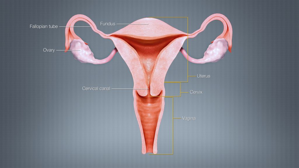
Female reproductive system. Credit: Scientific Animations CC4.0 BY-SA

Researchers at the Icahn School of Medicine at Mount Sinai, in collaboration with other leading institutions across the country, have published an innovative study that provides radiation oncologists with practical guidance to identify and protect female sexual organs during pelvic cancer treatment.  

Published in the latest issue of Practical Radiation Oncology, this study addresses a long-standing gap in cancer care by bringing key female sexual anatomy into consideration during routine radiotherapy planning and survivorship research. 

The study, “Getting c-literate: Bulboclitoris functional anatomy and its implications for radiotherapy,” synthesises current scientific knowledge and pairs it with original anatomic dissection, histology, and advanced imaging analysis. The work focuses on the bulboclitoris, a female erectile organ (consisting of the clitoris and the vestibular bulbs) that plays a central role in sexual arousal and orgasm and can be exposed to radiation during treatment for pelvic cancers. 

“Pelvic radiotherapy can be life-saving, but it can also affect sexual function and quality of life,” said Deborah Marshall, MD, MAS, Assistant Professor, Departments of Radiation Oncology and Population Health Science and Policy at the Icahn School of Medicine at Mount Sinai; Division Chief of Women’s Health, Department of Population Health Science and Policy; and senior author of the study. “Compared to male sexual anatomy, female erectile structures have been largely invisible in standard radiation workflows. Our goal was to provide clinicians with a practical anatomy-grounded way to change that.” 

Using detailed anatomic and radiologic correlation, the research team demonstrates how the bulboclitoris and related neurovascular structures can be identified on standard CT and MRI scans and consistently outlined (or “contoured”) for radiotherapy planning. This step-by-step guidance makes it feasible for clinicians to measure radiation dose to these tissues and begin linking exposure to patient-reported outcomes related to arousal and orgasm. 

“This work builds upon our previous knowledge that the clitoris is not just an external structure,” Dr. Marshall said. “It includes an entire internal organ comprised of erectile tissues located just outside the pelvis, and those tissues matter for sexual health and, in particular, for female sexual pleasure. Once clinicians can reliably see and measure them, we can begin to ask better questions, have better conversations with patients, and ultimately deliver better care.” 

Sexual function outcomes after pelvic radiotherapy have historically been understudied in women, limiting counselling, toxicity prevention strategies, and equitable survivorship care. By establishing a shared, standardised approach to identifying the bulboclitoris, the study lays the groundwork for future research to develop dose-volume constraints and mitigation strategies, as other organs at risk are managed in radiation oncology. 

For clinicians, the framework enables routine contouring and dose reporting using CT alone when necessary, with MRI improving soft-tissue visualization when available. In the absence of prospective dose-response data, the authors recommend minimising radiation dose to the bulboclitoris when oncologically appropriate, using an “as low as reasonably achievable” approach. 

For patients, the work supports more informed conversations about potential sexual side effects of pelvic radiotherapy, including changes in arousal, sensation, orgasm, lubrication, or pain. This research also promotes more personalized treatment planning that considers female sexual health and pleasure as a legitimate and important component of cancer survivorship. 

Next steps include prospective research through Mount Sinai’s STAR program, deeper mapping of neurovascular anatomy relevant to sexual function, expanded educational resources for oncology and radiology teams, and improved patient-reported outcome measures that reflect diverse sexual practices and experiences. 

Source: Mount Sinai
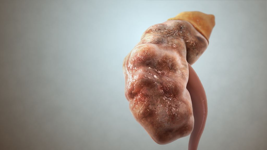
Simple Method for Early Detection of Chronic Kidney Disease

Chronic kidney disease (CKD). Credit: Scientific Animations CC4.0

Subtle abnormalities in kidney function – even within the range considered normal – may help identify people at risk of developing chronic kidney disease. This is shown in a new study from Karolinska Institutet, published in Kidney International. The researchers have therefore developed a web-based tool that could aid in early detection and thus primary prevention.

Chronic kidney disease is a growing global health concern afflicting 10−15% of adults worldwide, and is projected to become one of the top five leading causes of years of life lost by 2040. In the absence of effective screening programmes, patients are often diagnosed late, when more than half of their kidney function has already been lost. 

To address this gap, researchers at Karolinska Institutet have constructed population-based distributions for estimated glomerular filtration rate (eGFR), the most widely used measure of kidney function. The aim is to help doctors identify people at risk, thus enabling early preventive action.

“We were inspired by the growth and weight charts used in paediatrics, which intuitively help clinicians identify children at risk of obesity or undergrowth,” says the first author of the study, Yuanhang Yang, Postdoctoral Researcher at the Department of Clinical Science and Education, Södersjukhuset, Karolinska Institutet.

A web-based calculator

The researchers have made their eGFR distribution charts openly available to healthcare professionals and developed a web-based calculator, developed by PhD student Antoine Creon, that can help assess how a patient’s eGFR compares with population norms for their age.

The study included over 1.1 million adults in the region of Stockholm, covering roughly 80 per cent of the population aged between 40 and 100 years. Nearly seven million eGFR tests collected between 2006 and 2021 were used to construct age- and sex-specific distributions. 

The findings show that departures from the median eGFR for one’s age and sex are associated with worse outcomes. Individuals with an eGFR below the 25th percentile had a markedly higher risk of developing kidney failure requiring dialysis or transplantation. Mortality also displayed a U-shaped relationship; both low and high percentile extremes were linked to increased risk of death. 

Ability to act earlier 

The study also illustrates this lack of awareness in healthcare, according to the researchers. Among those with a seemingly normal eGFR above 60 ml/min/1.73 m², but below the 25th percentile, only one fourth had received additional testing for urinary albumin, which is important for detecting early kidney damage.

“For example, consider a 55-year-old woman with an eGFR of 80. Most clinicians would not react to such a seemingly normal value. However, our charts show that this corresponds to the 10th percentile for women of that age – and that she has a three-fold higher risk of starting dialysis in the future. This signals an opportunity to act earlier,” says Juan Jesús Carrero, Professor at the Department of Medical Epidemiology and Biostatistics, Karolinska Institutet.

Source: Karolinska Institutet

Disagreement Between Two Kidney Function Tests Predicts Disease Risk

Photo by National Cancer Institute on Unsplash

A mismatch between two common tests for kidney function may indicate a higher risk for kidney failure, heart disease, and death, a new study shows.

Healthcare providers for decades have measured blood levels of creatinine to track the rate at which kidneys filter waste from muscle breakdown in the bloodstream. According to more recent guidelines, levels of cystatin C, a small protein made by all cells in the body, can also be used to measure kidney function. Since these two tests are influenced by different factors – including some related to disease or aging – using both markers together can provide a better measure of kidney function and risk of organ failure than either one alone.

Led by NYU Langone Health researchers, the new work reveals that many people, especially those who are sick, often have a large gap between the two readings, which may be a signal of future disease. Specifically, the global study shows that more than a third of hospitalised participants had a cystatin C-based readout of kidney function that was at least 30% lower than one based on their creatinine levels.

“Our findings highlight the importance of measuring both creatinine and cystatin C to gain a true understanding of how well the kidneys are working, particularly among older and sicker adults,” said study co-corresponding author Morgan Grams, MD, PhD. “Evaluating both biomarkers may identify far more people with poor kidney function, and earlier in the disease process, by covering the blind spots that go with either test.”

The study published online November 7 in the Journal of the American Medical Association and is simultaneously being presented at the American Society of Nephrology’s annual Kidney Week conference.

Beyond detecting signs of disease, assessing patients’ kidney function is important for calculating the appropriate dosage for cancer medicines, antibiotics, and many other drugs, says Dr Grams, Professor of Medicine at the NYU Grossman School of Medicine.

During another investigation, the results of which were published the same day, the same research team found that a record number of people worldwide have chronic kidney disease, which is now the ninth leading cause of death globally. Having new ways to spot the condition early can help ensure that patients receive swift treatment and avoid more-dramatic interventions such as dialysis and organ transplantation, says Dr Grams.

For the recent investigation, the research team analysed healthcare records, blood tests, and demographic data collected from 860, 66 men and women of a half-dozen nationalities. All participants had their creatinine and cystatin C levels measured on the same day and received follow-ups 11 years later, on average. The team considered factors unrelated to kidney function that influence the biomarkers’ readings, such as smoking, obesity, and history of cancer.

Performed as part of the international Chronic Kidney Disease Prognosis Consortium, the study is the largest to date to explore differences between the two tests and whether they may signal potential health problems, the authors say. Established to better understand and treat the condition, the consortium provides evidence for global definitions of chronic kidney disease and related health risks.

According to the new findings, those whose cystatin C-based measures of kidney filtration were at least 30% lower than their creatinine-based measures were at higher risk for death, heart disease, and heart failure than those who had a smaller difference between the two metrics. The former group was also more likely to be diagnosed with severe chronic kidney disease that required dialysis or an organ transplant. The same was found for 11% of outpatients and seemingly healthy volunteers.

Dr. Grams notes that while cystatin C testing was first recommended in 2012 by the international organization Kidney Disease—Improving Global Outcomes, a 2019 survey revealed that less than 10 percent of clinical laboratories in the United States performed it in-house. The two largest laboratories, Quest Diagnostics and Labcorp, now offer the test.

“These results underscore the need for physicians to take advantage of the fact that more hospitals and healthcare providers are starting to offer cystatin C testing,” said study co-corresponding author Josef Coresh, MD, PhD, director of NYU Langone’s Optimal Aging Institute. “Physicians might otherwise miss out on valuable information about their patients’ wellbeing and future medical concerns.”

Dr Coresh cautions that among the hospitalised Americans in the study, less than 1% were tested for cystatin C.

Source: NYU Langone Health

Promising New Breakthrough for Preventing Kidney Damage in Type 1 Diabetes

Chronic kidney disease (CKD). Credit: Scientific Animations CC4.0

A new gene therapy approach aimed at protecting people with type 1 diabetes from developing diabetic kidney disease – a serious and common complication of the condition, has shown promising results in a University of Bristol study.

One in three people with type one diabetes will develop kidney damage during their lifetime, which can develop silently over many years, often going undetected until it becomes severe.

Current treatments can slow kidney damage but there are none that act on the root cause: a tiny filter called the glomerulus. A new study published in Molecular Therapy, demonstrated a 64% reduction in a damage indicator for kidney disease, paving the way for a potential new treatment.

The study, driven by first author, Dr Aldara Martin Alonso and led by Dr Rebecca Foster, Associate Professor of Microvascular Medicine at Bristol Medical School: Translational Health Sciences, explored the potential of delivering a protein called VEGF-C directly into kidney cells.

Previous studies have shown VEGFC could protect against kidney disease as it helps keeps blood vessels in the kidney filter healthy, repairing early signs of diabetes-related kidney damage.

To test whether this new approach could be used to treat or slow down kidney disease, the team used a harmless virus to deliver VEGF-C directly into the kidney cells of diabetic mice.

Their results showed that this approach not only helped the kidneys work better, but also protected a key part of the kidney filter that normally helps prevent damage. It led to a 64% reduction in albuminuria. Importantly, this reduction is more than twice the reduction recommended by the American Diabetes Association to slow the progression of chronic kidney disease.

Dr Foster, the study’s senior author, explained: “Currently, there are no drugs specifically available to protect people with type 1 diabetes from kidney disease, despite their higher risk of developing kidney disease. This gap in treatment highlights the urgent need for new therapeutic approaches. Our goal was to investigate whether gene therapy could offer a viable solution by delivering VEGFC in a more targeted way.”

Dr Foster added: “This gene therapy approach has not been explored before in pre-clinical models and offers a long-term solution for these patients who are at risk of developing kidney disease.”

Source: University of Bristol

Clinical Trial Shows a Constipation Drug Aids Kidney Function

Human kidney. Credit: Scientific Animations CC0

Chronic kidney disease (CKD) is a major health issue worldwide. Many patients end up requiring regular dialysis to avoid kidney failure and stay alive. Despite the severity of the condition, there are currently no drugs available that improve kidney function. A research group led by Tohoku University Graduate School of Medicine’s Professor Takaaki Abe has found a remarkable solution to treat patients with CKD by co-opting a drug typically used for constipation. This is the first time that this drug (lubiprostone) was shown to prevent the decline of renal function in patients with CKD.

“We noticed that constipation is a symptom that often accompanies CKD, and decided to investigate this link further,” explains Abe. “Essentially, constipation disrupts the intestinal microbiota, which worsens kidney function. Working backwards, we hypothesised that we could improve kidney function by treating constipation.”

To address this issue, the group conducted a multicentre Phase II clinical trial (LUBI-CKD TRIAL) at nine Japanese medical institutions, enrolling 150 patients with moderate CKD. This study evaluated the effects of lubiprostone on kidney function. The results showed that, compared to the placebo group, the decline in kidney function (defined as the estimated glomerular filtration rate: eGFR) was suppressed in a dose-dependent manner in patients treated with 8µg or 16µg of lubiprostone.

The researchers also investigated the mechanism underlying how this effect occurred. They found that lubiprostone increases spermidine production, which improves mitochondrial function by promoting bacterial growth in the gut. The improved mitochondrial function was seen to exert a renoprotective effect – suppressing further kidney damage.

Going forward, the research team has plans to validate the trial results in a larger population (Phase 3 clinical trial) and advance the exploration of biomarkers that predict treatment efficacy. Their goal is to provide each patient with CKD the optimal treatment plan tailored to their needs. This discovery has the potential to significantly transform the conventional approach to CKD treatment, which primarily focuses on reducing uremic toxins.

These findings suggest a new therapeutic strategy in which laxatives suppress renal function decline. This strategy is expected to contribute to the development of treatments for not only CKD, but also mitochondrial dysfunction disorders. The results of this study were published in Science Advances on August 30, 2025.

Source: Tohoku University

Study Ranks Kidney Stone Surgical Interventions for Children and Adolescents

Human kidney. Credit: Scientific Animations CC0

Researchers at Children’s Hospital of Philadelphia (CHOP), along with several academic partners, announced the primary results of the Pediatric KIDney Stone (PKIDS) trial, the largest comparative effectiveness study of surgical interventions for children and adolescents with kidney stones. The CHOP-led PKIDS trial, with two published studies, marks a significant breakthrough by offering stronger evidence for treating stones of varying sizes, including new information on patient experiences after surgery, thus reducing uncertainty and empowering informed decision-making for patients, caregivers, and physicians.

Kidney stones were once largely a disease that affected adults. However, kidney stones in children have been on the rise in recent decades, doubling the likelihood that a child will develop a kidney stone. As a result, CHOP founded the PKIDS Care Improvement Network in 2019, which now includes 31 sites in the United States and Canada.   

Ureteroscopy (an endoscopic outpatient procedure), shockwave lithotripsy (a noninvasive outpatient procedure) and percutaneous nephrolithotomy (a minimally invasive surgery with a short hospital stays) are the procedures used to treat children and adults with kidney stones. Most children and adolescents with kidney and ureteral stones are treated with ureteroscopy despite uncertainty of which procedure is more effective and their impact on patients’ lives.  As pioneers in urology, CHOP leaders aimed to enhance pediatric patient and caregiver decision-making for kidney stone surgeries while enabling urologists to adopt techniques for optimal outcomes, including patient-selected experiences.

In the first study in JAMA Network Open, Tasian and his team enrolled 1142 patients aged 8 to 21 with kidney and/or ureteral stones between 2020 and 2023 at all 31 sites in the United States and Canada. Researchers evaluated ureteroscopy against shockwave lithotripsy and found that shockwave lithotripsy was associated with less pain and fewer urinary symptoms compared with those who had ureteroscopy. No meaningful differences were detected in stone-free rates for the procedures. 

In the other CHOP-led study in JAMA Network Open, Jonathan S. Ellison, MD, an Associate Professor of Urology at the Medical College of Wisconsin and Pediatric Urologist at Children’s Wisconsin, and the PKIDS team compared percutaneous nephrolithotomy (PCNL) and ureteroscopy. That study found that for children with larger stones, PCNL not only cleared more stones effectively but also led to a better overall recovery experience than ureteroscopy.

Overall, the authors emphasised the post-surgery experiences of children, noting that quality of life factors, such as the loss of school time for children and work time for caregivers, are crucial in determining effective treatment options. While the authors are planning further research, they also hope these findings will lead to immediate improvements for families. 

“The PKIDS trial demonstrated that ureteroscopy and shockwave lithotripsy remove stones equally well and that patients having shockwave lithotripsy recover more quickly after surgery with less pain and fewer urinary symptoms. Our findings provide new information that allow for tailored approaches to kidney stone treatment for children and their families,” said Gregory E. Tasian, MD, MSc, MSCE, Director of the PKIDS network and an attending pediatric urologist in the Division of Urology at Children’s Hospital of Philadelphia. “Although future clinical trials are important, we hope that clinical practice guidelines will consider outcomes that matter to patients.” 

Source: Children’s Hospital of Philadelphia

New Discovery Reveals the Spinal Cord’s Role in Bladder Control

Urinary incontinence. Credit: Scientific Animations CC4.0

Urinary incontinence is a devastating condition, leading to significant adverse impacts on patients’ mental health and quality of life. Disorders of urination are also a key feature of all neurological disorders.

A USC research team has now made major progress in understanding how the human spinal cord triggers the bladder emptying process. The discovery could lead to exciting new therapies to help patients regain control of this essential function.

In the pioneering study, a team from USC Viterbi School of Engineering and Keck School of Medicine of USC has harnessed functional ultrasound imaging to observe real-time changes in blood flow dynamics in the human spinal cord during bladder filling and emptying.

The work was published in Nature Communications and was led by Charles Liu, the USC Neurorestoration Center director at Keck School of Medicine of USC and professor of biomedical engineering at USC Viterbi, and Vasileios Christopoulos, assistant professor at the Alfred E. Mann Department of Biomedical Engineering.

The spinal cord regulates many essential human functions, including autonomic processes like bladder, bowel, and sexual function. These processes can break down when the spinal cord is damaged or degenerated due to injury, disease, stroke, or aging. However, the spinal cord’s small size and intricate bony enclosure have made it notoriously challenging to study directly in humans.

Unlike in the brain, routine clinical care does not involve invasive electrodes and biopsies in the spinal cord due to the obvious risks of paralysis.

Furthermore, fMRI imaging, which comprises most of human functional neuroimaging, does not exist in practical reality for the spinal cord, especially in the thoracic and lumbar regions where much of the critical function localises.

“The spinal cord is a very undiscovered area,” Christopoulos said. “It’s very surprising to me because when I started doing neuroscience, everybody was talking about the brain. And Dr. Liu and I asked, “What about the spinal cord?”

“For many, it was just a cable that transfers information from the brain to the peripheral system. The truth was that we didn’t know how to go there—how to study the spinal cord in action, visualize its dynamics and truly grasp its role in physiological functions.”

Functional ultrasound imaging: A new window into the spinal cord

To overcome these barriers, the USC team employed functional ultrasound imaging (fUSI), an emerging neuroimaging technology that is minimally invasive. The fUSI process allowed the team to measure where changes in blood volume occur on the spinal cord during the cycle of urination.

However, fUSI requires a “window” through the bone to image the spinal cord. The researchers found a unique opportunity by working with a group of patients undergoing standard-of-care epidural spinal cord stimulation surgery for chronic low back pain.

“During the implantation of the spinal cord stimulator, the window we create in the bone through which we insert the leads gives us a perfect and safe opportunity to image the spinal cord using fUSI with no risk or discomfort to the study volunteers,” said co-first author Darrin Lee, associate director of the USC Neurorestoration Center, who performed the surgeries.

“While the surgical team was preparing the stimulator, we gently filled and emptied the bladder with saline to simulate a full urination cycle under anaesthesia while the research team gathered the fUSI data,” added Evgeniy Kreydin from the Rancho Los Amigos National Rehabilitation Center and the USC Institute of Urology, who was already working closely with Liu to study the brain of stroke patients during micturition using fMRI.

“This is the first study where we’ve shown that there are areas in the spinal cord where activity is correlated with the pressure inside the bladder,” Christopoulos said.

“Nobody had ever shown a network in the spinal cord correlated with bladder pressure. What this means is I can look at the activity of your spinal cord in these specific areas and tell you your stage of the bladder cycle – how full your bladder is and whether you’re about to urinate.”

Christopoulos said the experiments identified that some spinal cord regions showed positive correlation, meaning their activity increased as bladder pressure rose, while others showed negative (anti-correlation), with activity decreasing as pressure increased. This suggests the involvement of both excitatory and inhibitory spinal cord networks in bladder control.

“It was extremely exciting to take data straight from the fUSI scanner in the OR to the lab, where advanced data science techniques quickly revealed results that have never been seen before, even in animal models, let alone in humans,” said co-first author Kofi Agyeman, biomedical engineering postdoc.

New hope for patients

Liu has worked for two decades at the intersection of engineering and medicine to develop transformative strategies to restore function to the nervous system. Christopoulos has spent much of his research career developing neuromodulation techniques to help patients regain motor control.

Together, they noted that for patients, retaining control of the autonomic processes that many of us take for granted is more fundamental than even walking.

“If you ask these patients, the most important function they wanted to restore was not their motor or sensory function. It was things like sexual function and bowel and bladder control,” Christopoulos said, noting that urinary dysfunction often leads to poor mental health. “It’s a very dehumanising problem to deal with.”

Worse still, urinary incontinence leads to more frequent urinary tract infections (UTIs) because patients must often be fitted with a catheter. Due to limited sensory function, they may not be able to feel that they have an infection until it is more severe and has spread to the kidneys, resulting in hospitalisation.

This study offers a tangible path toward addressing this critical need for patients suffering from neurogenic lower urinary tract dysfunction. The ability to decode bladder pressure from spinal cord activity provides proof-of-concept for developing personalised spinal cord interfaces that could warn patients about their bladder state, helping them regain control.

Currently, almost all neuromodulation strategies for disorders of micturition are focused on the lower urinary tract, largely because the neural basis of this critical process remains unclear.

“One has to understand a process before one can rationally improve it,” Liu said.

This latest research marks a significant step forward, opening new avenues for precision medicine interventions that combine invasive and noninvasive neuromodulation with pharmacological therapeutics to make neurorestoration of the genitourinary system a clinical reality for millions worldwide.

Source: University of Southern Carolina

Microplastics Detected in Human Reproductive Fluids

Photo by FLY:D on Unsplash

New research presented today at the 41st Annual Meeting of the European Society of Human Reproduction and Embryology (ESHRE) reveals the presence of microplastics in human reproductive fluids, raising important questions about their potential risks to fertility and reproductive health.[1]

The study abstract appears in Human Reproduction, one of the world’s leading reproductive medicine journals.

Researchers examined follicular fluid from 29 women and seminal fluid from 22 men, both of which play critical roles in natural conception and assisted reproduction.

A range of commonly used microplastic polymers, including polytetrafluoroethylene (PTFE), polystyrene (PS), polyethylene terephthalate (PET), polyamide (PA), polypropylene (PP) and polyurethane (PU), were identified in both groups.

Microplastics were present in 69% of the follicular fluid samples analysed. Notably, the most frequently detected polymer was PTFE, found in 31% of the samples. This was followed by PP (28%), PET (17%), PA (14%), polyethylene (PE) (10%), PU (10%) and PS (7%), in descending order of prevalence.

In male seminal fluid samples, microplastics were found in 55% of those analysed. PTFE again emerged as the most prevalent polymer, identified in 41% of the samples. Other polymers detected included PS (14%), PET (9%), PA (5%), and PU (5%), though in lower concentrations.

To prevent contamination, all samples were collected and stored in glass containers and underwent chemical treatment before analysis using laser direct infrared microscopy.

Lead researcher Dr. Emilio Gomez-Sanchez commented, “Previous studies had already shown that microplastics can be found in various human organs. As a result, we weren´t entirely surprised to find microplastics in fluids of the human reproductive system, but we were struck by how common they were – found in 69% of the women and 55% of the men we studied.”

Microplastics are defined as plastic particles under 5mm in size, and there is evidence that they pose a threat to environmental and public health.[2] While this research did not directly assess how microplastics affect fertility, their detection highlights the need to explore possible implications for human reproductive health.

“What we know from animal studies is that in the tissues where microplastics accumulate, they can induce inflammation, free radical formation, DNA damage, cellular senescence, and endocrine disruptions”, continued Dr Gomez-Sanchez. “It’s possible they could impair egg or sperm quality in humans, but we don’t yet have enough evidence to confirm that.”

The research team plans to expand their analysis to a larger cohort, alongside detailed lifestyle and environmental exposure questionnaires. Further phases of the project will also explore the potential relationship between the presence of microplastics and oocyte and sperm quality.

Dr Gomez-Sanchez stressed that fertility is influenced by many factors, including age, health, and genetics, and that the findings should not cause alarm among those trying to conceive. “There’s no need for alarm at this point. Microplastics are just one of many elements that may play a role in fertility. However, it is sensible to consider ways of reducing our exposure to them. Simple steps, such as using glass containers to store and heat food, or limiting the amount of water we consume from plastic bottles, can help minimise our intake.”

Source: Focus on Reproduction

References:

[1] Gomez-Sanchez, E., et al. (2025) Unveiling the Hidden Danger: Detection and characterisation of microplastics in human follicular and seminal fluids. Human Reproduction. [insert link when available]

[2] Wang, L., Yin, Y., & He, X. (2024). The hidden threat: Unraveling the impact of microplastics on reproductive health. Science of the Total Environment, 912, 173177.

Brain Stimulation and Mindfulness Exercises Could Reduce ‘Latchkey Incontinence’

Urinary incontinence. Credit: Scientific Animations CC4.0

Arriving home after a long day may be a relief, but for some people, seeing their front door or inserting a key into the lock triggers a powerful urge to pee. Known as “latchkey incontinence,” this phenomenon is the subject of a new study by researchers at the University of Pittsburgh who found that mindfulness training and/or non-invasive brain stimulation could reduce bladder leaks and feelings of urgency evoked by these cues. 

The findings of the pilot study, the first evaluation of brain-based therapies for urinary incontinence, are published in the latest issue of the journal Continence. 

“Incontinence is a massive deal,” said senior author Dr. Becky Clarkson, research assistant professor in the Pitt School of Medicine Division of Geriatrics and co-director of the Continence Research Center. “Bladder leaks can be really traumatizing. People often feel like they can’t go out and socialise or exercise because they’re worried about having an accident. Especially for older adults, this feeds into social isolation, depression and functional decline. Our research aims to empower people with tools to get back their quality of life.” 

Latchkey incontinence, or situational urgency urinary incontinence, is bladder leakage triggered by specific environments or scenarios. Common cues include one’s front or garage door, running water, getting into a car or walking past public restrooms. 

According to lead author Dr. Cynthia Conklin, associate professor in the Pitt Department of Psychiatry, latchkey incontinence is a type of Pavlovian conditioning. Like Pavlov’s dogs, which salivated upon hearing a bell that they associated with food, years of going to the bathroom immediately upon entering the house can condition one to feel strong bladder urgency when seeing the front door.

In a previous study, Clarkson and Conklin showed participants pictures of their own front doors or other triggers versus “safe” images of things that did not evoke urgency while they had an MRI of their brain. A part of the brain called the dorsolateral prefrontal cortex was more active when participants viewed urgency-related images.  

“The prefrontal cortex is the seat of cognitive control,” said Clarkson. “It’s the executive function center of the bladder, the bit that is telling you, ‘Okay, it’s time to go. You should find somewhere to go.’” 

The researchers hypothesised that activating this part of the brain during exposure to urgency cues, through mindfulness and/or with transcranial direct current stimulation (tDCS) of the brain, could improve participants’ ability to regulate responses to these cues and control urgency and leakage.  

They recruited 61 women aged over 40 who reported regular situationally triggered bladder leaks and randomly assigned them to one of three groups: Participants either listened to a 20-minute mindfulness exercise, received tDCS or both while viewing personal trigger photos.  

The mindfulness exercise, developed by coauthor Dr Carol Greco, associate professor of psychiatry and physical therapy at Pitt, was like a typical body scan practice that instructs participants to move through their body, bringing attention to each part in turn. But unlike most body scans, it included specific acknowledgment of bladder sensation.  

After completing four in-office sessions over five to six days, participants in all three groups experienced reduced urgency when they viewed trigger cues. Women in all three groups also reported an improvement in the number of urgency episodes and leaks after completing the sessions. 

Although this pilot study did not have a control group, for comparison, the researchers say that the magnitude of improvement from tDCS and mindfulness was similar to what other research has reported for interventions such as medications and pelvic floor therapy.  

“Although we need to do more research, these results are really encouraging because they suggest that a behavioral tool like mindfulness can be an alternative or additional way to improve symptoms,” said Conklin. “Balancing multiple prescriptions is a big issue among older adults, and a lot of people are reluctant to take another medication, so I think that’s one of the reasons that we saw such high acceptability of non-pharmacologic interventions in this study.” 

More than 90% of recruited participants completed the study.  

“Participants loved it,” said Clarkson. “Almost everyone who started the study finished it, even though coming into the office four days within one week was quite a big commitment. We got really great feedback, and a lot of women told us that they continue to use the mindfulness exercise in their daily lives.” 

“For the first time in 20 years of doing research, we got thank you cards!” added Conklin. “I think that incontinence is such a taboo subject, and a lot of people find it difficult to talk about, so they often don’t even realize that there are treatments out there. But you don’t have to suffer in silence.” 

Now, the researchers are planning to explore whether the mindfulness component of the study could be helpful in independent living facilities to reach a wide range of older adults. They also hope to eventually develop an app-based tool for smartphones. 

Source: University of Pittsburgh

Surgeons Perform World-first Bladder Transplant

A UCLA Health surgical team has performed the first-in-human bladder transplant. 

The surgery was successfully completed at Ronald Reagan UCLA Medical Center on May 4, 2025. The team was led by Dr Nima Nassiri, a urologic transplant surgeon and director of the UCLA Vascularized Composite Bladder Allograft Transplant Program, with assistance from Dr Inderbir Gill, founding executive director of USC Urology.

“Bladder transplantation has been Dr Nassiri’s principal academic focus since we recruited him to the UCLA faculty several years ago,” said Dr Mark Litwin, UCLA Urology Chair, “It is incredibly gratifying to see him take this work from the laboratory to human patients at UCLA, which operates the busiest and most successful solid-organ transplant program in the western United States.”

“This first attempt at bladder transplantation has been over four years in the making,” Nassiri said. “For the appropriately selected patient, it is exciting to be able to offer a new potential option.” 

The patient had lost most of his bladder during a tumour removal, leaving the remainder too small and compromised to work. Both of his kidneys were also subsequently removed due to renal cancer in the setting of pre-existing end-stage kidney disease. As a result, he was on dialysis for seven years. 

The biggest risks of organ transplantation are the body’s potential rejection of the organ and side-effects caused by the mandatory immune suppressing drugs given to prevent organ rejection.

“Because of the need for long-term immunosuppression, the best current candidates are those who are already either on immunosuppression or have an imminent need for it,” Nassiri said. 

Nassiri and Gill collaborated for several years to develop the surgical technique. Numerous pre-clinical procedures were performed at USC and OneLegacy, Southern California’s organ procurement organisation, to prepare for the first human bladder transplant. 

During the complex procedure, the surgeons transplanted the donated kidney, following that with the bladder. The new kidney was then connected to the new bladder using the technique that Nassiri and Gill pioneered. The entire procedure lasted approximately eight hours. 

“The kidney immediately made a large volume of urine, and the patient’s kidney function improved immediately,” Nassiri said. “There was no need for any dialysis after surgery, and the urine drained properly into the new bladder.”

Current treatment for severe terminal cases of bladder dysfunction or a bladder that has been removed due to various conditions includes replacement or augmentation of the urinary reservoir. These surgeries use a portion of a patient’s intestine to create a new bladder or a pathway for the urine to exit the body. 

While these surgeries can be effective, they come with many short-and long-term risks that compromise a patient’s health such as internal bleeding, bacterial infection and digestive issues.

“A bladder transplant, on the other hand, results in a more normal urinary reservoir, and may circumvent some short- and long-term issues associated with using the intestine,” Nassiri said.

As a first-in-human attempt, there are naturally many unknowns associated with the procedure, such as how well the transplanted bladder will function immediately and over time, and how much immunosuppression will ultimately be needed. 

Bladder transplants have not been done previously, in part because of the complicated vascular structure of the pelvic area and the technical complexity of the procedure. As part of the research and development stage, Nassiri and Gill successfully completed numerous practice transplantation surgeries at Keck Medical Center of USC, including the first-ever robotic bladder retrievals and successful robotic transplantations in five recently deceased donors with cardiac function maintained on ventilator support.

The two surgeons also undertook several non-robotic trial runs of bladder recovery at OneLegacy, allowing them to perfect the technique while working closely with multi-disciplinary surgical teams. 

The bladder is strictly within the domain of urologists. At UCLA, kidney transplantation is also housed within the department of urology. This is why the combined kidney and bladder transplant was ultimately performed at UCLA, which has the necessary infrastructure, clinical expertise, and multidisciplinary support to carry out the procedure and manage the patient from pre-transplant evaluation through post-transplant care, all within the one department.

The procedure was performed as part of a UCLA clinical trial. Nassiri hopes to perform more bladder transplants in the near future. 

UCLA Urology has long been at the frontier of urologic transplantation, with pioneering research in kidney transplantation and now, bladder transplantation. 

Source: UCLA Health