Category: Respiratory Diseases

Why People with Asthma Get Fewer Brain Tumours

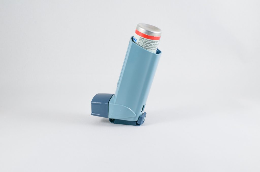
Photo by Mart Production on Pexels

A new study reveals why people with asthma seem to be less likely to develop brain tumours than others.

Asthma causes T cell activation, and researchers discovered in a mouse study that asthma causes the T cells to behave in a way that induces lung inflammation but prevents the growth of brain tumours.

The findings, appearing in Nature Communications, suggest that reprogramming T cells in brain tumour patients to act more like T cells in asthma patients could be a new approach to treating brain tumours.

“Of course, we’re not going to start inducing asthma in anyone; asthma can be a lethal disease,” said senior author David H. Gutmann, MD, PhD, at Washington University School of Medicine. “But what if we could trick the T cells into thinking they’re asthma T cells when they enter the brain, so they no longer support brain tumor formation and growth? These findings open the door to new kinds of therapies targeting T cells and their interactions with cells in the brain.”

Based on epidemiologic observations, 15 years ago it was first proposed that people with inflammatory diseases, such as asthma or eczema, are less prone to developing brain tumours. However, there was no explanation for the link between the two very different kinds of diseases, and some scientists questioned whether the association was real.

Gutmann is an expert on neurofibromatosis (NF), a set of complex genetic disorders that cause tumours to grow on nerves in the brain and throughout the body. Children with NF type 1 (NF1) can develop an optic pathway glioma, where tumours grow within the optic nerves. Gutmann, director of the Washington University NF Center, noted an inverse association between asthma and brain tumours among his patients more than five years ago but didn’t know what to make of it. When more recent studies from his lab began to reveal the crucial role that immune cells play in the development of optic pathway gliomas,  he began to wonder whether immune cells could account for the asthma–brain tumour link.

Jit Chatterjee, PhD, a postdoctoral researcher and the paper’s first author, took up the investigation. Working with co-author Professor Michael J. Holtzman, MD, Dr Chatterjee studied mice genetically modified to carry a mutation in their NF1 genes and form optic pathway gliomas by three months of age.

Dr Chatterjee exposed groups of mice to asthma-inducing irritants at age four  weeks to six weeks, and treated a control group with saltwater. Then, he checked for optic pathway gliomas at three months and three months of age. The mice with asthma did not form these brain tumours.

Further experiments revealed that inducing asthma in tumour-prone mice changes the behaviour of their T cells. After the mice developed asthma, their T cells began secreting decorin, a protein that asthma researchers are well acquainted with.

Decorin is a problem in the airways, acting on lining tissues and exacerbating asthma symptoms. But the researchers found that in the brain, decorin is beneficial. There, the protein acts on microglia immune cells, blocking their activation by interfering with the NFkappaB activation pathway. Activated microglia promote brain tumour growth and development.

Treatment with either decorin or caffeic acid phenethyl ester (CAPE), a compound that inhibits the NFkappaB activation pathway, protected mice with NF1 mutations from developing optic pathway gliomas. The findings suggest that blocking microglial activation may be a potentially useful therapeutic approach for brain tumours.

“The most exciting part of this is that it shows that there is a normal communication between T cells in the body and the cells in the brain that support optic pathway glioma formation and growth,” said Prof Gutmann. “The next step for us is to see whether this is also true for other kinds of brain tumours. We’re also investigating the role of eczema and early-childhood infections, because they both involve T cells. As we understand this communication between T cells and the cells that promote brain tumours better, we’ll start finding more opportunities to develop clever therapeutics to intervene in the process.”

Source: Washington University School of Medicine

Melatonin May be The Culprit for Nocturnal Asthma

Source: Pixabay

Why patients with asthma find their condition worsens at night has remained largely unknown, but now researchers have found that the sleep hormone melatonin may be the culprit.

In ‘nocturnal asthma’ , patients with asthma often experience a worsening of asthmatic symptoms at night. More than 50% of asthma deaths occur at night, showing a link between nocturnal asthma symptoms and asthma deaths. Though numerous triggers that explain the pathogenesis of nocturnal asthma have been described, the precise mechanisms regulating this asthma phenotype remain obscured until now. Now, a study published in the American Journal of Physiology Lung Cellular and Molecular Physiology may have explained the relationship via melatonin.

Asthma patients suffer from bronchoconstriction which may be eased with a bronchodilator. However, melatonin, which is often prescribed for insomnia, favours a state of bronchoconstriction and weakens the relaxing effect of a bronchodilator through the activation of the melatonin MT2 receptor.

To elucidate this, the research group identified the expression of the melatonin MT2 receptor in human airway smooth muscle. They observed that the activation of the melatonin MT2 receptor with higher doses of melatonin or melatonin receptor agonist ramelteon greatly potentiated the bronchoconstriction. In addition, melatonin attenuated the relaxing effects of the widely used bronchodilator β-adrenoceptor agonist.

“Although serum concentration of melatonin did not significantly induce the airway constriction, greater doses of melatonin, which is clinically used to treat insomnia, jet lag, or cancer, worsened asthma symptoms and impaired the therapeutic effect of bronchodilators,” said study leader Kentaro Mizuta from Tohoku University Graduate School of Dentistry .

First author Haruka Sasaki added, “The pharmacological therapy that blocks the melatonin MT2 receptor could inhibit the detrimental effects of melatonin on airways.”

Source: Tohoku University

Small RNA Molecule Holds COPD Treatment Potential

Photo by Robina Weermeijer on Unsplash

Researchers have identified a small RNA molecule called microRNA-21 as a therapeutic target and its inhibition as a potential treatment for chronic obstructive pulmonary disease (COPD). An inflammatory lung disease that makes it hard to breathe, COPD is the third most common cause of death in the world. It is often caused by smoking tobacco products or inhaling air pollution.

In the pre-clinical study which was published in Science Translational Medicine, the researchers found elevated levels of microRNA-21 in mouse models of COPD. MicroRNA-21 inhibitor (antagomir-21) as a therapeutic treatment reduced inflammation and improved lung capacity and function in these models.

The researchers said antagomir-21 both reduced microRNA-21 expression and also suppressed the influx of inflammatory cells – macrophages, neutrophils and lymphocytes – into the airways and lungs. Lung cytokine production, that amplifies inflammatory responses, was also inhibited through use of antagomir-21.

The paper’s senior author, Professor Phil Hansbro, said that their findings offered up a completely new understanding of COPD.

“MicroRNA-21 is a common molecule that is expressed in most cells in the human body and regulates many critical biological processes. Our findings demonstrate, however, that microRNA-21 levels increase when it comes to COPD,” said Professor Hansbro.

“We believe that the development of new drugs that inhibit microRNA-21 may offer up an entirely new therapeutic approach when it comes to COPD treatment.”

Professor Hansbro said that the findings may address the limited effectiveness that current therapies have in controlling COPD or halting its progression.

“The development of effective COPD treatments has been hampered by a lack of understanding of the disease’s underlying mechanisms. Our data defines microRNA-21 as a novel therapeutic target and its inhibitors as a potential new treatment for this major, currently intractable lung disease.”

Source: News-Medical.Net

Fermented Soy Products Found to Reduce Asthma Inflammation

A bowl of tofu, a fermented soy food. Photo by Polina Tankilevitch from Pexels

Fermented soy products are common in the Japanese diet, and one brand known as ImmuBalance has been found to suppress airway inflammation in animal models of asthma.

Bronchial asthma causes symptoms such as wheezing and cough due to chronic airway inflammation, but there is no fundamental treatment for it, leaving a desire for new prevention and treatment methods. Osaka University researchers found that in a ImmuBalance-treated group of asthma model mice, eosinophils associated with asthma were significantly reduced in the bronchoalveolar lavage fluid (BALF). As well as a decrease in inflammation and mucus around the bronchi, the team observed a suppression of proteins that induce eosinophilic inflammation.

“The relationship between soy intake and allergic diseases has been epidemiologically reported in the past,” explained first author Hideaki Kadotani, “suggesting that the components of soy may have some anti-allergic effects”

“It was reported that imbalances in the gut microbiota may be involved in immune system and allergic diseases, and fermented dietary fiber, like that found in soy, might have beneficial effects in allergic asthma models.” continues Associate Professor Kazuhisa Asai, supporting author of the study.

In the study, which appears in the journal Nutrients, such a gut imbalance’s effect on asthma were examined by giving ImmuBalance-enriched feed to asthma model mice. In the ImmuBalance-treated group, there was a significant drop in the number of eosinophils in BALF, and inflammation around the bronchi and mucus production in the bronchial epithelium was suppressed. Additionally, the expression of Th2 cytokines and the immunoglobulin serum IgE that induce eosinophilic inflammation in BALF were found to be significantly suppressed.

“In clinical practice, steroid inhalants are the basis of asthma treatments, yet they are known to have adverse side effects“, stated lead advisor to the study, Professor Tomoya Kawaguchi. “Our results suggest that the intake of fermented soybean products should be recommended as a complementary coping strategy to asthma with fewer side effects”

Source: Osaka University

Distinct Lower Airways Bacterial Profile inChildren with Cystic Fibrosis

Phot by Ben Wicks on Unsplash

In children with cystic fibrosis (CF), their lower airways have a higher burden of infection, more inflammation and lower diversity of microorganisms, compared to children with other illnesses who also have lung issues, researchers have found. They noted a clear divergence in these bacterial communities in toddlers, before progressive lung disease manifests in CF patients. 

Their findings, published in the journal PLOS ONE, could help providers target specific pathogens earlier, treat them and potentially prevent more severe lung disease.

As lead author Jack O’Connor, at Ann & Robert H. Lurie Children’s Hospital of Chicago explained, “We compared lower airway samples from bronchoscopy in children with CF and disease controls across the age spectrum, and used genetic sequencing to identify microorganisms, finding that a few common cystic fibrosis pathogens begin to dominate at very early ages. Such a clear split from disease controls in this young age group has not been shown before. Our findings deepen our understanding of the disease trajectory in cystic fibrosis and could help improve outcomes through earlier intervention.”

Chronic airway infection and inflammation which leads to progressive, obstructive lung disease is the main cause of illness and death in people with cystic fibrosis.

Researchers tested lower airway samples from 191 patients (63 with cystic fibrosis) aged 0-21 years. The disease controls included patients with diverse conditions, such as cancer, immune deficiency and pneumonia. Using genetic sequencing, researchers were able to identify distinct pathogens that are more dominant at different ages in patients with cystic fibrosis.

“Establishing key age-related differences in lower airway bacterial communities and inflammation in patients with CF, especially during early childhood, may give us a window of opportunity for earlier and more precise treatment,” said senior author Theresa Laguna, MD, MSCS, Division Head of Pulmonary and Sleep Medicine at Lurie Children’s and Associate Professor of Pediatrics at Northwestern University Feinberg School of Medicine. “If we can prevent worse infections, we could improve the quality of life and potentially expand the life expectancy of patients with CF.”

Source: News-Medical.Net

Surprising Finding Links Asthma Risk to Meat Consumption as Infants

Source: PIxabay/CC0

Researchers looking for post-breastfeeding dietary patterns in two prospective birth cohorts, were surprised to discover meat consumption as a predictive factor.

Alexander Hose, MA, MPH, of Ludwig Maximilian University in Munich presented the study’s findings at the virtual European Respiratory Society annual meeting. 

After switching from breast milk, babies up to age 1 whose protein intake came largely from meat products, rather than dairy, fish, or egg proteins, had a more than eight-fold greater chance of developing asthma by age 6 versus non-meat protein consumption. Asthma prevalence reached 30% in some subgroups.

Wheezing was more common in this diet pattern, which Hose and colleagues termed “unbalanced meat consumption” (UMC); this continued up to age 10, with a five times higher odds.

The duration of breastfeeding was an important factor, likely because switching to baby foods prolonged the exposure. Odds of developing asthma by age 6 increased nearly 12-fold in UMC-fed infants whose breastfeeding stopped by week 19, versus about four-fold in those continuing longer on breast milk.

In addition, UMC was also linked to a certain intestinal microbiome profile featuring unusually high levels of Lactococcus, Granulicatella, and Acinetobacter species.

This type of microbiome scavenges iron in the gut, Hose said, which could explain why the children became especially susceptible to asthma. Additionally, milk proteins may exert an opposite effect on asthma risk by generating a type of “nutritional immunity.”

While the mechanism connecting the gut microbiome to respiratory disease is unknown, the existence of a ‘gut-lung axis‘ is well established; a recent trial showed that probiotics can prevent coughs and wheezing in older adults. The phenomenon has also been considered for COVID’s gastrointestinal symptoms.

A pair of European birth cohort studies, PASTURE and LUKAS2, provided the data for the study. In these, about 1400 infants were followed through age 10 and parents kept detailed records of their infants’ feeding, and other environmental factors, and children’s medical records were accessed as well.

However, a key limitation is the cohorts being from rural areas since investigating asthma’s relationship to animal exposure was a key goal for the studies. Partly because of this, Hose and colleagues were able to separate out ‘industrial’ meat, milk, and yoghurt from that produced at home. A trend toward greater asthma risk was observed with store-bought protein products.

Source: MedPage Today

Circadian Rhythm Contributes to Asthma Severity

Source: Pixabay/CC0

By pinning down the influence of the circadian system on nocturnal asthma, researchers have uncovered a key role for the biological clock in asthma.

Asthma severity has long been observed to worsen in the nighttime. Lung function is highest at around 4pm and worst around 4am. One longstanding question has been to what degree the body’s internal circadian clock contributes to worsening of asthma severity, as opposed to behaviours such as sleep. Using two circadian protocols, researchers have delineated the influence of the circadian system. Understanding the mechanisms behind asthma severity could have important implications for both studying and treating asthma. 

“This is one of the first studies to carefully isolate the influence of the circadian system from the other factors that are behavioral and environmental, including sleep,” said co-corresponding author Frank AJL Scheer, PhD, director of the Medical Chronobiology Program in the Division of Sleep and Circadian Disorders at the Brigham.

As many as 75 percent of people with asthma report experiencing worsening asthma severity at night. Asthma severity is influenced by behavioural and environmental factors, such as exercise, air temperature, posture, and sleep environment. The researcher sought to understand the internal circadian system’s contributions to this problem. The circadian system is composed of a central pacemaker in the brain (the suprachiasmatic nucleus) and “clocks” throughout the body and is critical for the coordination of bodily functions and to anticipate the daily cycling environmental and behavioral demands.

To isolate the influence of the circadian system from that of sleep and other behavioural and environmental factors, the researchers enrolled 17 participants with asthma into two complementary laboratory protocols where lung function, asthma symptoms and bronchodilator use were continuously assessed. In the “constant routine” protocol, participants spent 38 hours continuously awake, in a constant posture, and under dim light conditions, with identical snacks every two hours. In the “forced desynchrony” protocol, participants were placed on a recurring 28-hour sleep/wake cycle for a week under dim light conditions, with all behaviours scheduled evenly across the cycle.

Co-corresponding author Steven A. Shea, Ph.D., professor and director at Oregon Institute of Occupational Health Sciences said, “We observed that those people who have the worst asthma in general are the ones who suffer from the greatest circadian-induced drops in pulmonary function at night, and also had the greatest changes induced by behaviours, including sleep. We also found that these results are clinically important because, when studied in the laboratory, symptom-driven bronchodilator inhaler use was as much as four times more often during the circadian night than during the day.”  

The study was published in Proceedings of the National Academy of Sciences.

Source: Medical Xpress

Bisexual People Have Over Twice the Rates of Lung Diseases

Photo by Robina Weermeijer on Unsplash

A new national study found that bisexual Americans had over two times the rates of lung diseases including asthma as heterosexual adults.

The study, published in Annals of the American Thoracic Society, analysed data from 12 209 adults in the National Longitudinal Study of Adolescent to Adult Health. Overall, 29% of bisexual adults reported experiencing lung disease compared to 14% of heterosexual adults. There is consistent evidence that bisexual individuals are at increased risk for negative health outcomes (such as mental health, substance use, and sexual health problems) compared to monosexual (heterosexual and gay/lesbian) individuals.

“Higher levels of discrimination experienced by bisexual people could lead to more stress and lead to inflammation or stress hormones which would worsen asthma,” said lead author, Jason Nagata, MD, assistant professor of paediatrics at the University of California, San Francisco. “Bisexual adults have been shown to have worse health outcomes across a number of physical and mental health domains, and we add to this literature by showing disparities in asthma and other lung diseases.”

Even people who identified as ‘mostly’ heterosexual had higher rates of asthma than those who identified as exclusively heterosexual. Mostly heterosexual individuals may also face discrimination but may not be ‘out’, with access to the social support and communities ‘out’ LGBTQ+ people have available.

“Medical professionals, social workers, and clinicians need to be aware of these sexual orientation disparities in health outcomes,” said co-author Kyle T Ganson, PhD, assistant professor at the University of Toronto’s Factor-Inwentash Faculty of Social Work. “Providing appropriate and tailored care is needed to address these disparities.”

“Some sexual minorities may be less likely to seek care due to barriers to accessing health care or experiences of discrimination at a clinician’s office. Doctors should offer materials on LGBTQ health, publicise nondiscrimination statements and have inclusive forms for sexual minorities,” Prof Nagata added, “so that they’re not discouraged from seeking care.”

Source: EurekAlert!

CT Scans Reveal Lung Destruction in Asthmatics

Source: Pixabay CC0

A new study using CT scans have revealed significant changes indicating lung destruction in some asthmatics.

Clinicians have long thought that some people with asthma experience declines in their lung function, called fixed airflow obstruction (FAO), due to changes to their airways. In this study published in The Journal of Allergy and Clinical Immunology, researchers have found that this issue could extend to the surrounding lung tissue.

Respirologist Kaoruko Shimizu at Hokkaido University said, “Bronchial asthma is considered to be mainly due to inflammation and remodeling of the larger respiratory airways. But not all asthmatics improve with the typical treatments prescribed to alleviate this condition. We wanted to know if changes to the surrounding lung tissue induced a decline in pulmonary function over time in this subgroup of patients.”

Shimizu and her colleagues applied a novel computer tomography (CT)-based approach to detect changes in lung tissue. In this approach, the scientists examined CT scans employing an index called “exponent D” for areas of reduced lung density with increasing coalescence of neighbouring airspaces, which indicates emphysema, or the destruction of air sacs. Airway obstruction was measured by testing the ability of people with asthma to forcefully exhale air in one second. This ability is reduced when the airways are narrower.

The tests examined around 200 smokers and non-smokers with varying degrees of asthma, who were then followed up annually for five years.

People with asthma who experienced persistent airflow limitation, regardless of the severity of their asthma or their smoking status, exhibited constricted airways and also showed signs of lung tissue destruction, the researchers found.

The observed changes to lung tissue in this subgroup of asthmatic patients were not associated with the typical inflammatory markers linked to bronchial asthma. This is important, because it could explain why conventional anti-inflammatory treatments are not as successful in this group.

Future studies should investigate lung destruction in asthma, enabling more personalised management, said Shimizu.

Source: Hokkaido University

80% of Childhood Asthma Hospital Presentations are Preventable

Image by Bob Williams from Pixabay

Based on a comprehensive Australian survey, approximately 80 percent of asthma-related hospital presentations in school-aged children are potentially avoidable through a standardised comprehensive care pathway for children with asthma.

These preventative measures include using evidence-based clinical guidelines, ensuring that there is an asthma action plan in place; regular follow-up with GP; provision of asthma education to parents/carers; and establishing a community-based approach for continuity of care.

Senior author Dr Nusrat Homaira, respiratory epidemiologist at UNSW Sydney said, “During our research, we surveyed 236 nurses and 266 doctors across 37 hospitals in all 15 local health districts (LHDs) across New South Wales (NSW) to identify the existing care pathway following discharge from hospital for children with asthma.”

This study by researchers at UNSW Sydney identified major variations in the existing asthma care pathway, including:

Use of asthma clinical guidelines and Asthma Action Plan: Although clinical guidelines and Asthma Action Plans (AAPs) were used across all hospitals, on average, there were four to six different types of documents used in each (LHD), between hospitals in the same LHD and within departments in the same hospital. Such variations can be confusing for clinicians, as noted by a survey participant: “Conflicting advice given to asthma patients between general practitioners, emergency departments and sometimes paediatricians; patients are then confused about what to do in exacerbation of symptoms.”

GP follow-up: In most LHDs (75 percent) parents/carers were advised to have their child followed up with their GP within two to three days after hospital discharge, but in some areas, follow-up appointments could be recommended for over six days post-hospitalisation. Parents/carers were reportedly responsible for organising follow-up with their GP with no system to ensure they in fact attended.

Asthma education for parents/carers of asthmatic children: Formal asthma education (27 percent of respondents) were seldom provided to parents/carers during hospital stays; limited to asthma device techniques and rarely involved key topics such as basic knowledge of asthma, asthma control and the importance of regular medical review.

Communication with schools/childcare services: When children with asthma were discharged from hospitals, only four percent of the surveyed staff reported that schools or childcare services were notified of the child’s recent hospital presentation.

Community services integration: The majority of participants (55 percent) were unaware of any community services for children with asthma in their local areas.

The survey identified marked variations in asthma care and management for children within different health districts, different hospitals in the same district and different departments within the same hospital in. The findings highlight opportunities to improve the health outcomes in children with asthma and reduce unnecessary burden on health systems from preventable asthma hospital presentations.

Source: EurekAlert!

Journal information: Chan, M., et al. (2021) Assessment of Variation in Care Following Hospital Discharge for Children with Acute Asthma. Journal of Asthma and Allergy. doi.org/10.2147/JAA.S311721.