A breakthrough study, led by scientists at Waipapa Taumata Rau, University of Auckland, has uncovered how daylight can boost the immune system’s ability to fight infections.
The circadian clock is a 2.5-billion-year-old cellular timekeeper that allows organisms to adapt to the rhythms of day and night. Evidence has shown that disruption of our internal body clock through the likes of shift work or jet lag makes people more susceptible to infections, so the researchers wanted to find out what in the body contributed to that susceptibility.
“In earlier studies, we had observed that immune responses to infection peaked in the morning, during the animals’ early active phase,” says lead researcher Associate Professor Christopher Hall, from the Department of Molecular Medicine and Pathology.
“We think this represents an evolutionary response such that during daylight hours the host is more active so more likely to encounter bacterial infections,” says Hall.
However, the scientists wanted to find out how the immune response was being synchronised with daylight.
They focused on ‘neutrophils’ the most abundant immune cells in our bodies. These cells move quickly to the site of an infection and kill invading bacteria.
The scientists used zebrafish, a small freshwater fish, as a model organism, because its genetic make-up is similar to ours and they can be bred to have transparent bodies, making it easy to observe biological processes in real time.
With this new study, published in Science Immunology, neutrophils were found to possess a circadian clock that alerted them to daytime, and boosted their ability to kill bacteria.
Circadian clocks are present in almost every cell and tissue in the body, telling them what is going on in the outside world and coordinating physiological processes like metabolism, hormone release, and sleep-wake cycles.
Light has the biggest influence on resetting these circadian clocks.
“Given that neutrophils are the first immune cells to be recruited to sites of inflammation, our discovery has very broad implications for therapeutic benefit in many inflammatory diseases,” Hall says.
“This finding paves the way for development of drugs that target the circadian clock in neutrophils to boost their ability to fight infections.”
Immune molecules called cytokines play important roles in the body’s defence against infection, helping to control inflammation and coordinating the responses of other immune cells. A growing body of evidence suggests that some of these molecules also influence the brain, leading to behavioural changes during illness.
Two new studies from MIT and Harvard Medical School, focused on a cytokine called IL-17, now add to that evidence. The researchers found that IL-17 acts on two distinct brain regions — the amygdala and the somatosensory cortex — to exert two divergent effects. In the amygdala, IL-17 can elicit feelings of anxiety, while in the cortex it promotes sociable behaviour.
These findings suggest that the immune and nervous systems are tightly interconnected, says Gloria Choi, an associate professor of brain and cognitive sciences, a member of MIT’s Picower Institute for Learning and Memory, and one of the senior authors of the studies.
“If you’re sick, there’s so many more things that are happening to your internal states, your mood, and your behavioural states, and that’s not simply you being fatigued physically. It has something to do with the brain,” she says.
Jun Huh, an associate professor of immunology at Harvard Medical School, is also a senior author of both studies, which appear today in Cell. One of the papers was led by research scientists Byeongjun Lee and Jeong-Tae Kwon, and the other was led by postdocs Yunjin Lee and Tomoe Ishikawa.
Behavioral effects
Choi and Huh became interested in IL-17 several years ago, when they found it was involved in a phenomenon known as the fever effect. Large-scale studies of autistic children have found that for many of them, their behavioural symptoms temporarily diminish when they have a fever.
In a 2019 study in mice, Choi and Huh showed that in some cases of infection, IL-17 is released and suppresses a small region of the brain’s cortex known as S1DZ. Overactivation of neurons in this region can lead to autism-like behavioral symptoms in mice, including repetitive behaviours and reduced sociability.
“This molecule became a link that connects immune system activation, manifested as a fever, to changes in brain function and changes in the animals’ behaviour,” Choi says.
IL-17 comes in six different forms, and there are five different receptors that can bind to it. In their two new papers, the researchers set out to map which of these receptors are expressed in different parts of the brain. This mapping revealed that a pair of receptors known as IL-17RA and IL-17RB is found in the cortex, including in the S1DZ region that the researchers had previously identified. The receptors are located in a population of neurons that receive proprioceptive input and are involved in controlling behaviour.
When a type of IL-17 known as IL-17E binds to these receptors, the neurons become less excitable, which leads to the behavioural effects seen in the 2019 study.
“IL-17E, which we’ve shown to be necessary for behavioural mitigation, actually does act almost exactly like a neuromodulator in that it will immediately reduce these neurons’ excitability,” Choi says. “So, there is an immune molecule that’s acting as a neuromodulator in the brain, and its main function is to regulate excitability of neurons.”
Choi hypothesises that IL-17 may have originally evolved as a neuromodulator, and later on was appropriated by the immune system to play a role in promoting inflammation. That idea is consistent with previous work showing that in the worm C. elegans, IL-17 has no role in the immune system but instead acts on neurons. Among its effects in worms, IL-17 promotes aggregation, a form of social behaviour. Additionally, in mammals, IL-17E is actually made by neurons in the cortex, including S1DZ.
“There’s a possibility that a couple of forms of IL-17 perhaps evolved first and foremost to act as a neuromodulator in the brain, and maybe later were hijacked by the immune system also to act as immune modulators,” Choi says.
Provoking anxiety
In the other Cell paper, the researchers explored another brain location where they found IL-17 receptors — the amygdala. This almond-shaped structure plays an important role in processing emotions, including fear and anxiety.
That study revealed that in a region known as the basolateral amygdala (BLA), the IL-17RA and IL-17RE receptors, which work as a pair, are expressed in a discrete population of neurons. When these receptors bind to IL-17A and IL-17C, the neurons become more excitable, leading to an increase in anxiety.
The researchers also found that, counterintuitively, if animals are treated with antibodies that block IL-17 receptors, it actually increases the amount of IL-17C circulating in the body. This finding may help to explain unexpected outcomes observed in a clinical trial of a drug targeting the IL-17-RA receptor for psoriasis treatment, particularly regarding its potential adverse effects on mental health.
“We hypothesise that there’s a possibility that the IL-17 ligand that is upregulated in this patient cohort might act on the brain to induce suicide ideation, while in animals there is an anxiogenic phenotype,” Choi says.
During infections, this anxiety may be a beneficial response, keeping the sick individual away from others to whom the infection could spread, Choi hypothesises.
“Other than its main function of fighting pathogens, one of the ways that the immune system works is to control the host behaviour, to protect the host itself and also protect the community the host belongs to,” she says. “One of the ways the immune system is doing that is to use cytokines, secreted factors, to go to the brain as communication tools.”
The researchers found that the same BLA neurons that have receptors for IL-17 also have receptors for IL-10, a cytokine that suppresses inflammation. This molecule counteracts the excitability generated by IL-17, giving the body a way to shut off anxiety once it’s no longer useful.
Distinctive behaviours
Together, the two studies suggest that the immune system, and even a single family of cytokines, can exert a variety of effects in the brain.
“We have now different combinations of IL-17 receptors being expressed in different populations of neurons, in two different brain regions, that regulate very distinct behaviours. One is actually somewhat positive and enhances social behaviours, and another is somewhat negative and induces anxiogenic phenotypes,” Choi says.
Her lab is now working on additional mapping of IL-17 receptor locations, as well as the IL-17 molecules that bind to them, focusing on the S1DZ region. Eventually, a better understanding of these neuro-immune interactions may help researchers develop new treatments for neurological conditions such as autism or depression.
“The fact that these molecules are made by the immune system gives us a novel approach to influence brain function as means of therapeutics,” Choi says. “Instead of thinking about directly going for the brain, can we think about doing something to the immune system?”
Scanning electron micrograph of a T cell lymphocyte. Credit: NIH / NIAID
When an infection is prolonged and severe, T cell exhaustion comes into play to prevent to reign in the immune system and prevent damage to the body. A study published in Nature reveals that right from the beginning of mild illness, the body also produces these special T cells previously known only from chronic, severe infections and tumours.
There are different types of T cells in the body, all of which play a crucial role in the immune system. They fight pathogens and control the immune response. However, some subtypes become less effective or even cease their activity altogether as the disease progresses. This has a protective function: in persisting infections, it would harm the body if the immune system continued to fight the pathogens aggressively. But in cancer treatment, T cell exhaustion means that therapeutic measures may no longer be effective.
Until now, it was assumed that the body only produces such T cells in severe and persisting infections. The results of the researchers, from Helmholtz Munich and the Technical University of Munich, show that this is not the case. “We were able to show that the body prepares T cell subtypes that are predisposed to exhaustion even in early infection phases of moderate diseases,” says Dietmar Zehn, Professor of Animal Physiology and Immunology at TUM and last study author.
Different T Cells for Different Purposes
The team deduces from the discovery that the body assembles a range of different T cells early on at the onset of the disease to arm itself for different disease progressions. Depending on the course of the disease, it then has cells at its disposal to make the immune response more aggressive or more gentle — and in some circumstances, even to abort it.
“Our results expand the classic idea of the development of T cell exhaustion,” says Dietmar Zehn. “We therefore assume that our observations will help to further decipher the mechanisms behind T cell exhaustion.” A better understanding of these processes could help in the future to control the immune response in a targeted manner — for example, to strengthen the immune system in cancer patients or to weaken excessive defences, as is typical in severe cases of COVID-19, for instance.
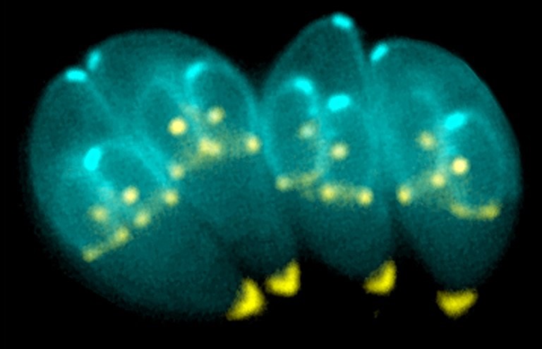
Most humans have long-lived infections in various tissues, including in the nervous system, that typically do not result in disease. The microbes associated with these infections, such as Toxoplasma gondii, enter a latent stage during which they quietly hide in cells, playing the long game to evade capture and ensure their own survival. But a lack of natural models to study these quiescent stages has led to gaps in scientists’ understanding of how latency contributes to pathogen persistence and whether these stages can be targeted by the immune system.
Now, a team led by University of Pennsylvania School of Veterinary Medicine researchers shows that the immune system indeed recognises the latent stage of the parasite Toxoplasma gondii, which causes toxoplasmosis. The work, published in Nature Microbiology, challenges some common assumptions about how the immune system deals with infections in the brain. Senior author Christopher A. Hunter, professor at PennU Vet, says this knowledge supports the idea that Toxoplasma gondii cysts can be targeted and perhaps even cleared, and the findings have implications for other infections and potential future therapies. The paper also demonstrates how cysts promote the mutual survival of the parasite and host.
In its latent stage, Toxoplasma gondii forms long-lived cysts in neurons in the brain, which helps the parasite evade the host’s immune response. In this study, the researchers found that certain T cells can target neurons containing cysts, thereby promoting parasite control. But there’s a tradeoff: They also found that when cysts are not formed, there is an even higher parasite burden and increased damage to the brain. The study is published in Nature Microbiology.
“There’s this balance of the pathogen needing to take hold in the host but not expand so much that it’s detrimental to the host, because if the host dies, the pathogen may not survive,” says author Lindsey A. Shallberg, who at the time of the research was a doctoral student in Hunter’s lab.
Toxoplasma gondii causes toxoplasmosis, an infection that is asymptomatic for most healthy people but poses a greater risk for those who are immunocompromised or pregnant. It is caused by eating contaminated, poorly cooked meat and by exposure to infected cat faeces, as felines are the only animal in which the parasite can sexually reproduce.
Co-author Julia N. Eberhard, an immunology doctoral student, points to two findings that run counter to preexisting literature and common notions among immunologists. She says scientists long thought that Toxoplasma gondii cysts could hide out in neurons to prevent immune recognition, but this study showed that “neurons aren’t this complete refuge for pathogens.”
This image shows Toxoplasma gondii (red) and a neuron (green) in a mouse brain. (Image: Courtesy of Anita Koshy)
Eberhard says another commonly held belief was that the parasite needs to form cysts to be able to persist, but in looking at a parasite strain that couldn’t convert to the cyst stage, the researchers found that the immune system did not clear the parasite. They could still identify parasites in mice six months later, which Eberhard found very surprising.
Mathematical modelling independently confirmed experimental findings and indicated that immune pressure on the latent stage of Toxoplasma gondii could explain the observed rise and fall in cyst numbers. This was done by Aaron Winn, a doctoral student in the Department of Physics and Astronomy.
Shallberg says this paper came about because co-author Sebastian Lourido, an associate professor of biology at MIT, had identified the key molecular mechanism that allows the parasite to become latent and wanted to know what would happen if the parasite could not form cysts. In addition, co-author Anita Koshy, a neurologist and scientist at the University of Arizona, had evidence that some neurons could rid themselves of this infection.
While Toxoplasma gondii is a relevant microorganism to study in and of itself, it is also useful in furthering scientists’ understanding of nervous system infections with latent stages in humans that don’t have mouse models, such as cytomegalovirus. “What makes it special is the fact that it’s a tractable model that we can use in the lab and then apply what we’ve learned to other infections,” Shallberg says.
Looking ahead, Hunter says that his laboratory continues to investigate whether T cells directly recognise the neurons and to study the T cell response in more detail.
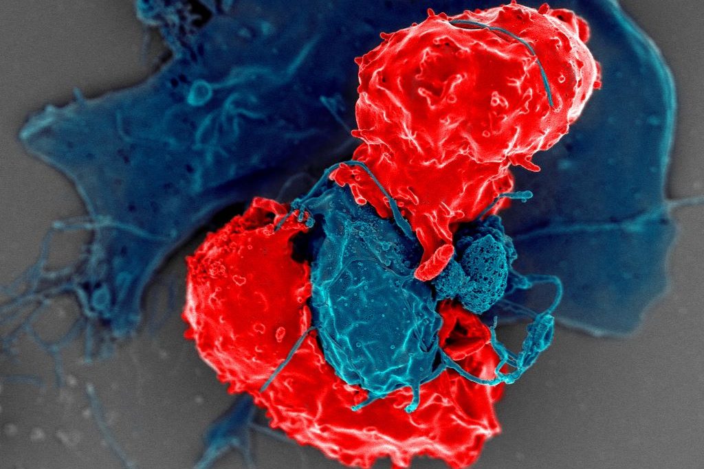
Scanning electron microscope image of T regulatory cells (red) interacting with antigen-presenting cells (blue). T regulatory cells can suppress responses by T cells to maintain homeostasis in the immune system. Credit: National Institute of Allergy and Infectious Diseases/NIH
In the flurry of immune activity in an infection, immune cells need to be prevented from mistakenly attacking each other. New research from the University of Chicago shows how a specially trained population of immune cells keeps the peace by preventing other immune cells from attacking their own. The study, published in Science, provides a better understanding of immune regulation during infection and could provide a foundation for interventions to prevent or reverse autoimmune diseases.
Several groups of white blood cells help coordinate immune responses. Dendritic cells take up proteins from foreign pathogens, chop them up into peptides called antigens, and display them on their surface. CD4+ conventional T (Tconv) cells, or helper T cells, inspect the peptides presented by dendritic cells. If the peptides are foreign antigens, the T cells expand in numbers and transform into an activated state, specialized to eradicate the pathogen. If the dendritic cell is carrying a “self-peptide,” or peptides from the body’s own tissue, the T cells are supposed to lay off.
During an autoimmune response, the helper T cells don’t distinguish between foreign peptide antigens and self-peptides properly and go on the attack no matter what. To prevent this from happening, another group of T cells called CD4+ regulatory T (Treg) cells, are supposed to intervene and prevent friendly fire from the Tconv cells.
“You can think of them [Treg cells] as peacekeeper cells,” said Pete Savage, PhD, Professor of Pathology at UChicago and senior author of the new study. Tregs obviously do their job well most of the time, but Savage said that it has never been clear how they know when to intervene and prevent helper T cells from starting an autoimmune response, and when to hold back and let them fight an infection.
So, Savage and his team, led by David Klawon, PhD, a former graduate student in his lab who is now a postdoctoral fellow at the Massachusetts Institute of Technology (MIT), wanted to explore this property of the immune system, known in the field as self-nonself discrimination. T cells are produced in the thymus, a specialised organ of the immune system. During development, Treg cells are trained to recognise specific peptides, including self-peptides from the body. When dendritic cells present a self-peptide, the Treg cells trained to spot them intervene to stop helper T cells from getting triggered.
For the study, Savage and Klawon worked in close collaboration with co-first author Nicole Pagane, a graduate student at MIT, as well as co-corresponding authors Harikesh Wong at the Ragon Institute of the Massachusetts General Hospital, MIT and Harvard University, and Ron Germain at the National Institutes of Health.
T cell specificity is what the team found makes a crucial difference in self-nonself discrimination. The researchers experimentally depleted Treg cells in mice that were specific to a single self-peptide from the prostate. In healthy mice in the absence of infection, this change did not trigger autoimmunity to the prostate. When the researchers infected mice with a bacterium that expressed the prostate self-peptide, however, the absence of matched, prostate-specific Treg cells triggered prostate-reactive T helper cells and introduced autoimmunity to the prostate.
Interestingly though, this alteration did not impair the ability of helper T cells to control the bacterial infection by responding to foreign peptides.
“It’s like a doppelganger population of T cells. The CD4 helper cells that could induce disease by attacking the self share an equivalent, matched population of these peacekeeper Treg cells,” Savage said. “When we removed Treg cells reactive to a single self-peptide, the T helper cells reactive to that self-peptide were no longer controlled, and they induced autoimmunity.”
The root causes of autoimmune disease are a complex interaction of genetics, the environment, lifestyle, and the immune system. Classic, conventional thinking in the immunology field promoted the idea that the immune system establishes self-nonself discrimination by purging the body of helper T cells that are reactive to self-peptides, thereby preventing autoimmunity. Savage said this study shows that purging is inefficient though, and that specificity matching by Treg cells may be equally as important.
“The idea is that specificity matters, and for a fully healthy immune system, you need to have a good collection of these doppelganger Treg cells,” he said. As long as the immune system generates enough matched Treg cells, they can prevent autoimmune responses without impacting responses to infections.
“It’s like flipping the idea of self-nonself discrimination upside down. Instead of having to delete all helper T cells reactive to self-antigens, you simply generate enough of these Treg peacekeeper cells instead,” Savage said.
A new study on psoriasis has determined that a protein called NF-kB c-Rel can intensify the condition’s symptoms when activated by signals from the body’s immune system. Understanding how “c-Rel” affects skin inflammation could lead to new treatments, said the researchers at Case Western Reserve University School of Medicine.
The study, published recently in eBioMedicine, examined how c-Rel contributes to the function of dendritic cells (DCs), a type of immune cell. The study examined how c-Rel responds to specific immunological signals through Toll Like Receptor 7 (TLR7), which regulates innate immunity and inflammation, exacerbating psoriasis.
The researchers also found the absence of c-Rel alleviates inflammation that causes red, scaly patches on the skin. TLR7 meanwhile is known to be activated by diseases such as HIV and HPV, which are also linked to the development psoriasis.
“We believe that by focusing on c-Rel and TLR7, scientists might be able to create more targeted treatments that reduce inflammation and help psoriasis symptoms,” said Parameswaran Ramakrishnan, associate professor of pathology, member of the Case Comprehensive Cancer Center and researcher at Louis Stokes Cleveland VA Medical Center, the study’s principal investigator.
“This may help relieve the discomfort millions of people live with skin inflammation.”
The researchers examined skin samples from psoriasis patients and a mouse model with similar skin changes.
They analysed c-Rel levels and its behaviour in specially engineered cells lacking the protein; they also examined the mouse model lacking c-Rel.
Their goal: to better understand how c-Rel impacts the immune response in psoriasis.
“Our research shows that c-Rel plays a major role in psoriasis inflammation,” said Angela Liu, lead author and a recent graduate of the School of Medicine’s pathology department.
“We saw higher levels of c-Rel in psoriasis; mice lacking c-Rel were significantly protected from developing psoriasis and showed less inflammation.”
Ramakrishnan said their study revealed the potential role for TLR7 and c-Rel signalling in human psoriasis. A range of viruses that activates TLR7, including human immunodeficiency virus (HIV), human papilloma virus (HPV) and hepatitis C virus (HCV), are linked to the development of psoriasis.
“The research warrants future studies on TLR7-c-Rel-dependent molecular mechanism regulating DC function as a potential link for how viral TLR7 activation is involved in worsening psoriatic disease,” Ramakrishnan said. “From a broad perspective, it would be interesting to further explore the role of c-Rel and TLR7 in other biologically relevant diseases involving these proteins, such as systemic lupus erythematosus and wound-healing in diabetes.”
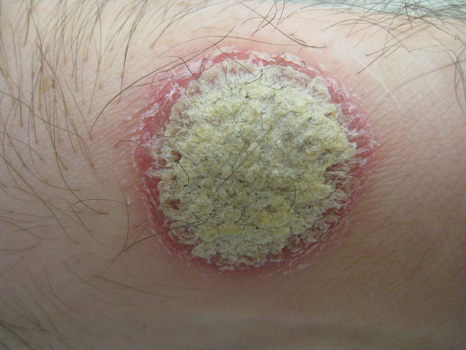
A woman with Systemic Lupus Erythematosus. Source: Wikimedia CC0
Researchers have found that topical mupirocin is effective in reducing rashes caused by systemic lupus erythematosus. Instead of directly lowering inflammation, the treatment kills bacteria that promote it. The findings are published in Arthritis & Rheumatology.
Cutaneous lupus erythematosus is a common manifestation of systemic lupus erythematosus, caused by the autoimmune conditions. The condition is characterised by rashes on various parts of the body including the face and scalp, hair loss and scarring of the skin.
The standard treatment for cutaneous lupus erythematosus is using immunosuppressants and biologic drugs to reduce inflammation. While the medications can be helpful, many patients with systemic lupus erythematosus already take several drugs and are looking for alternatives to pills.
J. Michelle Kahlenberg, MD, PhD, a professor of internal medicine in the Division of Rheumatology at University of Michigan Health led a team of researchers investigating topical mupirocin which is one such alternatives.
This trial was based on Kahlenberg’s previous discovery that cutaneous lupus rashes are often colonised with a common skin bacteria, Staphyloccous areus, also known asstaph, and contributes to inflammation in the rashes. Mupirocin kills this type of bacteria.
The study randomly selected systemic lupus erythematosus patients currently experiencing cutaneous lupus erythematosus flares to treat their skin lesions with mupirocin or with an inactive control, petrolatum jelly.
Samples from the nose and lesional skin were used to determine baseline and post treatment Staphylococcus abundance and microbial community profiles. Paired samples collected prior to treatment with the topical solution and seven days after treatment showed decreases in lesional staphylococcus aureusin the mupirocin treated samples.
Importantly, the reduction in staph also was accompanied by a reduction in inflammatory signals, including interferon-driven gene expression, in the lesions.
“In addition to decreasing the inflammation by decreasing lesional staphylococcus aureus, the mupirocin treatment also lowered skin monocyte levels, which are important in driving cutaneous lupus,” said Kahlenberg.
Mupirocin is a prescription treatment, and while this early study showed signs of decreasing inflammation, the study wasn’t designed to see if it can decrease the rash of cutaneous lupus erythematosus.
“Additional larger studies are needed to determine whether topical antibiotics will be helpful to make rashes go away,” Kahlenberg said.
“However, this is an exciting first step to show that there may be additional treatments that can improve inflammation beyond our usual immunosuppressant and biologic drugs.”
McGill University scientists have discovered that pregnancy may trigger a natural immunity to boost protection against severe flu infection. Contrary to the common belief that pregnancy increases vulnerability to infections, researchers found that it strengthened an immune defence in mice, blocking the Influenza A virus from spreading to the lungs, where it can cause severe infection.
“Our results are surprising because of the current dogma, but it makes sense from an evolutionary perspective,” said co-lead author Dr Maziar Divangahi, Professor in McGill’s Faculty of Medicine and Health Sciences and Senior Scientist at the Research Institute of the McGill University Health Centre (The Institute).
“A mother needs to stay healthy to protect her developing baby, so the immune system adapts to provide stronger defenses. This fascinating response in the nasal cavity is the body’s way of adding an extra layer of protection, which turns on during pregnancy.”
Exploring benefits for pregnancy and beyond
The researchers used a mouse model to observe how a certain type of immune cell activates in the nasal cavity of mice during pregnancy, producing a powerful molecule that boosts the body’s antiviral defenses, especially in the nose and upper airways.
“Influenza A virus remains among the deadliest threats to humanity,” said first author Julia Chronopoulos, who carried out the research while completing her PhD at McGill. “This natural immunity in pregnancy could change the way we think about flu protection for expectant mothers.”
The Public Health Agency of Canada recommends pregnant women and pregnant individuals get the flu vaccine, as they are at high risk of severe illness and complications like preterm birth. The new insights offer promise for more targeted vaccines for influenza, which is among the top 10 leading causes of death in Canada.
“The broader population could also benefit, as our findings suggest the immune response we observed could be replicated beyond pregnancy,” said co-lead author Dr James Martin, Professor in McGill’s Faculty of Medicine and Health Sciences and Senior Scientist at the RI-MUHC. This could mean new nasal vaccines or treatments that increase protective molecules, known as Interleukin-17.
The team’s next focus is on finding ways to reduce lung damage during viral infections like the flu or COVID. Rather than targeting the virus, as previous research has done, they aim to prevent dysregulated immune systems from overreacting, an approach that could lower the risk of serious complications associated with flu infection.
Killer T cells about to destroy a cancer cell. Credit: NIH
Researchers have discovered a genomic ‘brake’ in a subset of immune cells that could help advance immunotherapy for cancer and autoimmune disease. The findings, led by a team at the Peter MacCallum Cancer Centre in collaboration with researchers at the Garvan Institute and Kirby Institute, provide new insights into how the body’s immune defence mechanisms can go awry in these diseases and open a new class of potential drug targets that could activate immune cells in tumour tissue.
The research was published in the journal Immunity.
Discovering an immune brake
Specialised killer T cells are released during an infection, trained to recognise and destroy the threat. When unchecked, these cells can cause autoimmune diseases such as type 1 diabetes or rheumatoid arthritis if they mistakenly attack the body’s own healthy tissues.
The immune system employs a mechanism to prevent autoimmune attacks called ‘tolerance’ – a process that can be exploited by cancer cells to shield them from the body’s natural defences.
“Until now, it was not fully understood how tolerance works at a molecular level. We used advanced sequencing techniques to identify a unique genomic signature in ‘tolerant’ T cells that differentiated them from killer T cells that were activated in response to viral infection,” says Dr Timothy Peters, co-first author from Garvan.
“These precise genome locations have never before been observed and allowed us to track precisely how killer T cells progress through the tolerance pathway, and how specific gene networks enable tolerance to be abused.”
Breakthrough for cancer and autoimmune disease
Dr Ian Parish, who led the research at the Peter MacCallum Cancer Centre said this breakthrough helps to understand why cancer treatments fail and opens the door to developing new treatments in the future.
“Current cancer immunotherapy treatments target the exhaustion phase of the immune response,” he said. “Our research suggests that a second, earlier ‘off-switch’ called tolerance may explain how many cancers resist current immunotherapies by blocking anti-cancer immunity from getting off the ground. We’re excited as these findings can be exploited for new treatments. Our next step is to understand if we can disrupt tolerance and engage the immune system to restart and attack those cancers resistant to treatment.”
Professor Chris Goodnow, Head of the Immunogenomics Lab at Garvan, says this new understanding of T cell tolerance opens up opportunities to develop new drugs that could selectively alter this pathway.
“The discovery of these gene locations provides us with a roadmap for developing future drugs, which could block the tolerance mechanisms to boost cancer-killing ability for immunotherapies. Conversely, for autoimmune diseases, enhancing tolerance could prevent harmful autoimmune attack,” he says.
In next steps, the researchers will focus on understanding how to disrupt the tolerance mechanism and engage the immune system to restart.
“These findings have revealed around 100 new potential targets for drugs to target the tolerance mechanism in T cells, which have until now been largely developed by trial and error,” says Professor Goodnow. “This could lead to a whole new class of treatments for autoimmunity and cancer.”
Professor Chris Goodnow is The Bill and Patricia Ritchie Foundation Chair and Director of the Cellular Genomics Futures Institute, UNSW Sydney. Dr Tim Peters is a Conjoint Lecturer at St Vincent’s Clinical School, UNSW Medicine and Health.
Along with defending against pathogens, the body’s innate immune system helps to protect the stability of our genomes in unexpected ways that have important implications for the development of cancer, researchers at Memorial Sloan Kettering Cancer Center (MSK) are discovering.
In a pair of recent papers, scientists in the lab of molecular biologist John Petrini, PhD, showed that innate immune signaling plays a key role in maintaining genome stability during DNA replication. Furthermore, the researchers showed that chronic activation of these immune pathways can contribute to tumour development in a mouse model of breast cancer.
Not only do the findings add vital insights to our understanding of fundamental human biology, says Dr Petrini, they may also shed new light on tumour initiation and present potential opportunities for new therapies.
“Living organisms have evolved complex pathways to sense, signal, and repair damaged DNA,” he says. “Here we’re learning new things about the role of the innate immune system in responding to that damage – both in the context of cancer and also in human health more generally.”
How Chronic Activation of the Innate Immune System Can Lead to Cancer
The newest paper, led by first author Hexiao Wang, PhD, a postdoctoral fellow in the Petrini Lab, and published in Genes & Development, reveals a connection between innate immune signaling and tumour development in breast tissue. And, Dr Petrini says, the data suggest that when instability arises in the genome, chronic activation of the innate immune system can greatly increase the chances of developing cancer.
The study focused on a protein complex called the Mre11 complex, which plays a pivotal role in maintaining the stability of the genome by sensing and repairing double-strand breaks in DNA.
To study how problems with the Mre11 complex can lead to cancer, the team manipulated copies of the protein in mammary tissue organoids (miniature lab-grown model organs) and then implanted them into laboratory animals.
When oncogenes (genes known to drive cancer) were activated in these mice, tumors arose about 40% of the time, compared with about 5% in their normal counterparts. And the tumors in the mice with mutant Mre11 organoids were highly aggressive.
The research further showed that the mutant Mre11 led to higher activation of interferon-stimulated genes (ISGs). Interferons are signaling molecules that are released by cells in response to viral infections, immune responses, and other cellular stressors.
They also found that the normally tightly controlled packaging of DNA was improperly accessible in these organoids — making it more likely that genes will get expressed, when they otherwise would be inaccessible for transcription.
“We actually saw differences in the expression of more than 5600 genes between the two different groups of mice,” Dr Petrini says.
And strikingly, these profound effects depended on an immune sensor called IFI205.
When the organoids were further manipulated so they would lack IFI205, the packaging of DNA returned almost to normal, and the mice developed cancer at essentially the same rate as normal mice.
“So what we learned is that problems with Mre11 – which can be inherited or develop during life like other mutations – can create an environment where the activation of an oncogene is much more likely to lead to cancer,” Dr Petrini says. “And that the real lynch pin of this cascade is this innate immune sensor, IFI205, which detects that there’s a problem and starts sending out alarm signals. In other words, when problems with Mre11 occur, chronic activation of this innate immune signaling can significantly contribute to the development of cancer.”
New Understandings Could Pave the Way for Future Treatments
The work builds on a previous study, led by Christopher Wardlaw, PhD, a former senior scientist in the Petrini Lab, that appeared in Nature Communications.
That study focused on the role of the Mre11 complex in maintaining genomic integrity. It found that when the Mre11 complex is inactive or deficient, it results in the accumulation of DNA in the cytoplasm of cells and in the activation of innate immune signaling. This research primarily looked at the involvement of ISG15, a protein made by an interferon-stimulating gene, in protecting against replication stress and promoting genomic stability.
“Together, these studies shed new light on how the Mre11 complex works to protect the genome when cells replicate, and how, when it’s not working properly, it can trigger the innate immune system in ways that can promote cancer,” D. Petrini says.
By shedding light on the interrelationships between these complex systems and processes, the researchers hope to identify new strategies to prevent or treat cancer, he adds, such as finding ways to short-circuit the increased DNA accessibility when Mre11 isn’t working properly.