

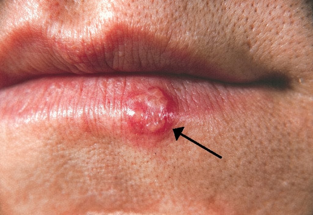
Using skin organoids, researchers in Germany have traced how herpes simplex virus 1 infection progresses among neighbouring skin cells. They found that the virus did not infect every type of skin cell in the same way, with fibroblasts in the dermis carrying large amounts of virus early on in the infection, progressing to hair follicles and sensory neurons only much later. The work, published in Science Advances, might ultimately help researchers understand why a common virus sometimes has serious consequences.
Most people think of herpes simplex virus 1 (HSV‑1) as a mere cosmetic problem: The virus can cause outbreaks of cold sores on the lips or face, which usually heal on their own within two weeks. But HSV‑1 infection can be more sinister. The virus infects sensory nerve cells in the face, where it usually remains dormant. In rare cases, it travels to the central nervous system and causes life-threatening brain inflammation. HSV‑1 is also being investigated for potentially playing a role in Alzheimer’s disease — its lifelong persistence in our nervous system may eventually damage nerve cells.
Dr Emanuel Wyler, a scientist in the RNA Biology and Posttranscriptional Regulation lab at the Berlin Institute for Medical Systems Biology of the Max Delbrück Center (MDC-BIMSB), has had a longstanding interest in understanding how the virus causes disease. Now he and collaborators at University Medical Center Hamburg-Eppendorf (UKE) have traced step by step how HSV‑1 infects the multiple layers of skin cells. Using human hair-bearing skin organoids as a model, the researchers mapped how the virus spreads and which signaling molecules cells release to their neighbors upon being infected.
The research team included Dr Manja Czech-Sioli, senior author of the paper at the UKE, Dr Silvia Albertini, co-first author with Wyler, and Dr Nicole Fischer. Albertini and Fischer are also scientists at UKE.
“It was fascinating to see how the virus spreads through the organoid tissue, from one cell type to another, and also how each cell type responds to it,” adds Czech-Sioli.
Tracking HSV‑1 in 3D skin
Skin is built like a layered sandwich. The protective outermost layer is the epidermis, which primarily consists of keratinocytes. Beneath it sits the dermis, a support layer rich in fibroblasts. Hair follicles pass through these layers, which also contain sensory nerve fibres.
The skin organoids provided by the groups of Manja Czech-Sioli and Nicole Fischer at UKE enabled the team to study infection in a complex, three-dimensional tissue model. While flat cell cultures can reveal how viruses behave in one cell type, these organoids contain many of the features of actual human skin: epidermal cells, dermal cells, hair follicles and nerve-like cells. Consequently, they serve as a better model of how HSV‑1 infection progresses.
The team found that the virus did not infect every type of skin cell in the same way. Fibroblasts in the dermis carried large amounts of virus early during infection. Basal keratinocytes, which sit in the lower epidermis, and several hair follicle cell types carried relatively lower amounts, even when infected cells were nearby. Only later did the virus reach hair follicles and sensory neurons, indicating that both cell types might have some intrinsic resistance to the virus.
How neighbouring cells react to infection
The researchers then combined imaging with spatial transcriptomics, which measures gene activity in individual cells while preserving their place in tissues. This allowed them to study the leading edge of infection – the zone where HSV‑1 had just reached new cells – and identify which genes were switched on.
At the infection front, dermal fibroblasts switched on IER3, a gene known to respond to HSV‑1, for example, and basal keratinocytes increased expression of IRF3, a regulator of antiviral defences. They also found that members of the Tumour Necrosis Factor (TNF) family of signaling molecules are activated in a spatially coordinated manner.
“TNFs activate the immune system and may prove to be a major player in determining how well our bodies can control the infection,” explains Wyler.
Future organoid models that include immune cells and blood vessels could help researchers study how the body controls HSV‑1 before it reaches nerves, why some cells slow the virus while others support replication, and how the infection and the subsequent inflammation damage tissue.
Text: Gunjan Sinha
Source: Max Delbrück Center
