Tag: trauma

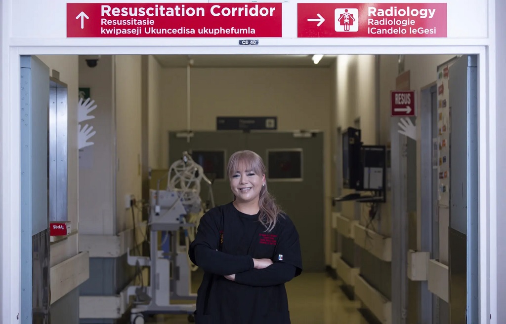
Between Silence and Sirens: Cape Town Trauma Surgeon Dr Deidre McPherson’s Midnight Vigils

Dr Deidre McPherson is one of ten women trauma surgeons in South Africa’s public healthcare sector. (Photo: Discovery Foundation)

By Biénne Huisman

Groote Schuur Hospital in Cape Town has one of the busiest emergency centres in the Western Cape. As it turns to the public to raise R20 million for the opening of a new emergency centre, Dr Deidre McPherson chats to Spotlight about the hospital’s trauma frontline.

Deep into the night while most of Cape Town is asleep, trauma surgeon Dr Deidre McPherson slips into work scrubs, hitting the highway to Groote Schuur Hospital to save the lives of critically injured patients.

In a boardroom next to the hospital’s Trauma Centre, McPherson details her solitary early-morning drives along the deserted N1 highway to perform life-saving surgery on people hurt in road accidents, gang violence, and other incidents.

She says she is called out from her home in Bellville past midnight at least once or twice a week. “It’s a surreal feeling,” she says. “I mean driving alone while the rest of the world is sleeping. By now, my husband is used to me leaving at weird times and coming back at like 03:00 or 05:00.”

In South Africa, trauma surgery only became a defined sub-speciality in 2008, meaning a formal training pathway for trauma surgery as its own discipline was created. Trauma surgeons are trained to manage multi-system injuries.

McPerson explains: “We are there at the most crucial moments, when life hangs in the balance. For me, there is nothing more rewarding than seeing a patient arrive critically injured, and walk out the hospital three weeks later, back to their lives.”

R20 million to equip new emergency centre

A new state-of-the-art emergency centre, which includes a new trauma centre, is being constructed at Groote Schuur, beside the existing facility. While it is set to open in 2026, hospital executives are turning to the public for R20 million in additional funding to fit the new premises with upgraded equipment.

As part of the fundraising drive, healthcare professionals recently took journalists on a candid tour of the existing facilities. Inside, corridors are clean but with linoleum floors peeling in places; some patients on trolley beds are stationed against walls, indicating wards filled to capacity.

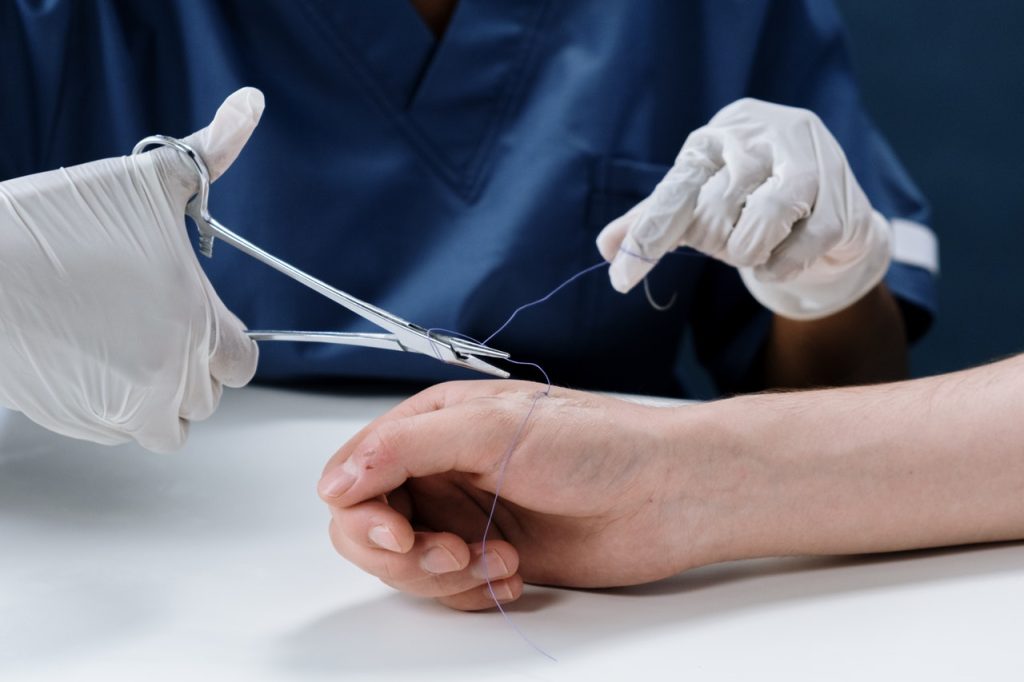
Just beyond a sign that reads “C14 Welcome to Trauma Centre” – with translations in Afrikaans and isiXhosa – McPherson points out the trauma centre’s resuscitation ward, which can hold six intubated patients, she says. One recent admission can be seen on life support.

Increased capacity and privacy for critically injured patients

McPherson says the new facility will have a more spacious assessment or triage area, where staff decide which patients require immediate life-saving care and which can safely wait.

She says the new trauma centre will expand capacity across all three colour-coded wards. The resuscitation ward (red) will increase from six to ten beds. “This is severe trauma, for example [patients involved in] a motor vehicle accident, with head injury, chest injury and fractures needing life support”. The intermediate ward (yellow) will increase from 12 to 16 beds. “This is moderate trauma, for example, [patients with] multiple fractures, but stable”. The minor ward (green) will increase from 12 to 14 beds. “This is minor trauma for example, [patients with] cuts, bites and bruises – the walking wounded”.

Through the public funding drive, they hope to upgrade the computer system, buy more mobile ventilators for critically injured patients, and get a new full-body X-ray machine for rapid imaging in seconds, which McPherson says is “critical for assessing multiple gunshot or high-impact injuries”.

She says that the centre’s current computer has been in use for over 15 years and frequently stalls. “Sandy, our secretary, is on the phone to IT every second week,” McPherson says, adding that it isn’t necessarily dangerous but that it’s very frustrating. “Time matters so much in trauma,” she emphasises.

In addition, there are lighting issues in some of the examination rooms, with doctors occasionally having to do sutures by headlamp or the flashlight on their phone, McPherson says.

A woman in a male dominated field

During our follow-up interview in the boardroom, McPherson’s gestures are soft, framing her words. Her eyes are level, her cadence precise and unaffected. At present, she is one of ten women trauma surgeons in South Africa’s public sector, compared to 22 men. She is the only woman of four trauma surgeons at Groote Schuur’s trauma centre, which is led by Professor Andrew Nicol.

“Surgery has always been male-dominated and even more so sub-specialties like trauma,” says McPherson. “I was discouraged from following this path by colleagues and even family. This is not a career for women, they said. What if I have a family? The hours are so unpredictable. And there are the violent things we see each day…”

But she was determined. For McPherson, it was a calling, a job she loves. “it doesn’t feel like work,” she says.

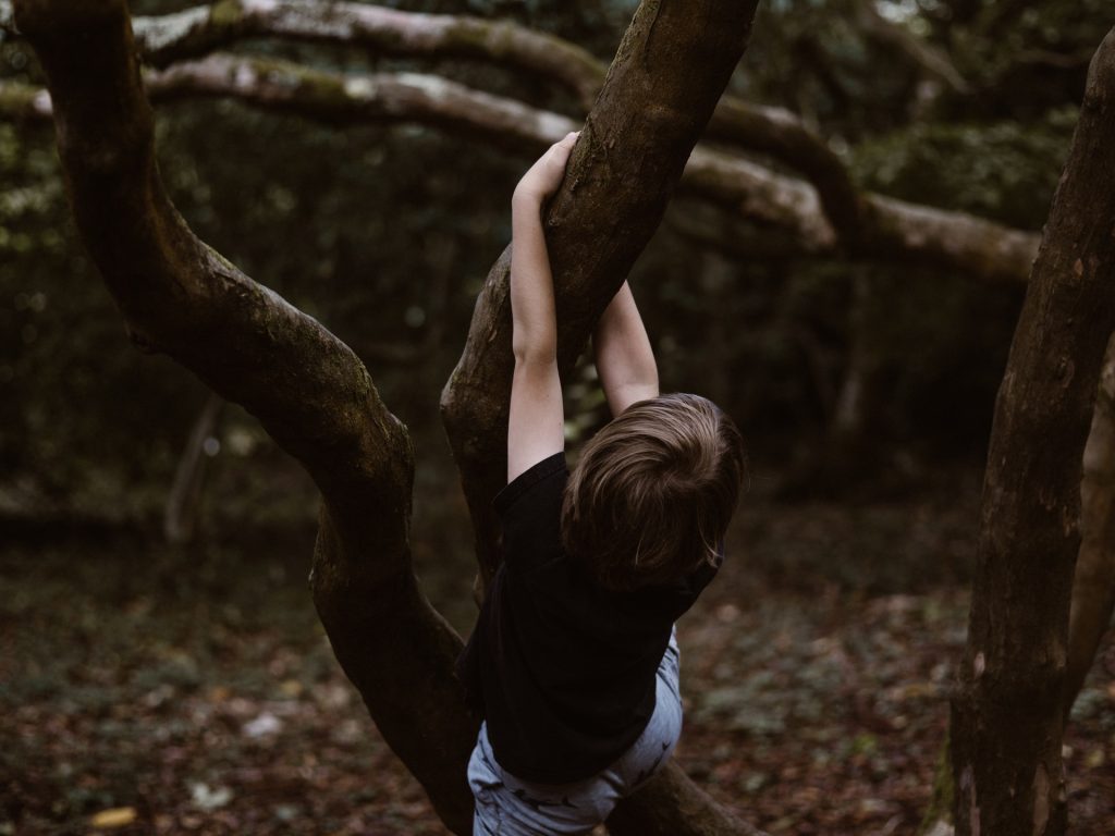
Road accidents and gang violence

On average, 1000 patients are admitted to Groote Schuur’s trauma centre per month. Critical injuries, particularly road accidents, spike around Easter, on public holidays, on pay day, and in December, she says. She suggests semigration to Cape Town has seen an additional traffic burden and increased road accidents. Another major contributing factor is accidents involving delivery motorcycle drivers.

In addition, August and September this year have seen a marked month-on-month increase in gunshot wounds, McPherson says, with up to three patients with firearm injuries admitted each day.

“On particularly violent days, that number can rise to as many as 10 patients in 24 hours,” she says.

“What is particularly striking is not just the frequency, but the severity. These are not single gunshot wounds – we often see patients who have sustained multiple injuries, sometimes up to 20 bullet wounds at once.”

This echoes damning murder statistics recently quoted in The Guardian, which notes six people aged from 19 to 25 shot dead over two days in Wallacedene and Eikendal, on the Cape Flats.

Responding, McPherson says: “Sadly what is described in The Guardian is not an isolated incident – it is our daily reality. At Groote Schuur Hospital, we feel that burden first-hand. Every day we are treating teenagers and those in their twenties – who should be building their futures, not fighting for their lives – in our resuscitation bays.”

The latest crime statistics from the South African Police Service lists four precincts on the Cape Flats among the country’s five police stations with the highest murder rates. From January to March this year, Delft had 66 murders, Mfuleni had 65, Nyanga had 63, and Philippi East had 59. This is topped only by Inanda in KwaZulu-Natal which had 74 murders. In each of the last three years over 25 000 people were murdered in South Africa.

This constant cycle of violence is devastating and disheartening, she says, particularly “the high rate of recidivism – when patients return again and again with new injuries”.

For McPherson, cases linked to gender-based violence are especially disturbing. “And yet, as trauma surgeons, we try to focus on what we can do in those critical moments: stop the bleeding, repair the injuries, and give our patients a second chance at life.”

Are there any solutions?

Ultimately, McPherson says the real solution to trauma lies “upstream” in prevention.

“This means tackling the drivers of violence: unemployment, poor housing, failing schools, and the lack of opportunities that trap so many young people in cycles of crime and despair. It also means building safer communities through stronger policing, a justice system that works, and meaningful gun control laws to reduce the number of firearms circulating in our neighbourhoods,” she says.

Then there is preventable road accidents.

“Road traffic injuries remain one of the leading causes of admissions to our unit. As we move into the festive season, I want to urge the public to take responsibility for one another: do not drink and drive, wear seatbelts, and slow down on the roads. These are simple actions that can save lives,” she says.

To this end, she points out the importance of South Africa’s “Arrive Alive” campaign which aims to decrease the number of lives lost on the country’s roads through raising public awareness of road safety. Western Cape officials estimate that 139 people died in road accidents in the province between 1 December 2024 and 11 January 2025, with 627 arrests made for drunk driving.

Childhood inspiration

Born in Bellville to parents who worked in education, the eldest of three sisters, McPherson’s interest in medicine started early, fuelled by a weekly booklet series called How My Body Works. “It was out every Friday, I couldn’t wait for it to arrive. These booklets sparked my fascination with biology and science and it has stayed with me ever since. I still have them at home, packed away in a box,” she says.

McPherson matriculated at Settler’s High School in Parow and studied medicine at Stellenbosch University. She completed her internship at Tygerberg Hospital with a community service year in Atlantis, on the West Coast, where she first saw “how daily violence devastates young people”.

A mother to three-year-old twins, a boy and a girl, McPherson scrolls on her phone to her WhatsApp profile picture, which features her children dressed up in tiny doctor’s scrubs – pink and blue – each with a tiny stethoscope. “It was ‘career day’ so we chose outfits that was easy,” she says, smiling.

McPherson, who also counts a PhD on her resume, says she has processed pangs of “mum guilt” for her children. “My husband has been a constant pillar of support,” she says.

“Plus, I am happy and fulfilled, my children are growing up with a happy mother – but yes, it’s a juggling act, sometimes I have to decide which ball to drop. Is it a rubber ball, that will bounce back, or a glass ball that might shatter?”

To relax, she says she likes to read “sappy romantic fiction” like novels by Danielle Steele.

On her future radar? Becoming a full professor.

In the meantime, McPherson says she believes every encounter is an opportunity to make a difference. “We don’t just treat the injuries, we also try to offer support and counselling, hoping that this time might change the trajectory of a life,” she says.

Republished from Spotlight under a Creative Commons licence.

Read the original article.

Unlocking Fast, Targeted Treatment for Trauma Injuries

Photo by Mat Napo on Unsplash

A groundbreaking study by researchers at Rutgers Health has uncovered a way to precisely identify and target trauma sites in the body within minutes of injury. The findings, published in the journal Med (Cell Press), could revolutionise emergency care by enabling real-time diagnostics and site-specific treatments delivered within minutes of injury.

A team of scientists, led by Renata Pasqualini and Wadih Arap at the Rutgers Cancer Institute discovered something new about how the body reacts to injury. When cells are damaged, like in a major bone break, calcium levels shift, which causes certain proteins to change shape. These changed proteins, called the “traumome,” are only found in injured tissues and show up right after an injury happens. This discovery opens up a new way to treat injuries directly, without affecting healthy parts of the body.

“The moment trauma occurs, specific proteins undergo structural changes, creating a molecular footprint of injury,” said Arap. “This opens the door to delivering diagnostics or therapies directly to the site – without affecting healthy tissues.”

This discovery has relevance in emergency treatment because many medicines can affect healthy organs when they’re given too soon. With this new approach, doctors could deliver treatments like imaging agents, clotting factors or antibiotics directly to the injured area, which would help the body heal faster with fewer side effects.    

“Our long-term vision is a simple injection that autonomously finds and treats injury sites,” said Pasqualini. “This could be transformative for battlefield medicine and emergency trauma care, where every second matters.”

The team used advanced testing on a pig model with major injuries to find tiny protein pieces called peptides. These peptides are like guides that can find and stick to the specific proteins altered by injury. One of these peptides stands out because it can attach to a protein that changes shape when calcium levels rise after an injury. This makes it possible to use special scans, like PET or MRI, to see exactly where the injury is in the body.

The trauma-targeting peptide worked the same way in rats, which shows that this injury “signature” is similar in all mammals, including humans.

The work was supported by the Defense Advanced Research Projects Agency (DARPA), an agency of the U.S. Department of Defense, underscoring its strategic value in both civilian and military medical applications. “Non-compressible bleeding remains a leading cause of death among soldiers before they reach a hospital, and localised treatment could dramatically improve survival rates, which was the original impetus of this research,” said Jon Mogford, a study co-author and former DARPA official.

The next phase of research will involve linking therapeutic agents to the trauma site-homing peptides and testing them in animal models before moving to early human clinical trials. The team envisions translational applications ranging from battlefield medicine to civilian trauma response and possibly even sports injuries or surgical recovery.

“We are actively developing peptide-linked drugs and imaging agents based on this discovery,” said Arap. “The traumome concept may also have applications beyond trauma, including in surgery, inflammation and tissue regeneration.”

Source: Rutgers University

Glucocorticoid Levels Influence the Development of PTSD after Trauma

Photo by Caleb Woods on Unsplash

Posttraumatic stress disorder (PTSD) is a debilitating condition that arises after experiencing traumatic events. While many people experience trauma, only about 25–35% of them develop PTSD. Understanding the factors that make certain individuals more susceptible is crucial for both prevention and treatment.

A new study led by Carmen Sandi and Simone Astori at EPFL now reveals how the development of PTSD is influenced by glucocorticoids, which are stress hormones such as cortisol. The work, which is published in Biological Psychiatry, provides significant insights into the behavioural and biological traits associated with PTSD vulnerability.

“There are considerable differences in the levels of glucocorticoids that individuals release to the bloodstream when stressed,” says Carmen Sandi. “Low glucocorticoid levels are frequently observed in PTSD patients following trauma exposure and were initially suspected to be a consequence of trauma exposure.”

She continues: “The possibility that this could be a trait constituting a pre-existing PTSD risk factor has been an outstanding open question for many years, but tackling it has been challenging due to the difficulties of both collecting biological measures before trauma exposure, and having access to relevant animal models in which the causal role of these traits can be investigated.”

To explore how a reduced hormonal response to stress might be linked to PTSD symptoms, the researchers used a genetically selected rat model that mimics people with blunted responses to cortisol. To do this, the team used MRI scans to measure the volume of different brain regions, trained rats to associate a cue with fear, recorded their sleep patterns, and measured their brain activity.

By combining these methods, the researchers discovered that a blunted responsiveness to glucocorticoids led to a “correlated multi-trait response” that includes impaired fear extinction (in males), reduced hippocampal volume, and rapid-eye movement sleep disturbances.

To explain the terms: Fear extinction is a process by which a conditioned fear response diminishes over time; problems with fear extinction are a hallmark of PTSD. Rapid-eye movement is crucial for memory consolidation, and disturbances in this type of sleep pattern have long been associated with PTSD.

But the study didn’t end there: the researchers treated the rats with the equivalent of human cognitive and behavioral therapy to reduce their learned fears. After that, they gave the rats corticosterone. As a result, both excessive fear and disturbances in rapid-eye movement sleep receded. Not only that, but the increased levels of the stress-related neurotransmitter norepinephrine in the brain also returned to normal.

“Our study provides causal evidence of a direct implication of low glucocorticoid responsiveness in the development of PTSD symptomatology following exposure to traumatic experiences, i.e., impaired fear extinction,” says Carmen Sandi. “In addition, it shows that low glucocorticoids are causally implicated in the determination of other risk factors and symptoms that were until now only independently related to PTSD.”

Silvia Monari, the study’s first author, adds: “In a nutshell, we present mechanistic evidence – previously missing – that having low glucocorticoids such as cortisol in humans is a condition for causally predisposed individuals to present all to-date vulnerability factors for developing PTSD, and causally involved in deficits to extinguish traumatic memories.”

Source: Ecole Polytechnique Fédérale de Lausanne

Scans of Brain Connectivity in Veterans Yield Objective Pain Measures

MRI images of the brain
Photo by Anna Shvets on Pexels

A brain connectivity study of military veterans discovered three unique brain subtypes potentially indicating high, medium, and low susceptibility to pain and trauma symptoms. This could constitute an objective measurement of pain and trauma susceptibility, possibly leading to personalised treatments and new therapies based on neural connectivity patterns.  

Comorbidity Goes Unexplored

“Chronic pain is a major public health concern, especially among veterans,” said first author Prof Irina Strigo. “Moreover, chronic pain sufferers almost never present with a single disorder but often with multiple co-morbidities, such as trauma, posttraumatic stress, and depression.”

It is already understood that both pain and trauma can affect brain connections, but this had not been studied in the context of comorbid trauma and pain. Much pain and trauma research also relies on subjective measurements, such as questionnaires, rather than objective measurements like brain scans. This study, published in Frontiers in Pain Research, addresses these problems.

Theresearchers studied a group of 57 veterans with both chronic back pain and trauma, who had quite varied symptoms in terms of pain and trauma severity. Functional MRI scans of the veterans’ brains showed the strength of connections between brain regions involved in pain and trauma. The researchers then used a statistical technique to automatically group the veterans based on their brain connection signatures, regardless of their self-reported pain and trauma levels.

Based on the veterans’ brain activity, they were sorted into three groups. Strikingly, these divisions were comparable to the severity of the veterans’ symptoms, and they fell into a low, medium, or high symptom group.

The team hypothesised that the pattern of brain connections found in the low symptom group allowed veterans to avoid some of the emotional fallout from pain and trauma, and also included natural pain reduction capabilities. Conversely, the high symptom group demonstrated brain connection patterns that may have increased their chances of anxiety and catastrophising when experiencing pain.

Interestingly, based on self-reported pain and trauma symptoms, the medium symptom group was largely similar to the low symptom group. However, the medium symptom group showed differences in their brain connectivity signature, which suggested that they were better at focusing on other things when experiencing pain, reducing its impact.

Putting the findings into future practice

“Despite the fact that the majority of subjects within each subgroup had a co-morbid diagnosis of pain and trauma, their brain connections differed,” said Prof Strigo.

“In other words, despite demographic and diagnostic similarities, we found neurobiologically distinct groups with different mechanisms for managing pain and trauma. Neurobiological-based subgroups can provide insights into how these individuals will respond to brain stimulation and psychopharmacological treatments.”

Thus far, it’s not known whether these neural hallmarks represent a vulnerability to trauma and pain or a consequence of these conditions. The technique does however provide an objective and unbiased hallmark of pain and trauma susceptibility or resilience, not reliant on subjective measures such as the surveys. In fact, subjective measurements of pain in this study would not differentiate between the low and medium groups.

Techniques using objective measures like brain connectivity appear more sensitive and could provide a clearer overall picture of someone’s resilience or susceptibility to pain and trauma, thereby guiding personalised treatment and paving the way for new treatments.

Source: Frontiers

Injectable Nanoparticles That Could Slow Internal Bleeding

Photo by Camilo Jimenez on Unsplash

Researchers at MIT have found the ideal size for injectable nanoparticles that could slow traumatic internal bleeding, buying more time for a patient to reach a hospital for further treatment.

In a rat study, the researchers showed that polymer nanoparticles particles in an intermediate size range, (about 150nm in diameter) were the most effective at stopping bleeding. These particles also were much less likely to travel to the lungs or other off-target sites, which larger particles often do. The results were published in ACS Nano.

“With nano systems, there is always some accumulation in the liver and the spleen, but we’d like more of the active system to accumulate at the wound than at these filtration sites in the body,” said senior author Paula Hammond, Professor at MIT.

Nanoparticles that can stop bleeding, also called haemostatic nanoparticles, can be made in a variety of ways. One of the most commonly used strategies is to create nanoparticles made of a biocompatible polymer conjugated with a protein or peptide that attracts platelets, the blood cells that initiate blood clotting.

In this study, the researchers used a polymer known as PEG-PLGA, conjugated with a peptide called GRGDS, to make their particles. Most of the previous studies of polymeric particles to stop bleeding have focused on particles ranging in size from 300–500nm. However, few, if any studies have systematically analysed how size affects the function of the nanoparticles.

“We were really trying to look at how the size of the nanoparticle affects its interactions with the wound, which is an area that hasn’t been explored with the polymer nanoparticles used as haemostats before,” Hong says.

Studies in animals have shown that larger nanoparticles can help to stop bleeding, but those particles also tend to accumulate in the lungs, which can cause unwanted clotting there. In the new study, the MIT team analysed a range of nanoparticles, including small (< 100nm), intermediate (140–220nm), and large (500–650nm).

They first analysed the nanoparticles in the lab to see how how they interacted with platelets in various conditions, to see how well platelets bound to them. They found that, flowing through a tube, the smallest particles bound best to platelets, while the largest particles stuck best to surfaces coated with platelets. However, in terms of the ratio particles to platelets, the intermediate-sized particles were the lowest.

“If you attract a bunch of nanoparticles and they end up blocking platelet binding because they clump onto each other, that is not very useful. We want platelets to come in,” said lead author, Celestine Hong, an MIT graduate student. “When we did that experiment, we found that the intermediate particle size was the one that ended up with the greatest platelet content.”

The researchers injected the different size classes of nanoparticles into mice to see how long they would circulate for, and where they would end up in the body. As with previous studies, the largest nanoparticles tended accumulated in the lungs or other off-target sites.

The researchers then used a rat model of internal injury to study which particles would be most effective at stopping bleeding. They found that the intermediate-sized particles appeared to work the best, and that those particles also showed the greatest accumulation rate at the wound site.

“This study suggests that the bigger nanoparticles are not necessarily the system that we want to focus on, and I think that was not clear from the previous work. Being able to turn our attention to this medium-size range can open up some new doors,” Prof Hammond said.

The researchers now hope to test these intermediate-sized particles in larger animal models, to get more information on their safety and the most effective doses. They hope that eventually, such particles could be used as a first line of treatment to stop bleeding from traumatic injuries long enough for a patient to reach the hospital.

Source: Massachusetts Institute of Technology

New Insights into First Stages of Wound Healing

Photo by cottonbro from Pexels

A new study from Vanderbilt University researchers has revealed how cells detect and react to wounds.

The epithelial cells which cover the body and its organs, must be able to heal wounds, as they are constantly exposed to insults and abrasion. “When these cells detect a wound nearby, they change their behaviours,” said study co-leader Professor Andrea Page-McCaw in the Department of Cell and Developmental Biology. “They transition from stationary, nondividing, noninvasive cells to cells that migrate, divide and invade.” This also describes the behaviors of cancer cells, which adopt wound-healing behaviours without any wound.

The researchers began with focusing on epithelial cells’ first known reaction of to a nearby wound: an increase in calcium levels, which typically occurs within a minute of wounding.

“We were able to connect the response of these cells directly to the cellular damage inherent in wounding,” Prof Page-McCaw said. “We found that wounds destroy cells, causing them to leak or even burst, and some of their contents get out. Outside of cells, tissues have a detector molecule ready to sense these cellular contents. When they do, proteases in the cellular contents chop up the detector molecule into smaller pieces, which spread to nearby cells. This activates receptors on the cells’ surfaces, giving them the information that a wound is nearby.”

Successful and efficient wound healing is key for recovery from trauma or surgery, and this study improves the understanding of how wounds are recognised by epithelial cells and how this leads to wound healing. This will help develop therapeutics that can address this health issue.

Slow wound healing time can be caused by a number of factors, such as diabetes, and can lead to infection and declining health. By figuring out how to downregulate these wound-healing behaviours in combination with other cancer interventions, this work offers insights that could help combat cancer’s adoption of this mechanism.

The researchers will next focus on how cells use the information they receive about the presence of a wound, specifically how the information is encoded in the calcium signal dynamics and then converted into migration, proliferation and changes in cell- and tissue-level mechanics. “Now that we have a solid understanding of how the presence of a wound is first signaled to nearby cells, we can ask a lot of interesting follow-up questions,” said study co-leader Shane Hutson, chair of the Department of Physics and Astronomy and professor of physics and biological sciences. “How much information is present in those signals? Can cells interpret the signals to know how large the wound is or how far they are from the wound? Do they use the way the dynamic signals change with time to make that measurement? What are the detailed mechanisms by which the signals then get turned into cellular actions?”

Source: Vanderbilt University

Journal information: James T. O’Connor et al, Proteolytic activation of Growth-blocking peptides triggers calcium responses through the GPCR Mthl10 during epithelial wound detection, Developmental Cell (2021). DOI: 10.1016/j.devcel.2021.06.020

Artificial Sweetener Delivers a Protective Carbon Monoxide Dose

Photo by Sharon McCutcheon on Unsplash

An oral prodrug has been developed which uses artificial sweeteners to deliver a protective carbon monoxide dose which protects against acute kidney injury.

Although carbon monoxide (CO) gas is toxic in large doses, with some 50 000 people suffering CO poisoning each year in the US, scientists have discovered it can reduce inflammation and protect cells against injury. The  protective effects of CO against injury in the kidneys, lungs, gastrointestinal tract and liver, among other organs has been shown in previous research. For the past five years, Wang and his collaborators have worked to design a safe way to deliver CO to human patients via prodrugs, which are inactive compounds that must undergo a chemical process in the body to release the active pharmacological agent. Their paper was published in Chemical Science.

Using two common artificial sweeteners, saccharine and acesulfame, as ‘carrier’ molecules for a prodrug, Prof Wang’s team were able to create an oral administration route for CO. They designed the molecules to release CO as they decomposed from water exposure. These are the first examples of orally active, organic CO prodrugs using a benign carrier that is approved by the Food & Drug Administration with a demonstrated safety profile.

“It’s difficult to deliver a gas, much less a poisonous gas, as a therapeutic to patients, and this work represents a pivotal step forward in developing alternative delivery forms,” said Prof Wang, a Georgia Research Alliance Eminent Scholar. “We wanted to work with a carrier that has a very well characterized safety profile, which confers a higher degree of certainty that it will be safe to use in a pill for human consumption.”

The scientists tested one of the prodrugs, CO-306, for pharmacological efficacy against acute kidney damage. CO-306, which uses saccharine as a carrier molecule, was administered to mice and it was found that it reduced biomarkers for kidney injury, indicating it could be developed working therapy. The type of kidney injury modelled mimicked those in humans that occur with extensive muscle damage, sickle cell disease, a common type of malaria, cardiopulmonary bypass surgery and severe sepsis.

Further animal model studies and safety assessments on CO-306 are planned by Wang and colleagues before they progress to human clinical studies. They also plan to test CO-306 for efficacy against other types of organ injuries.

Additionally, CO-based therapies hold promise as a method of reducing the likelihood of organ damage during transplantation and improving outcomes for transplant patients, according to Prof Wang.

“Science shows that exposing organs to CO gas can help preserve organs and prevent them from deteriorating during the process of transplantation,” he said. “Now we need to demonstrate that these prodrugs can have a similar effect.”

Source: Georgia State University

Journal information: De La Cruz, L. K., et al. (2021) Adapting decarbonylation chemistry for the development of prodrugs capable of in vivo delivery of carbon monoxide utilizing sweeteners as carrier molecules. Chemical Science. doi.org/10.1039/D1SC02711E.

New Biomarker for Soft Tissue Infections

This illustration depicted a three-dimensional (3D), computer-generated image, of a group of Gram-positive, Streptococcus pneumoniae bacteria. The artistic recreation was based upon scanning electron microscopic (SEM) imagery. Photo by CDC on Unsplash
This illustration depicted a three-dimensional (3D), computer-generated image, of a group of Gram-positive, Streptococcus pneumoniae bacteria. The artistic recreation was based upon scanning electron microscopic (SEM) imagery. Image by CDC on Unsplash

Researchers have identified a new and very promising biomarker for bacterial soft tissue infections, which previously lacked one. 

In bacterial soft tissue infections, rapid diagnosis is crucial in reducing the risk of severe injury or amputation. Vague symptoms and a varied patient presentations increase the risk of misdiagnosis.The study, by  Researchers at Karolinska Institutet in Sweden  and other research institutions, and published in the Journal of Clinical Investigation, may have implications for both diagnosis and treatment.

Last author Anna Norrby-Teglund, Professor, Department of Medicine, Karolinska Institutet, said: “There are currently no tools for safe, rapid diagnosis in life-threatening soft tissue infections. Our findings are consequently very interesting as the biomarkers identified are possible candidates for improved diagnostics. The results are also relevant for individualised treatment in the future.”

Necrotising soft tissue infections (NSTI) are bacterial infections which are characterised by rapid tissue degradation. Such infections, often caused by streptococci, while relatively uncommon, are extremely serious. In most cases they necessitate intensive care and can quickly become life-threatening.

Extensive surgery, intravenous antibiotics are often required to prevent the infection from spreading, and amputation may be required in extreme situations. Many patients also develop sepsis, which further complicates the course of the condition.

Early, correct diagnosis is crucial to save lives and avoid amputation, but this is complicated by factors such as vague symptoms including vomiting, fever and severe pain, as well as the heterogeneous group of patients. Despite recommendations for surgical evaluation in suspected NSTI, there is a considerable risk of misdiagnosis.

Currently, various laboratory tests, including white blood cell counts, are used as diagnostic tools, but suffer from low sensitivity. NSTI-specific biomarkers are therefore needed. The condition is classified into four types depending on the infecting organism.

Researchers at Karolinska Institutet, Haukeland University Hospital, Norway, and Copenhagen University Hospital, Denmark, have now been able to identify biomarkers specific to different patient groups with soft tissue infections.

Using machine learning, the researchers analysed 36 soluble factors in blood plasma from the 311 NSTI patients included in the international INFECT study. Control groups included patients with suspected NSTI and sepsis, respectively.

The analyses showed a new biomarker that accurately identifies patients with tissue necrosis.

“The new biomarker, thrombomodulin, proved to be superior to the laboratory parameters used clinically today. The analyses also identified biomarkers for patients with soft tissue infection caused by different types of bacteria, as well as patients who developed septic shock,” said first author Laura Palma Medina, researcher at the Department of Medicine, Karolinska Institutet (Huddinge).

Source: Karolinska Institutet

Journal information: Palma Medina, L.M., et al. (2021) Discriminatory plasma biomarkers predict specific clinical phenotypes of necrotizing soft-tissue infections. Journal of Clinical Investigation. doi.org/10.1172/JCI149523.

Self-inflicted Firearm Injuries Among Rural Youth Three Times Urban Rates

Photo by Annie Spratt on Unsplash

Emergency Department visits by youth for self-inflicted firearm injuries were three times more common in rural areas compared to urban ones, a national study has found.
The study, published in the Journal of Pediatrics found that Emergency Department (ED) visits by youth for self-harm were nearly 40 percent higher in rural areas compared to urban settings. Youth from rural areas presenting to the ED for suicidal ideation or self-harm also were more likely to need to be transferred to another hospital for care, which underscores the insufficient mental health resources in rural hospitals.

“Our study used pre-pandemic data, and we know that increased attention to youth mental health is even more pressing now everywhere, but especially in rural settings to prevent self-harm in youth,” said lead author Jennifer Hoffmann, MD, pediatric emergency medicine physician at Ann & Robert H. Lurie Children’s Hospital of Chicago and Assistant Professor of Pediatrics at Northwestern University Feinberg School of Medicine. “We need universal screening for suicidal ideation for all children and adolescents age 10 and up who present in the ED to identify youth at risk and intervene before tragedy occurs.”

The study drew on national data on suicidal ideation or self-harm in youth (ages 5-19 years) from a sample of EDs across the country, including those in general hospitals and children’s hospitals. The researchers extrapolated the results to arrive at national estimates.

Dr Hoffmann explained that a number of factors contribute to higher suicide rates and self-harm in rural youth. One of these is access to mental healthcare, which she said is a huge challenge. A lack of paediatric mental health professionals in rural areas is another factor, forcing patients to travel long distances for help. In addition, poor insurance coverage resulting from lower family income and unemployment. Small towns also have anonymity concerns, possibly delaying seeking care until a crisis brings the child to the ED. Firearm ownership is higher in rural firearms, so increased access to firearms may account for the high degree of disparity in self-inflicted firearm injuries.

“We need to improve mental health training for ED providers, allocate more resources and implement policies in rural hospitals on managing young patients who present with suicidal ideation or self-harm,” said Dr Hoffmann. “More widespread use of tele-psychiatry also might help prevent unnecessary transfers to other hospitals. But even more importantly, we need to train primary care providers to help diagnose and treat mental health issues earlier, so we can prevent self-inflicted injuries and death.”

Source: Ann & Robert H. Lurie Children’s Hospital of Chicago

A New Snake Venom ‘Super Glue’ For Wound Closures

Photo by David Clode on Unsplash

A novel snake venom ‘super glue’ has been developed, that can stop life-threatening bleeding in under a minute.

Over the past 20 years, bioengineer Kibret Mequanint, a professor at the University, has developed an array of biomaterials-based medical devices and therapeutic technologies – some of which are either now licensed to medical companies or are in the advanced stage of preclinical testing.

This latest work focuses on a blood clotting enzyme called reptilase or batroxobin, which is found in the venom of lancehead snakes (Bothrops atrox), which are amongst the most venomous snakes in South America.

Prof Mequanint and the international research team designed a body tissue adhesive that takes advantage of the clotting property of this enzyme, incorporating it into a modified gelatin that can be packaged into a small, handy tube for easy application.

“During trauma, injury and emergency bleeding, this ‘super glue’ can be applied by simply squeezing the tube and shining a visible light, such as a laser pointer, over it for a few seconds. Even a smartphone flashlight will do the job,” said Prof Mequanint.

Compared to the industry gold standard for clinical and field surgeons, clinical fibrin glue, the new tissue sealant has 10 times the adhesive strength to resist detachment or washout from bleeding. The blood clotting time is also much shorter, halving the 90 seconds for fibrin glue to 45 seconds for this new adhesive.

This novel biotechnology could reduce blood loss and save more lives. Tests were performed in models of major bleeding, such as deep skin cuts, ruptured aortae, and severely injured livers.

“We envision that this tissue ‘super glue’ will be used in saving lives on the battlefield, or other accidental traumas like car crashes,” said Prof Mequanint. “The applicator easily fits in first aid kits too.”

Besides its trauma application, the new snake venom ‘super glue’ can be used in surgical wound closures.

The study was published in the journal Science Advances..

“The next phase of study which is underway is to translate the tissue ‘super glue’ discovery to the clinic,” said Prof Mequanint.

Source: University of Western Ontario

Journal information: Guo, Y., et al. (2021) Snake extract–laden hemostatic bioadhesive gel cross-linked by visible light. Science Advances.doi.org/10.1126/sciadv.abf9635.