Tag: REM sleep

What Does Caffeine Do to the Sleeping Brain?

Photo by Mike Kenneally on Unsplash

Caffeine is one of the most widely consumed psychoactive substances in the world, present in tea, coffee, chocolate and energy drinks. In a study published in Nature Communications Biology, a team of researchers from Université de Montréal shed new light on how caffeine can modify sleep and influence the brain’s recovery, both physical and cognitive, overnight.

The research was led by Philipp Thölke, a research trainee at UdeM’s Cognitive and Computational Neuroscience Laboratory (CoCo Lab), and co-led by the lab’s director Karim Jerbi, a psychology professor and researcher at Mila – Quebec AI Institute.

Working with sleep-and-aging psychology professor Julie Carrier and her team at UdeM’s Centre for Advanced Research in Sleep Medicine, the scientists used AI and electroencephalography (EEG) to study caffeine’s effect on sleep.

They showed for the first time that caffeine increases the complexity of brain signals and enhances brain “criticality” during sleep.  Interestingly, this was more pronounced in younger adults.

“Criticality describes a state of the brain that is balanced between order and chaos,” said Jerbi.

“It’s like an orchestra: too quiet and nothing happens, too chaotic and there’s cacophony. Criticality is the happy medium where brain activity is both organised and flexible. In this state, the brain functions optimally: it can process information efficiently, adapt quickly, learn and make decisions with agility.”

Added Carrier: “Caffeine stimulates the brain and pushes it into a state of criticality, where it is more awake, alert and reactive. While this is useful during the day for concentration, this state could interfere with rest at night: the brain would neither relax nor recover properly.”

Nocturnal brain activity

To study how caffeine affects the sleeping brain, Carrier’s team recorded the nocturnal brain activity of 40 healthy adults using an electroencephalogram.  They compared each participant’s brain activity on two separate nights, one when they consumed caffeine capsules three hours and then one hour before bedtime, and another when they took a placebo at the same times.

“We used advanced statistical analysis and artificial intelligence to identify subtle changes in neuronal activity,” said Thölke, the study’s first author. “The results showed that caffeine increased the complexity of brain signals, reflecting more dynamic and less predictable neuronal activity, especially during the non-rapid eye movement (NREM) phase of sleep that’s crucial for memory consolidation and cognitive recovery.”

The researchers also discovered striking changes in the brain’s electrical rhythms during sleep: caffeine attenuated slower oscillations such as theta and alpha waves (generally associated with deep, restorative sleep) and stimulated beta wave activity, which is more common during wakefulness and mental engagement.

“These changes suggest that even during sleep, the brain remains in a more activated, less restorative state under the influence of caffeine,” says Jerbi, who also holds the Canada Research Chair in Computational Neuroscience and Cognitive Neuroimaging. “This change in the brain’s rhythmic activity may help explain why caffeine affects the efficiency with which the brain recovers during the night, with potential consequences for memory processing.”

People in their 20s more affected

The study also showed that the effects of caffeine on brain dynamics were significantly more pronounced in young adults between ages 20 and 27 compared to middle-aged participants aged 41 to 58, especially during REM sleep, the phase associated with dreaming.

Young adults showed a greater response to caffeine, likely due to a higher density of adenosine receptors in their brains. Adenosine is a molecule that gradually accumulates in the brain throughout the day, causing a feeling of fatigue.

“Adenosine receptors naturally decrease with age, reducing caffeine’s ability to block them and improve brain complexity, which may partly explain the reduced effect of caffeine observed in middle-aged participants,” Carrier said.

And these age-related differences suggest that younger brains may be more susceptible to the stimulant effects of caffeine. Given caffeine’s widespread use, the researchers stress the importance of understanding its complex effects on brain activity across different age groups and health conditions.

They add that further research is needed to clarify how these neural changes affect cognitive health and daily functioning, and to potentially guide personalised recommendations for caffeine intake.

Source: University of Montreal

The Pupil as a Window into the Sleeping Brain

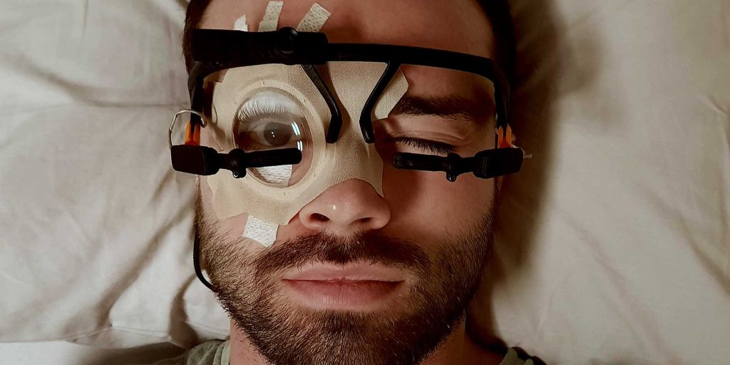
The eye of the sleeping subject was kept open with a special fixation device to record the pupil movements for several hours.  (Image: Neural Control of Movement Lab / ETH Zurich)

For the first time, researchers have been able to observe how the pupils react during sleep over a period of several hours. A look under the eyelids showed them that more happens in the brain during sleep than was previously assumed.

While eyes are typically closed in sleep, there is a flurry of activity taking place beneath the eyelids: a team of researchers, led by principal investigators Caroline Lustenberger, Sarah Meissner and Nicole Wenderoth from the Neural Control of Movement Lab at ETH Zurich, have observed that the size of the pupil fluctuates constantly during sleep. As they report in Nature Communications, sometimes it increases in size, sometimes it decreases; sometimes these changes occur within seconds, other times over the course of several minutes.

“These dynamics reflect the state of arousal, or the level of brain activation in regions that are responsible for sleep-wake regulation,” says Lustenberger. “These observations contradict the previous assumption that, essentially, the level of arousal during sleep is low.”

Instead, these fluctuations in pupil size show that even during sleep, the brain is constantly switching between a higher and lower level of activation. These new findings also confirm for humans what other research groups have recently discovered in studies on rodents, who also exhibit slow fluctuations in the activation level (known in the field as arousal).

New method for an old mystery

The regions of the brain which control the activation level are situated deep within the brainstem, making it previously difficult to directly measure these processes in humans during sleep. Existing methods are technically demanding and have not yet been established in this context. The ETH researchers’ study therefore relies on pupil measurements. Pupils are known to indicate the activation level when a person is awake. They can therefore be used as markers for the activity in regions situated deeper within the brain.

The ETH researchers developed a new method for examining the changes in people’s pupils while asleep: using a special adhesive technique and a transparent plaster, they were able to keep the eyes of the test subjects open for several hours.

“Our main concern was that the test subjects would be unable to sleep with their eyes open. But in a dark room, most people forget that their eyes are still open and they are able to sleep,” explains the study’s lead author, Manuel Carro Domínguez, who developed the technique.

Analysis of the data showed that pupil dynamics is related not just to the different stages of sleep, but also to specific patterns of brain activity, such as sleep spindles and pronounced deep sleep waves – brain waves that are important for memory consolidation and sleep stability. The researchers also discovered that the brain reacts to sounds with varying degrees of intensity, depending on the level of activation, which is reflected in the size of the pupil.

A central regulator of the activation level is a small region in the brainstem, known as the locus coeruleus. In animals, scientists have been able to show that this is important for the regulation of sleep stages and waking. The ETH researchers were unable to prove in this study whether the locus coeruleus is indeed directly responsible for pupil changes. “We are simply observing pupil changes that are related to the level of brain activation and heart activity,” Lustenberger explains.

In a follow-up study, the researchers will attempt to influence the activity of the locus coeruleus using medication, so that they can investigate how this affects pupil dynamics. They hope to discover whether this region of the brain is in fact responsible for controlling the pupils during sleep, and how changes in the level of activation affect sleep and its functions.

Using pupillary dynamics to diagnose illnesses

Understanding pupil dynamics during sleep could also provide important insights for the diagnosis and treatment of sleep disorders and other illnesses. The researchers therefore want to investigate whether pupil changes during sleep can provide indications of dysfunctions of the arousal system. These include disorders such as insomnia, post-traumatic stress disorder and possibly Alzheimer’s. “These are just hypotheses that we want to investigate in the future,” says Lustenberger.

Another goal is to make the technology usable outside of sleep laboratories, such as in hospitals where it could help to monitor waking in coma patients or to diagnose sleep disorders more accurately. The pupil as a window onto the brain could thus pave the way for new opportunities in sleep medicine and neuroscience.

Source: ETH Zurich

Why REM Sleep is Important in Animals

Photo by Bruce Christianson on Unsplash

Researchers in Japan have discovered that capillary blood flow in the brain is increased in mice during the dream-active REM phase of sleep, possibly preventing a buildup of waste products.

Scientists have long wondered why almost all animals sleep, despite the disadvantages to survival of being unconscious. Now, researchers led by a team from the University of Tsukuba have found new evidence of brain refreshing that takes place during a specific phase of sleep: rapid eye movement (REM) sleep, where dreaming occurs.

Previous studies have seen conflicting results when measuring differences in blood flow in the brain between REM sleep, non-REM sleep, and wakefulness using various methods. For this study, the investigators used a technique to directly visualise red blood cell movement in the brain capillaries of mice during awake and asleep states.

“We used a dye to make the brain blood vessels visible under fluorescent light, using a technique known as two-photon microscopy,” explained the senior study author, Professor Yu Hayashi. “In this way, we could directly observe the red blood cells in capillaries of the neocortex in non-anaesthetised mice.”

The researchers also measured electrical activity in the brain to identify REM sleep, non-REM sleep, and wakefulness, and looked for differences in blood flow between these phases.

“We were surprised by the results,” said Professor Hayashi. “There was a massive flow of red blood cells through the brain capillaries during REM sleep, but no difference between non-REM sleep and the awake state, showing that REM sleep is a unique state”

The research team then disrupted the mice’s sleep, resulting in ‘rebound’ REM sleep, which is a stronger form of REM sleep to compensate for the earlier disruption. During rebound REM sleep, blood flow was increased even further, suggesting an association between blood flow and REM sleep strength. However, when the researchers repeated the same experiments in mice without adenosine A2a receptors (blocking these receptors makes you feel more awake after a coffee), there was less of an increase in blood flow during REM sleep, even during rebound REM sleep.

“These results suggest that adenosine A2a receptors may be responsible for at least some of the changes in blood flow in the brain during REM sleep,” said Professor Hayashi.

Given that reduced blood flow in the brain and decreased REM sleep are correlated with the development of Alzheimer’s disease, in which waste products are seen to build up in the brain, this increased blood flow in the brain capillaries during REM sleep could be important for waste removal from the brain. This study highlights the role of adenosine A2a receptors in this process, perhaps leading to the development of new treatments for Alzheimer’s disease and other conditions.

Source: University of Tsukuba