Tag: hydrocephalus

Building Better Cerebrospinal Fluid Shunts for the Brain

Schematic of approach to simulating brain shunt fluid dynamics. Credit: Harvard SEAS

Millions of people worldwide suffer from hydrocephalus, a condition which recently received greater attention when Billy Joel announced his diagnosis. Treatment usually involves surgical placement of shunts to divert cerebrospinal fluid away, but this procedure often leads to complications, infections, and multiple re-treatments.  

Bioengineers in the Harvard John A. Paulson School of Engineering and Applied Sciences (SEAS) have now developed a new computational model to aid the creation of shunts tailored to individual patients’ anatomy and needs. The model combines brain anatomy, fluid flow, and biomolecular transport dynamics to simulate shunt performance with pinpoint accuracy.

The work was supported by federal funding from the National Science Foundation and published in Proceedings of the National Academy of Sciences. It was led by SEAS postdoctoral fellow Haritosh Patel, who works in the labs of Joanna Aizenberg, Professor of Materials Science at SEAS and Professor of Chemistry and Chemical Biology; and Venkatesh Murthy, Professor of Molecular and Cellular Biology and Director of the Center for Brain Science.

Repeat surgeries due to infection or obstruction

Tens of thousands of shunt procedures are performed annually in the U.S. — many of which are repeat surgeries due to the inserted devices becoming blocked or obstructed, or the patient suffering an infection.

“Some elderly patients told me they had had over 10 surgeries — one every two to three years,” Patel said. “We really wanted to understand why this was happening, and we realised that many of these obstructions and infections were tied to shunt designs that didn’t fully consider fluid dynamics as a fundamental part of their geometry. We noticed that the tubing geometry used in shunts closely resembles the kind of piping we rely on in household plumbing. While that simplicity has its advantages, we saw an opportunity to explore more creative, biomimetic solutions that better suit the complexity of the brain’s environment.”

Pursuing the problem from both a material and design perspective, the team quickly realized there was no universally accepted fluid flow model for the brain ventricle space to guide them. “Okay, well, we can’t test our devices in a model, so why don’t we first make a better model?” Patel said.

Computational tool simulates fluid flow in brain

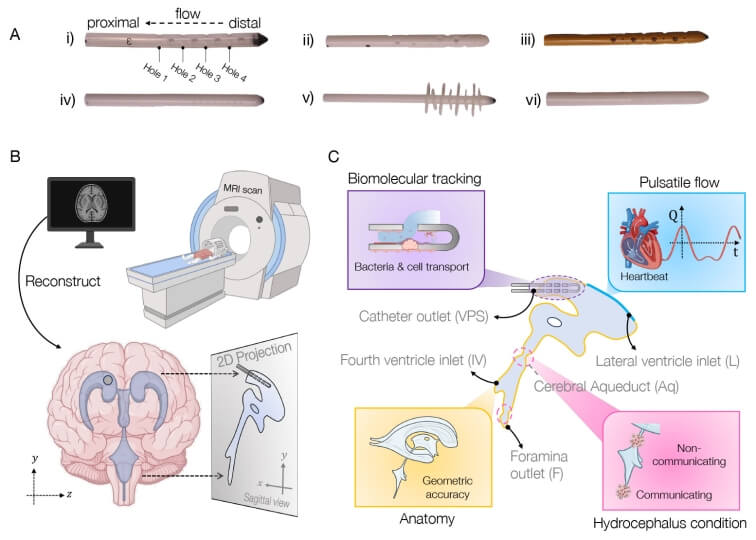
The result is their computational tool, called BrainFlow, which combines detailed anatomical and physiological features of the brain to simulate the flow of cerebrospinal fluid flow in the presence of shunt implants.

 The model incorporates patient-specific medical imaging data along with pulse-induced flow to mimic a patient’s cerebrospinal fluid dynamics, all to offer insight into optimal shunt design, placement, and even choice of materials.

“We believe that our model, combined with novel geometries and materials improvements such as anti-biofouling coatings developed in my lab, could lead to smoother integration of optimized, patient-specific medical devices into patients’ brains, with less likelihood of complications, and a better quality of life,” Aizenberg said.

The Harvard team is currently conducting studies that use the model to test different designs of shunts and calculate their efficacy.

Source: Harvard John A. Paulson School of Engineering and Applied Sciences

Diabetes Drug May Serve as Alternative Treatment Option for Hydrocephalus

Photo by Anna Shvets

A drug commonly used to treat type 2 diabetes may reduce excess fluid in the brains of patients with hydrocephalus, which could help treat the disease less invasively than current treatments, according to a Northwestern Medicine study published in the Journal of Clinical Investigation.

Normal pressure hydrocephalus occurs when excess cerebrospinal fluid builds up inside the skull and puts pressure on the brain. The cause of the condition is elusive and affects up to three percent of individuals over the age of 65, with symptoms including cognitive decline, difficulty walking and bladder problems.  

Patients are typically treated with permanent ventriculoperitoneal shunts, which are surgically implanted in the front or back of the skull and are connected to a valve that diverts excess cerebrospinal fluid away from the brain and into the abdomen where it is absorbed. The procedure has been shown to dramatically improve mobility, bladder control and cognitive functioning in patients with hydrocephalus, according to senior study author Stephen Magill, MD, PhD.

“It’s a great procedure because it’s one of the few things you can do that actually reverses these symptoms,” said Magill, who is assistant professor of Neurological Surgery.

There is, however, no pharmacological treatment currently approved to treat hydrocephalus. Additionally, nearly 20% of patients with normal pressure hydrocephalus also have type 2 diabetes and take sodium/glucose cotransporter 2 (SGLT2) inhibitors to manage their blood sugar, cardiovascular and kidney function, and weight loss.

Magill recently observed a reduction in the brain ventricle size in a patient with hydrocephalus who had a ventriculoperitoneal shunt surgically implanted and then began taking SGLT2 inhibitors to treat their type 2 diabetes. This phenomenon prompted Magill to further investigate the impact of SGLT2 inhibitors on ventricular size in patients with hydrocephalus.

“The medication inhibits a receptor found in the kidneys, which is where it works for diabetes. However, that receptor is also expressed in the choroid plexus, which is the structure in the brain that secretes the spinal fluid. Although this was known from animal studies, the clinical aspects of this biology have not been fully appreciated,” Magill said.

In the current study, three patients with hydrocephalus underwent CT scans both before and after surgery for ventriculoperitoneal shunts. After surgery, each patient began taking SGLT2 inhibitors for a medical indication and then underwent additional CT scans.

From analyzing these scans, Magill’s team discovered that all three patients showed a reduction in ventricle size as well as structural changes in their brains after starting SGLT2 therapy. One patient demonstrated dramatic ventricle size reduction due to ventricular collapse and required a shunt valve adjustment to reduce cerebrospinal fluid drainage.

“It’s a really interesting clinical observation because it raises the possibility that these medications could be used to treat normal pressure hydrocephalus in the future, which would normally require surgery,” Magill said.

Magill said the findings have sparked a new line of research in studying how SGLT2 inhibitors could help prevent hydrocephalus, adding that his team is now studying SGLT2 knockout mouse models to better understand the drug’s impact on ventricular size.

Their findings could ultimately inform new therapeutic strategies for treating normal pressure hydrocephalus as well as post-traumatic hydrocephalus, or the buildup of cerebrospinal fluid after traumatic brain injury, according to Magill.

“This sparks a new line of research on how normal pressured hydrocephalus develops, what causes it, how this protein works in creating and secreting spinal fluid, and has direct translational implications,” Magill said. “There’s a whole new avenue of potentially treating this disease that might save a patient from having surgery, and there’s always risks with surgery. It will also evolve our understanding of how these drugs work.”

Source: Northwestern Medicine

A Molecular Mechanism for Hydrocephalus may Enable a Non-surgical Treatment

MRI images of the brain
Photo by Anna Shvets on Pexels

Researchers at Massachusetts General Hospital have discovered a novel molecular mechanism behind the most common forms of acquired hydrocephalus – which could lead to the first non-surgical treatments for the life-threatening disease. Research in animal models uncovered a pathway through which infection or bleeding in the brain triggers inflammation, causing increased production of cerebrospinal fluid (CSF) by the choroid plexus and lead to swelling of the brain ventricles.

“Finding a nonsurgical treatment for hydrocephalus, given the fact neurosurgery is fraught with tremendous morbidity and complications, has been the holy grail for our field,” says Kristopher Kahle, MD, PhD, a paediatric neurosurgeon at MGH and senior author of the study in the journal Cell. “We’ve identified through a genome-wide analytical approach the mechanism that underlies the swelling of the ventricles which occurs after a brain bleed or brain infection in acquired hydrocephalus. We’re hopeful these findings will pave the way for approval of an anti-inflammatory drug to treat hydrocephalus, which could be a game-changer for populations in the US and around the world that don’t have access to surgery.”

Occurring in about 0.2% of births, acquired hydrocephalus is the most common cause of brain surgery in children, though it can affect people at any age. In underdeveloped regions where bacterial infection is the most prevalent form, hydrocephalus is often deadly for children due to the lack of surgical intervention. Brain surgery, where a shunt is implanted to drain fluid from the brain, is the only known treatment. But about half of all shunts in paediatric patients fail within two years of placement, according to the Hydrocephalus Association, requiring repeat neurosurgical operations and a lifetime of brain surgeries.

Pivotal to the process is the choroid plexus, the brain structure that routinely pumps cerebrospinal fluid into the four ventricles of the brain to keep the organ buoyant and injury-free within the skull. An infection or brain bleed, however, can create a dangerous neuroinflammatory response where the choroid plexus floods the ventricles with cerebral spinal fluid and immune cells from the periphery of the brain in a cytokine storm, swelling the brain ventricles.

“Scientists in the past thought that entirely different mechanisms were involved in hydrocephalus from infection and from haemorrhage in the brain,” explains co-author Bob Carter, MD, PhD, chair of the Department of Neurosurgery at MGH. “Dr Kahle’s lab found that the same pathway was involved in both types and that it can be targeted with immunomodulators like rapamycin, a drug that’s been approved by the US Food and Drug Administration for transplant patients who need to suppress their immune system to prevent organ rejection.”

MGH researchers are continuing to explore how rapamycin and other drugs which quell the inflammation seen in acquired hydrocephalus could be repurposed. “What has me most excited is that this noninvasive therapy could provide a way to help young patients who don’t have access to neurosurgeons or shunts,” says Kahle. “No longer would a diagnosis of hydrocephalus be fatal for these children.”

Source: Massachusetts General Hospital