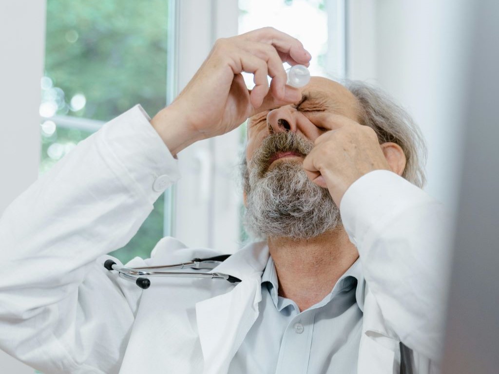
University of Pittsburgh School of Medicine researchers have developed an early-stage, experimental “living eye drop” that uses a naturally occurring eye bacterium to support corneal wound healing.
The proof-of-‑concept study, published in Cell Reports, demonstrates that the harmless eye-dwelling microbe Corynebacterium mastitidis can be genetically modified to secrete an anti-inflammatory therapeutic that promotes healing following corneal injury in a mouse model.
“This is the first demonstration that a microbe that lives on the ocular surface could be engineered to deliver a therapeutic that improves eye health,” said senior author Anthony St. Leger, associate professor of ophthalmology and of immunology and a faculty member of the UPMC Vision Institute. “It opens the door to the idea of ‘living medicine’ for the eye – something you apply once, and it stays, protects and helps the tissue heal.”
Because tears continually wash medications away, treating ocular surface disease often requires multiple daily applications of eye drops. This can limit the effectiveness of therapies for conditions such as corneal abrasions or dry eye disease.
To explore an alternative delivery method, the Pitt team engineered C. mastitidis, a benign bacterium that naturally resides under the eyelid, to continuously secrete cytokine interleukin10 (IL10). In mice, corneas that were gently scratched and treated with the engineered bacteria healed faster than those treated with regular bacteria or saline. When the IL10 receptor was blocked, this benefit disappeared – confirming the therapeutic effect was IL10-dependent.
The researchers also created a version of the microbe that releases human IL10, which improved wound closure in lab-grown cells that make up the outermost layer of human cornea and reduced inflammatory signaling in human immune cells. These studies offer an initial indication that the approach could eventually be adapted for use in people, though substantial development remains.
“What makes this exciting is that the system is modular,” St. Leger explained. “We built it so you can swap in different genes – different cytokines, growth factors or other proteins – to tailor the therapy to specific eye diseases.”
Though promising, the technology is still in early development. The researchers note that many steps must be completed before any clinical translation is possible, including developing built-in “off switches” to safely and reliably remove or deactivate the engineered bacteria after they are no longer needed.
Research shows that petrolatum-based eye ointments can cause the device to swell and potentially rupture, prompting an urgent update to clinical guidance.
Widely-used eye ointments can cause glaucoma implants to swell and potentially rupture, according to new research from Nagoya University in Japan. This study is the first to show, using clinical and experimental evidence, that petrolatum-based eye ointments can compromise the PRESERFLO® MicroShunt, an implant used in over 60 countries to treat glaucoma.
Glaucoma is an eye disease that damages the optic nerve and can lead to vision loss. It often results from increased intraocular pressure caused by blocked drainage of eye fluid. A recent study estimated that 76 million people globally are affected by glaucoma.
Progression of visual field loss (from left to right) due to glaucoma (Credit: Ryo Tomita)
MicroShunt is a small filtration device implanted in the eye to improve fluid drainage in glaucoma patients. Compared to traditional surgeries, it lowers post-operative complications and reduces reliance on additional medications.
MicroShunt is made from a styrenic thermoplastic elastomer based on a polystyrene-block-polyisobutylene-block-polystyrene (SIBS) block polymer, which is highly biocompatible, flexible, and less likely to cause inflammation or scarring. However, this material is vulnerable when it comes into contact with hydrocarbon- and oil-based materials. Due to its high oil affinity, exposure to petrolatum-based eye ointments may allow oil components to penetrate the device, causing swelling and potential changes in its shape and flexibility.
The MicroShunt manufacturer’s instructions state that “the MicroShunt should not be subjected to direct contact with petrolatum-based (ie, petrolatum jelly) materials, such as ointments and dispersions.” But this precaution is not widely recognised or consistently followed in clinical practice.
“Swollen MicroShunts can be structurally fragile,” said ophthalmologist and Assistant Professor Ryo Tomita of Nagoya University Graduate School of Medicine, the study’s first author. “During surgery, I observed a rupture in a swollen MicroShunt. If more clinicians are aware of this risk, they will be able to prevent similar problems.”
The clinical study examined seven glaucoma patients whose MicroShunt implants were later removed for different reasons. The results revealed a clear pattern. In three cases, the MicroShunt was exposed outside the conjunctiva, and patients received a petrolatum-based eye ointment. All three explanted devices showed significant swelling, and two of them ruptured.
In three other cases, the MicroShunt remained covered by the conjunctiva, and no ointment was administered. These devices retained their original structure. Crucially, in one additional case, the MicroShunt was exposed outside the conjunctiva, but no ointment was applied. The device did not swell. This indicates that direct contact with the ointment, rather than conjunctival rupture alone, is the primary cause of swelling.
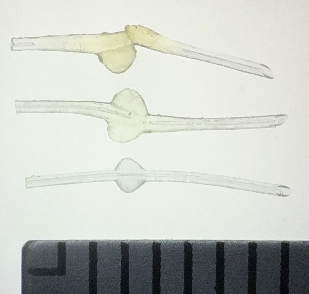
Photographic comparison of MicroShunt illustrating size changes Top: MicroShunt explanted from a patient, exhibiting diffuse swelling with fracture and loss of one fin Middle: MicroShunt explanted from another patient, showing localized swelling around the fin Bottom: Unused MicroShunt (control) Scale: 1 division = 1 mm (Credit: Ryo Tomita)
Laboratory confirmation
Laboratory experiments confirmed the clinical findings. The team immersed unused MicroShunts in petrolatum-based eye ointment to reproduce the swelling seen in clinical cases. Microscopic measurements showed significant changes. After 24 hours in the ointment, the MicroShunt’s outer diameter increased to 1.44 times its original size, and the fin-like portion widened to 1.29 times its initial value.
Chemical analysis identified the cause of this change. After 24 hours of immersion, oil components made up approximately 45% of the MicroShunt’s total weight, rising to 73% after three months. These results confirmed the primary cause of swelling to be the absorption of oil-based ointment constituents into the material.
Clinical implications
The research team emphasises that clinicians should avoid using petrolatum-based ointments on patients with MicroShunt implants, particularly when the device is exposed outside the conjunctiva. Alternative post-operative treatments should be considered, while further research is needed to assess whether swelling impacts MicroShunt performance even when rupture does not occur.
“Our study found that commonly used medical materials can cause unexpected complications if their chemical properties and usage environments are not fully understood,” Noro stated. “From both medical and engineering perspectives, we emphasise the importance of understanding the chemical properties of medical materials and appropriately managing their usage environments.”
Paper information:
Ryo Tomita, Taiga Inooka, Takato Kajita, Hideyuki Shimizu, Ayana Suzumura, Jun Takeuchi, Tsuyoshi Matsuno, Hidekazu Inami, Koji M. Nishiguchi, Atsushi Noro, and Kenya Yuki. (2026) Petrolatum-based ointment application induces swelling of the PRESERFLO MicroShunt. Graefe’s Archive for Clinical and Experimental Ophthalmology DOI: 10.1007/s00417-025-07075-2
Retina showing reticular pseudodrusen. Although they can infrequently appear in individuals with no other apparent pathology, their highest rates of occurrence are in association with age-related macular degeneration (AMD), for which they hold clinical significance by being highly correlated with end-stage disease sub-types, choroidal neovascularisation and geographic atrophy. Credit: National Eye Institute
A new compound potentially could offer an alternative to injections for the millions of people who suffer from wet age-related macular degeneration (AMD). The condition causes vision loss due to the uncontrolled growth and leakage of blood vessels in the back of the eye. A new paper in Cell Reports Medicine finds that a small-molecule inhibitor can reverse damage from AMD and promote regenerative and healing processes.
The drug can also be delivered via eyedrops – an improvement over current treatments for AMD, which require repeated injections into the eye.
“The idea was to develop something that can be more patient-friendly and doesn’t require a visit to the doctor’s office,” said lead researcher Yulia Komarova, associate professor of pharmacology at University of Illinois Chicago.
Komarova’s compound targets the protein End Binding-3 in endothelial cells, which line the inside of blood vessels. In the new study, the researchers looked at whether inhibiting EB3 function could stop the damaging leakage associated with wet AMD.
Using computational drug design methods, the team developed a small molecule drug, End Binding-3 inhibitor (EBIN), that could be delivered externally via eyedrops instead of by injection. They then tested its effectiveness in animal models of wet AMD, finding that twice-daily treatment reduced eye damage within 2–3 weeks.
Further investigation found that the inhibitor worked by rolling back aging-related genetic modifications. Aging causes inflammation and hypoxia in the eye that leads to changes in gene expression associated with the cellular effects and symptoms of wet AMD. Komarova and colleagues found that the EB3 inhibitor they developed reversed these epigenetic changes, restoring gene expression to a normal, healthy state.
“We reduce the effects of the stressor on endothelial cells and we improve regenerative processes, accelerating healing,” Komarova said. “That can be tremendous for the function of the cells.”
Because blood vessel leakage and hypoxic stress also drive many other medical conditions, Komarova’s group is interested in testing the inhibitor in models of acute lung injury, diabetic retinopathy, stroke, heart disease and even the general effects of aging on the brain. They are also exploring whether an implantable lens, similar to a contact lens, could deliver the drug to the eye more effectively than eyedrops.