Tag: bone fractures

‘Glue Gun’ Can 3D Print Bone Grafts Directly onto Fractures

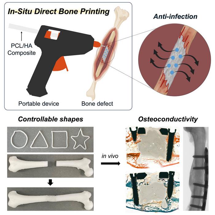
Graphical abstract. Credit: Device / Jeon et al. CC BY-SA

Scientists have developed a tool made from a modified glue gun that can 3D print bone grafts directly onto fractures and defects during surgery. The tool, described September 5th in the Cell Press journal Device, has been tested in rabbits to quickly create complex bone implants without the need for prefabricating in advance. What’s more, the team optimised the 3D-printed grafts for high structural flexibility, release of anti-inflammatory antibiotics, and promotion of natural bone regrowth at the grafting site.

Historically, bone implants have been made of metal, donor bone, or even more recently 3D-printed material. However, in cases involving irregular bone breaks, these implants must be designed and produced prior to surgery to allow for appropriate fitting.

“Our proposed technology offers a distinct approach by developing an in situ printing system that enables a real-time fabrication and application of a scaffold directly at the surgical site,” says Jung Seung Lee, co-author and associate professor of biomedical engineering at Sungkyunkwan University. “This allows for highly accurate anatomical matching even in irregular or complex defects without the need for preoperative preparation such as imaging, modelling, and trimming processes.”

The material fed into the glue gun is a filament comprised of two major components: a feature of natural bone known to promote healing called hydroxyapatite (HA) and a biocompatible thermoplastic called polycaprolactone (PCL). PCL can liquify in temperatures as low as 60°C, which when applied with a heat-modified glue gun, is cool enough to prevent tissue damage during surgical application while being able to conform to the jagged grooves of fractured bone. By adjusting the proportion of HA to PCL within the filament, the team can customise the hardness and strength of the grafts to fit different anatomical needs.

“Because the device is compact and manually operated, the surgeon can adjust the printing direction, angle, and depth during the procedure in real time,” says Lee. “Also, we demonstrated that this process could be completed in a matter of minutes. This highlights a significant advantage in terms of reducing operative time and improving procedural efficiency under real surgical conditions.”

Since infection is a common concern with surgical implants, the researchers incorporated vancomycin and gentamicin, two anti-bacterial compounds, into the filament. In both petri dish culture and liquid medium, the filament scaffold successfully inhibited the growth of E. coli and S. aureas, two common bacteria prone to cause infection post-surgery. Due to physical properties of HA and PCL within the filament, the drugs are released slowly and are able to diffuse directly onto the surgical site over several weeks.

“This localised delivery approach offers meaningful clinical advantages over systemic antibiotic administration by potentially reducing side effects and limiting the development of antibiotic resistance, while still effectively protecting against postoperative infection,” says Lee.

As a proof of concept, the device was tested on severe femoral bone fractures in rabbits. Within 12 weeks after surgery, the team found no signs of infection or necrosis and greater bone regeneration outcomes when compared to rabbits grafted with bone cement – a sealing compound commonly used for treating bone defects.

“The scaffold was designed not only to integrate biologically with surrounding bone tissue but also to gradually degrade over time and be replaced by newly formed bone,” says Lee. “The results showed that the printing group exhibited superior outcomes in key structural parameters such as bone surface area, cortical thickness, and polar moment of inertia, suggesting more effective bone healing and integration.”

Next, the team is setting their sights on optimising the anti-bacterial potential of the scaffold even further and preparing the procedure for human trials.

Source: EurekAlert!

Repurposed Multiple Sclerosis Drug Could Help Bones Heal Faster

Photo by Tima Miroshnichenko on Pexels

Researchers at the University of Arizona College of Medicine – Tucson found evidence that a drug that improves the ability to walk in people with multiple sclerosis can also make bone fractures heal faster.

The findings help further the understanding of specific factors involved in the bone healing process, and potentially open avenues for new therapeutic approaches.

“Broken bones are typically slow to heal in many people, and they can impact lives for months and in different ways. People lose time at work and daily activities at home with family and friends are impacted,” said senior author John Elfar, MD, professor, surgeon and chair of the Department of Orthopaedic Surgery at the U of A College of Medicine – Tucson. “This drug has the potential to change that.” 

Elfar partnered with Prem Kumar Govindappa, PhD, DVM, an assistant professor in the department, on the preclinical study that showed treatment with the drug 4-aminopyridine, or 4-AP, resulted in leg fractures healing faster and stronger than without the drug. The paper was published in The Journal of Bone and Joint Surgery.

“Mice with bone fractures healed quicker and were stronger after they healed after treatment with 4-AP,” said Elfar said, who is a member of the university’s BIO5 Institute. “We saw more bone mass and less intermediate cartilage, meaning there was accelerated bone healing.” 

The drug is approved for use in chronic neurological conditions, where it helps with walking by improving how signals from the brain and spinal cord reach limbs.

The team also saw improvements in bone mass and the ability to bear weight after treatment with 4-AP. Collagen deposition and bone mineralization, both of which are necessary for bone healing, also received a boost. Collagen forms the structural foundation of bones. In bone mineralization, minerals like calcium and phosphate join the newly forming bone matrix, strengthening and hardening the bone.

“We found that every fine-tuned measure of the strength of bone was better after administering 4-AP to mice,” Elfar said. “We also found more BMP2 protein in bone-forming cells at the fracture site, which again told us we found something that could accelerate the process.”

Examining human bone cells exposed to 4-AP in a dish, the scientists saw increased production of bone morphogenetic protein, or BMP2, a bone-building substance used clinically to help with some kinds of bone repair. BMP2 prompted the production of stem cells that become cells called osteoblasts, which are essential to form new bone.

The research team also measured 4-AP’s effects on human bone narrow mesenchymal stem cells and human osteoblast cells in the lab. 4-AP increased the conversion of the stem cells into osteoblasts and the latter’s ability to migrate and grow, which are essential to the healing process.

Elfar said that 4-AP’s role in driving BMP2 gene and protein activity is key to its bone healing effects, and using 4-AP to prompt BMP2 production in the body could be especially important.

“BMP2 is a hormone the body makes to accelerate bone healing,” Elfar said. 

BMP2 is known to modulate bone healing and is approved for use in certain medical procedures, including spinal fusion and sinus reconstruction surgery. An artificial version that has orthopedic medicine uses can have side effects, though, including bone resorption and cervical spine swelling. Finding a way to channel naturally produced BMP2 could improve bone healing while avoiding such problems.

The scientists previously showed that 4-AP could prevent bone and muscle loss in a mouse model of nerve damage. Similarly, they saw indications of 4-AP’s healing effects for wound, nerve and limb injuries. 

The researchers plan to eventually test 4-AP’s potential use in healing bones in a clinical trial. They also want to better understand the drug’s effects on BMP2 production, and more broadly, on the biology of healing bone.

Source: University of Arizona

A Revolutionary Coral-inspired Material for Bone Repair

(Left) An image of a 3D-printed material implanted in vivo for 4 weeks. The photo was taken using a scanning electron microscope. Credit: Dr Zhidao Xia.
(Right) A photo of coral. Credit: Jesus Cobaleda.

Researchers at Swansea University have developed a revolutionary bone graft substitute inspired by coral which not only promotes faster healing but dissolves naturally in the body after the repair is complete.

This groundbreaking research, led by Dr Zhidao Xia from Swansea University Medical School in collaboration with colleagues from the Faculty of Science and Engineering and several external partners, has been patented and published in the leading journal Bioactive Materials.

Bone defects caused by conditions like fractures, tumours, and non-healing injuries are one of the leading causes of disability worldwide. Traditionally, doctors use either a patient’s own bone (autograft) or donor bone (allograft) to fill these gaps. However, these methods come with challenges, including a limited supply, the risk of infection and ethical concerns.

By using advanced 3D-printing technology, the team have developed a biomimetic material that mimics the porous structure and chemical composition of coral-converted bone graft substitute, blending perfectly with human bone and offering several incredible benefits:

  • Rapid Healing – It helps new bone grow within just 2–4 weeks.
  • Complete Integration – The material naturally degrades within 6–12 months after enhanced regeneration, leaving behind only healthy bone.
  • Cost-Effective – Unlike natural coral or donor bone, this material is easy to produce in large quantities.

In preclinical in vivo studies, the material showed remarkable results: it fully repaired bone defects within 3–6 months and even triggered the formation of a new layer of strong, healthy cortical bone in 4 weeks.

Most synthetic bone graft substitutes currently on the market can’t match the performance of natural bone. They either take too long to dissolve, don’t integrate well, or cause side effects like inflammation. This new material overcomes these problems by closely mimicking natural bone in both structure and biological behaviour.

Dr Xia explained: “Our invention bridges the gap between synthetic substitutes and donor bone. We’ve shown that it’s possible to create a material that is safe, effective, and scalable to meet global demand. This could end the reliance on donor bone and tackle the ethical and supply issues in bone grafting.”

Innovations like this not only promise to improve patient quality of life but also reduce healthcare costs and provide new opportunities for the biomedical industry.

The Swansea University team is now looking to partner with companies and healthcare organisations to bring this life-changing technology to patients around the world.

Source: Swansea University

AI Helps Clinicians to Assess and Treat Leg Fractures

Photo by Tima Miroshnichenko on Pexels

By using artificial intelligence (AI) techniques to process gait analyses and medical records data of patients with leg fractures, researchers have uncovered insights on patients and aspects of their recovery.

The study, which is published in the Journal of Orthopaedic Research, uncovered a significant association between the rates of hospital readmission after fracture surgery and the presence of underlying medical conditions. Correlations were also found between underlying medical conditions and orthopaedic complications, although these links were not significant.

It was also apparent that gait analyses in the early postinjury phase offer valuable insights into the injury’s impact on locomotion and recovery. For clinical professionals, these patterns were key to optimising rehabilitation strategies.

“Our findings demonstrate the profound impact that integrating machine learning and gait analysis into orthopaedic practice can have, not only in improving the accuracy of post-injury complication predictions but also in tailoring rehabilitation strategies to individual patient needs,” said corresponding author Mostafa Rezapour, PhD, of Wake Forest University School of Medicine. “This approach represents a pivotal shift towards more personalised, predictive, and ultimately more effective orthopaedic care.”

Dr. Rezapour added that the study underscores the critical importance of adopting a holistic view that encompasses not just the mechanical aspects of injury recovery but also the broader spectrum of patient health. “This is a step forward in our quest to optimize rehabilitation strategies, reduce recovery times, and improve overall quality of life for patients with lower extremity fractures,” he said.

Source: Wiley

Vitamin D Supplements don’t Prevent Fractures in Children

Photo by cottonbro studio

A major clinical trial has found that vitamin D supplements do not increase bone strength or prevent bone fractures in children with vitamin D deficiency. The findings, published in Lancet Diabetes & Endocrinology, challenge widely held perceptions relating to the effects of vitamin D on bone health.

Around one-third of children have at least one fracture before the age of 18. This is a major global health issue, as childhood fractures can lead to life years of disability and/or poor quality of life. The potential for vitamin D supplements to improve bone strength has attracted growing interest in recent years, based on vitamin D’s role in promoting bone mineralisation. But there have been no clinical trials to test whether vitamin D supplements can prevent bone fractures in children.

Working with partners in Mongolia, a setting with a particularly high fracture burden and where vitamin D deficiency is highly prevalent, researchers from led by Queen Mary University of London and the Harvard T.H. Chan School of Public Health conducted a clinical trial to determine if vitamin D supplementation would decrease risk of bone fractures or increase bone strength in schoolchildren.

This study is also the largest randomised controlled trial of vitamin D supplementation ever conducted in children. Over the course of three years, 8851 schoolchildren aged 6-13 living in Mongolia received a weekly oral dose of vitamin D supplementation.

Testing revealed that 95.5% of participants had vitamin D deficiency at baseline, and study supplements were highly effective in boosting vitamin D levels into the normal range. No effect was seen on fracture risk or on bone strength, measured in a subset of 1438 participants using quantitative ultrasound.

The trial findings are likely to prompt scientists, doctors and public health specialists to re-consider the effects of vitamin D supplements on bone health.

Dr Ganmaa Davaasambuu, Associate Professor at the Harvard T.H. Chan School of Public Health, said:

“The absence of any effect of sustained, generous vitamin D supplementation on fracture risk or bone strength in vitamin D deficient children is striking. In adults, vitamin D supplementation works best for fracture prevention when calcium is given at the same time – so the fact that we did not offer calcium alongside vitamin D to trial participants may explain the null findings from this study.”

Professor Adrian Martineau, Lead of the Centre for Immunobiology at Queen Mary University of London, added:

“It is also important to note that children who were found to have rickets during screening for the trial were excluded from participation, as it would not have been ethical to offer them placebo (dummy medication). Thus, our findings only have relevance for children with low vitamin D status who have not developed bone complications. The importance of adequate vitamin D intake for prevention of rickets should not be ignored, and UK government guidance recommending a daily intake of 400 IU vitamin D remains important and should still be followed.”

Source: Queen Mary University of London

Aspirin as Effective as Heparin for Clot Prevention in Bone Fracture Hospitalisation

Source: Mat Napo on Unsplash

Patients hospitalised with fractures typically receive low-molecular-weight heparin to prevent life-threatening blood clots. A new clinical trial, however, found that inexpensive over-the-counter aspirin is just as effective. The findings, published today in the New England Journal of Medicine, may lead surgeons to change their practice and administer aspirin to these patients.

With more than 12 000 patients, the multi-centre randomised clinical trial is the largest trial ever conducted on orthopaedic trauma patients. This multidisciplinary collaboration between orthopaedic surgeons and trauma surgeons points to the importance of evaluating techniques used to prevent post-surgical complications, like blood clots and infections, through high-quality, head-to-head comparison studies.

“Many patients with fractures will likely strongly prefer to take a daily aspirin over receiving injections after we found that both give them similar outcomes for prevention of the most serious outcomes from blood clots,” said the study’s principal investigator Robert V. O’Toole, MD. “We expect our findings from this large-scale trial to have an important impact on clinical practice that may even alter the standard of care.”

Patients who experience fractures that require surgery are at increased risk of developing blood clots, including life-threatening pulmonary embolisms. Current guidelines recommend prescribing low-molecular-weight heparin (enoxaparin) to prevent these clots, although smaller clinical trials in total joint replacement surgery suggested a potential benefit of aspirin as a less-expensive, widely available option.

The study enrolled 12 211 patients with leg or arm fractures that necessitated surgery or pelvic fractures regardless of the treatment. Half were randomised to 30mg of injectable enoxaparin twice daily. The other half received 81mg of aspirin twice daily. Patients were followed for 90 days to measure health outcomes from the two treatments.

The main finding of the study was that aspirin was “non-inferior,” or no worse than low molecular-weight heparin in preventing death from any cause – 47 patients in the aspirin group died, compared with 45 patients in the heparin group. For other important complications, the researchers also found no differences in pulmonary embolisms between the two groups. The incidence of bleeding complications, infection, wound problems, and other adverse events from the treatments was also similar in both groups.

Of all the outcomes studied, the only potential difference noted was in deep vein thrombosis. This condition was relatively uncommon in both groups as it occurred in 2.5% of patients in the aspirin group, and in 1.7% of patients in the heparin group.

“This relatively small difference was driven by clots lower in the leg, which are thought to be of less clinical significance and often do not require treatment,” said study co-principal investigator Deborah Stein, MD, MPH.

“Many patients don’t like giving themselves injections. It’s not fun in terms of giving the actual injection because it burns, and your stomach tends to bruise more easily compared to aspirin,” said Debra Marvel, a 53-year-old from Columbia, MD, who served as a patient advisor on the study. She received Lovenox (low-molecular-weight heparin) after her legs were crushed in a 2015 pedestrian accident, requiring multiple surgeries at the University of Maryland Shock Trauma Center. “Patients also prefer aspirin because Lovenox can be expensive based on insurance.”

“An estimated one million Americans are hospitalised each year with extremity fractures, and this new finding could help prevent potentially fatal blood clots in these patients using a medication that is cheaper and far easier to administer,” said Mark T. Gladwin, MD, Vice President for Medical Affairs, University of Maryland, Baltimore, and the John Z. and Akiko K. Bowers Distinguished Professor and Dean, University of Maryland School of Medicine. “Given these important results, we can expect the guidelines for the prevention of blood clots to be revised to include the option of aspirin for patients with traumatic bone fractures.”

Source: University of Maryland School of Medicine

Obesity in Women Linked to Increased Fracture Risk

Obesity
Image source: Pixabay CC0

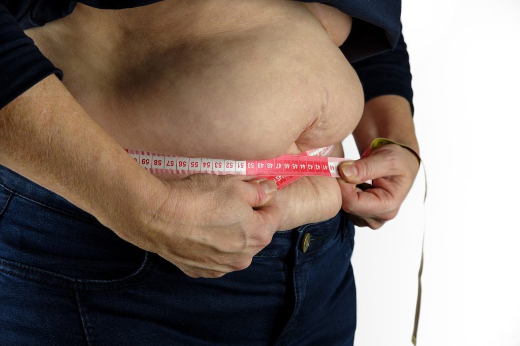
Women with obesity and overweight, particularly women with high waist circumference, are more susceptible to fractures than those with normal weight, according to new research presented at the European Congress on Obesity (ECO). In men, however, underweight, not overweight, is associated with a greater risk of broken bones.

Obesity has long been thought to help protect against fractures. This is because mechanical loading on bones, which increases with body weight, helps increase bone mineral density, an important determinant of bone strength.

However, recent studies have suggested that the relationship between obesity and fracture risk varies depending on sex, the skeletal site studied and definition of obesity used (body mass index [BMI] vs waist circumference).

To find out more, Dr Anne-Frederique Turcotte, Endocrinology and Nephrology Unit, CHU de Quebec Research Centre, Quebec City, Canada, and colleagues, analysed data from CARTaGENE, a prospective population-based cohort of almost 20 000 individuals aged 40-70 years from Quebec, Canada.

In women, greater waist circumference (WC) was linearly associated with an increased risk of fracture. For each 5cm (two inch) increase in WC, the risk of fracture at any site was 3% higher and the risk of a distal lower limb fracture was 7% higher.  The association between WC and ankle fractures was particularly strong.

In women, greater BMI was associated with a greater risk of distal lower limb fractures. Compared with women with a BMI of 25 kg/m², those with a BMI of 27.5-40 kg/m² showed a greater risk of distal lower limb fractures.  The increase in risk rose linearly from 5% in those with a BMI of 27.5 kg/m², to 40% in those with a BMI of 40 kg/m².

Women with a BMI of 22.5 kg/m² had a 5% lower risk of distal lower limb fractures than those with a BMI of 25 kg/m².

It isn’t known why obesity is associated with a higher risk of fractures in women.  However, most fractures are a result of a fall and falls are more common in people with obesity.  The ankle, unlike the hip and thighbone, is not protected by soft tissue, which could make it more prone to breaking during a fall.

Dr Turcotte added: “Waist circumference was more strongly associated with fractures in women than BMI.  This may be due to visceral fat – fat that is very metabolically active and stored deep within the abdomen, wrapped around the organs – secreting compounds that adversely affect bone strength.

“We also know that people with obesity take longer to stabilise their body, when they trip, for example.  This is particularly pronounced when weight is concentrated at the front of the body, suggesting that individuals with distribution of body fat in the abdominal area may be at higher risk of falling.” 

In men, increases in BMI and WC were not significantly associated with fractures.  However, men with underweight were at higher risk of distal upper limb fractures than those with normal weight.  Men with a BMI ≤17.5 kg/m² were twice as likely to have distal upper limb fracture as men with a BMI of 25 kg/m².

The researchers say a larger number of fractures in men is needed to determine whether this is a true result or whether the pattern for men follows that for women.

The analyses were adjusted for a number of potential confounders: age, menopausal status, ethnicity, marital status, education, income, area of residence, smoking status, alcohol consumption, physical activity level, supplemental calcium and vitamin D intake, history of fracture and comorbidities and medications known to influence fracture risk.

The study authors said: : “Our findings show that the relationship between obesity and fractures is complex and varies by sex. In women, there was a linear relationship between waist circumference and the incidence of fracture at any site and at the distal lower limb, particularly at the ankle.

“Similar results were observed for women with a BMI between 27 and 40 kg/m². In men however, there was no relationship between obesity and the risk of fracture, although a BMI in the underweight range was associated with a higher risk of some fractures.”

Source: EurekAlert!

PFAS and Phthalates Linked to Reduced Bone Density in Teen Boys

Photo by Gayatri Malhotra on Unsplash

Per- and polyfluoroalkyl substances (PFAS) and phthalates (two types of endocrine-disrupting chemicals) may be associated with lower areal bone mineral density (aBMD) in teenage boys, according to a new study published in the Journal of Clinical Endocrinology and Metabolism.

Endocrine disrupting chemicals (EDCs) and potential EDCs are mostly man-made found in various materials. By interfering with the body’s endocrine system, endocrine disruptors produce adverse developmental, reproductive, neurological, and immune effects in humans, abnormal growth patterns and neurodevelopmental delays in children. These include per- and polyfluoroalkyl substances (PFAS) are used in non-stick pots and pans, clothing and food packaging, and are increasingly being found in US water supplies. Phthalates are used in medical devices, personal care products, food processing and children’s toys.

“Adolescence is an important time when our bodies build up bone. Almost all US children and adolescents are exposed to PFAS and phthalates, but few studies have looked at how these chemicals could be impacting our bone health,” said Abby F. Fleisch, MD, MPH, of the Maine Medical Center Research Institute and Maine Medical Center. “Our research found an association between certain PFAS and phthalates and reduced bone mineral density in adolescent males. Because bone accrual primarily occurs during adolescence, if replicated, this finding may have implications for lifelong bone health.”

The researchers accessed data on urine and blood samples from 453 boys and 395 girls from the National Health and Nutrition Examination Survey (NHANES). Participants were on average 15.1 years old, and found that higher levels of PFAS and phthalates may be associated with lower aBMD in adolescent males. The same effect was not found in girls; rather a slight increase in aBMD was observed for certain PFAS and phthalates.

The researchers noted that bone mineral density tracks across a lifetime, so if the same results are seen in longitudinal cohorts, this finding may have implications for lifelong skeletal health.

Source: The Endocrine Society

Atmospheric Plasma Device Boosts Bone Regeneration

Photo by Zoltan Tasi on Unsplash

Scientists in Japan have developed a plasma device that promotes bone regeneration in fractures.

Unlike blood plasma, plasma here refers to the fourth state of matter, effectively a highly ionised gas, which has been long investigated as an effective surgical scalpel which cauterises tissue as it cuts. Other recent applications of plasma technology include surface sterilisation.

Now, a new type of plasma device, termed non-thermal atmospheric pressure plasma (NTAPP), was successfully tested in healing of bone fractures in animal bone defect models. It is cooler than most plasmas that are typically used. In a study published in PLOS ONE, researchers from Osaka City University detailed their findings using the technology in this world-first application.

Acceleration of cell growth
“NTAPP is considered a new therapeutic method,” said first author Akiyoshi Shimatani, “as it has been shown to accelerate cell growth when applied at low enough levels.” He explained that in an ambient atmosphere it can generate highly reactive oxygen and nitrogen species (RONS) which can be directly exposed to tissues.

Indirect treatments have shown the potential advantages of plasma in supporting the creation of stem cells that cause reactive oxygen species and in inducing osteogenic differentiation and bone formation, however, as the team points out there is no report on directly using NTAPP for bone fracture therapy. “Direct exposure of NTAPP is a key part of this study” states Jun-Seok Oh, professor at the OCU Graduate School of Engineering and advisor to the study, “It required a device specifically designed to generate and deliver RONS to areas of the bone defect ‘effectively’.”

The research group developed a pencil-like plasma device that can effectively generate and deliver RONS to an animal model with a well-established critical bone defect, allowing the team to search for the optimal exposure conditions. Comparing groups that were treated with NTAPP for 5, 10, and 15 minutes to control groups with no plasma administered, micro-CT images at eight weeks showed the 10-minute treatment time as the most successful bone regeneration with 1.51 times larger bone volume than the control group.

Since micro-CT images could not determine whether a bone defect has been filled with new bone, tissue or both, the team also ran a histological analysis and confirmed bone defects in the groups treated with plasma were in fact filled with new bone, and had no tissue or gaps like the control groups.

Precision therapy
The biological effect of plasma, like other therapies, depends on the treatment dose delivered into the targets. Although future research will be needed to clarify why the study saw the most bone regeneration during the 10-minute treatment period, surface wettability is understood to promote greater cell spreading and adhesion to biomaterials and implants. Hiroaki Nakamura, professor at the Graduate School of Medicine explained: “We wondered if something similar was occurring where we saw a strong generation of new bone. And we found that compared to the control group, bone surface of the plasma-treated group as statistically and significantly more hydrophilic.”

The research team hopes the plasma device they developed can be applied for surgical use.

Source: Osaka City University