Tag: 4/11/25

New Nanomedicine Turns Toxic Chemo Drug into a Cancer Assassin

By restructuring a common chemo drug, scientists boost its power by 10 000-fold

Spherical nucleic acid. Credit: Northwestern University

In a promising advance for cancer treatment, Northwestern University scientists have re-engineered the molecular structure of a common chemotherapy drug, making it dramatically more soluble and effective and less toxic.

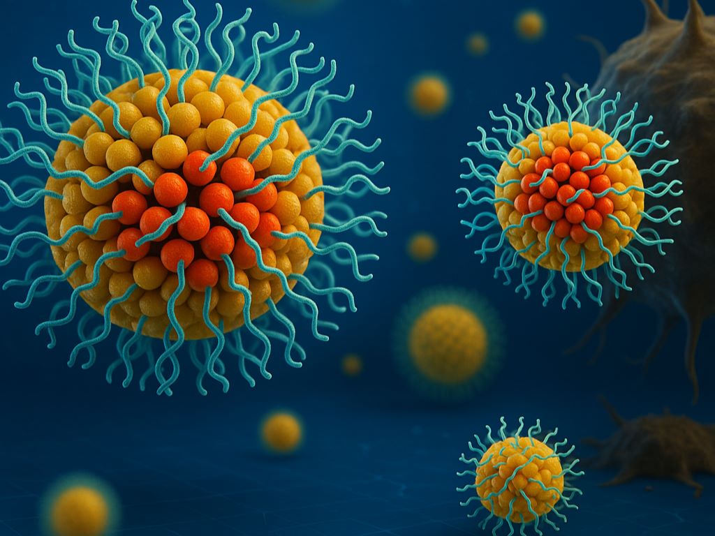
In the new study, the team designed a new drug from the ground up as a spherical nucleic acid (SNA): a nanostructure that weaves the drug directly into DNA strands coating tiny spheres. This design converts a poorly soluble, weakly performing drug into a powerful, targeted cancer killer that leaves healthy cells unharmed.

After developing the new therapy, the team tested it in a small animal model of acute myeloid leukaemia (AML), a fast-moving, difficult-to-treat blood cancer. Compared to the standard chemotherapy drug, the SNA-based drug entered leukaemia cells 12.5 times more efficiently, killed them up to 20 000 times more effectively and reduced cancer progression 59-fold – all without detectable side effects. 

This work is another example of the potential of structural nanomedicine, a new field in which scientists use precise structural, as well as compositional, control to fine-tune how nanomedicines interact with the human body. With seven SNA-based therapies currently in clinical trials, the new approach could lead to potent vaccines and treatments for cancers, infectious diseases, neurodegenerative diseases and autoimmune diseases. 

The study was published in the journal ACS Nano

“In animal models, we demonstrated that we can stop tumors in their tracks,” said Northwestern’s Chad A. Mirkin, who led the study. “If this translates to human patients, it’s a really exciting advance. It would mean more effective chemotherapy, better response rates and fewer side effects. That’s always the goal with any sort of cancer treatment.” 

For the new study, Mirkin and his team focused on the traditional chemotherapy drug 5-fluorouracil (5-Fu), which often fails to reach cancer cells efficiently. And, because it also attacks healthy tissue, 5-Fu causes myriad side effects, including nausea, fatigue and, in rare cases, even heart failure. 

According to Mirkin, the drug itself is not the problem – it’s how the body processes it. 5-Fu is poorly soluble, meaning less than 1% of it dissolves in many biological fluids. Most drugs need to dissolve in the bloodstream before they can travel through the body to enter cells. If a drug is poorly soluble, it clumps or retains a solid form, and the body cannot absorb it efficiently. 

“We all know that chemotherapy is often horribly toxic,” Mirkin said. “But a lot of people don’t realise it’s also often poorly soluble, so we have to find ways to transform it into water soluble forms and deliver it effectively.” 

To develop a more effective delivery system, Mirkin and his team turned to SNAs. Invented and developed by Mirkin at Northwestern, SNAs are globular nanostructures with a nanoparticle core surrounded by a dense shell of DNA or RNA. In previous studies, Mirkin discovered that cells recognise SNAs and invite them inside. In the new study, his team built new SNAs with the chemotherapy chemically incorporated into the DNA strands. 

“Most cells have scavenger receptors on their surfaces,” Mirkin said. “But myeloid cells overexpress these receptors, so there are even more of them. If they recognise a molecule, then they will pull it into the cell. Instead of having to force their way into cells, SNAs are naturally taken up by these receptors.” 

As Mirkin and his team suspected, the structural redesign completely changed how 5-Fu interacted with the cancer cells. Unlike with free-floating, unstructured chemotherapy molecules, the myeloid cells easily recognised and absorbed the SNA form. Once inside, enzymes broke down the DNA shell to release the drug molecules, which killed the cancer cell from within. 

In the mouse experiments, the therapy eliminated the leukaemia cells to near completion in the blood and spleen and significantly extended survival. And, because the SNAs selectively targeted AML cells, healthy tissues remained unharmed. 

“Today’s chemotherapeutics kill everything they encounter,” Mirkin said. “So, they kill the cancer cells but also a lot of healthy cells. Our structural nanomedicine preferentially seeks out the myeloid cells. Instead of overwhelming the whole body with chemotherapy, it delivers a higher, more focused dose exactly where it’s needed.” 

Next, Mirkin’s team plans to test the new strategy in a larger cohort of small animal models, then move to a larger animal model and, eventually, in human clinical trials, once funding is secured. 

Source: Northwestern University

EthiQal Invites You to an Ethics Webinar

Date: 20th November

Time: 18h00 to 19h45

Link: https://webinar.ethiqal.co.za/

During this webinar, attendees will review the HPCSA Booklet 10: General Ethical Guidelines for Good Practice in Telehealth where the pertinent South Africa laws and basic principles around ethical practice for patient consultations and the accompanying administration will be explored. The webinar will offer insights into different types of telehealth and ethical guidelines with specific focus on the quality, security, and safety of patient records.

The audience will have an opportunity to listen and engage with clinical, legal, medical malpractice insurance, cybersecurity, and telehealth subject matter experts. During the webinar, a range of learning opportunities will be offered including short lectures, interactive case studies with a series of multiple choices questions, panel discussions, and audience Q&A.

This webinar will focus on specialist practices. Administrative staff working in these practices are welcome to join the discussion.

Click here to register for the webinar

In Sleep Deprivation, Attention Lapses Correspond with CSF Cleaning Flushes

New research shows attention lapses due to sleep deprivation coincide with a flushing of fluid from the brain — a process that normally occurs during sleep.

Anne Trafton | MIT News

Photo by Tim Gouw on Unsplash

Nearly everyone has experienced it: After a night of poor sleep, you don’t feel as alert as you should. Your brain might seem foggy, and your mind drifts off when you should be paying attention.

A new study from MIT reveals what happens inside the brain as these momentary failures of attention occur. The scientists found that during these lapses, a wave of cerebrospinal fluid (CSF) flows out of the brain – a process that typically occurs during sleep and helps to wash away waste products that have built up during the day. This flushing is believed to be necessary for maintaining a healthy, normally functioning brain.

When a person is sleep-deprived, it appears that their body attempts to catch up on this cleansing process by initiating pulses of CSF flow. However, this comes at a cost of dramatically impaired attention.

“If you don’t sleep, the CSF waves start to intrude into wakefulness where normally you wouldn’t see them. However, they come with an attentional tradeoff, where attention fails during the moments that you have this wave of fluid flow,” says Laura Lewis, the Athinoula A. Martinos Associate Professor of Electrical Engineering and Computer Science, a member of MIT’s Institute for Medical Engineering and Science and the Research Laboratory of Electronics, and an associate member of the Picower Institute for Learning and Memory.

Lewis is the senior author of the study, which appears today in Nature Neuroscience. MIT visiting graduate student Zinong Yang is the lead author of the paper.

Flushing the brain

Although sleep is a critical biological process, it’s not known exactly why it is so important. It appears to be essential for maintaining alertness, and it has been well-documented that sleep deprivation leads to impairments of attention and other cognitive functions.

During sleep, the cerebrospinal fluid that cushions the brain helps to remove waste that has built up during the day. In a 2019 study, Lewis and colleagues showed that CSF flow during sleep follows a rhythmic pattern in and out of the brain, and that these flows are linked to changes in brain waves during sleep.

That finding led Lewis to wonder what might happen to CSF flow after sleep deprivation. To explore that question, she and her colleagues recruited 26 volunteers who were tested twice — once following a night of sleep deprivation in the lab, and once when they were well-rested.

In the morning, the researchers monitored several different measures of brain and body function as the participants performed a task that is commonly used to evaluate the effects of sleep deprivation.

During the task, each participant wore an electroencephalogram (EEG) cap that could record brain waves while they were also in a functional magnetic resonance imaging (fMRI) scanner. The researchers used a modified version of fMRI that allowed them to measure not only blood oxygenation in the brain, but also the flow of CSF in and out of the brain. They also measured each subject’s heart rate, breathing rate, and pupil diameter.

The participants performed two attentional tasks while in the fMRI scanner, one visual and one auditory. For the visual task, they had to look at a screen that had a fixed cross. At random intervals, the cross would turn into a square, and the participants were told to press a button whenever they saw this happen. For the auditory task, they would hear a beep instead of seeing a visual transformation.

Sleep-deprived participants performed much worse than well-rested participants on these tasks, as expected. Their response times were slower, and for some of the stimuli, the participants never registered the change at all.

During these momentary lapses of attention, the researchers identified several physiological changes that occurred at the same time. Most significantly, they found a flux of CSF out of the brain just as those lapses occurred. After each lapse, CSF flowed back into the brain.

“The results are suggesting that at the moment that attention fails, this fluid is actually being expelled outward away from the brain. And when attention recovers, it’s drawn back in,” Lewis says.

The researchers hypothesise that when the brain is sleep-deprived, it begins to compensate for the loss of the cleansing that normally occurs during sleep, even though these pulses of CSF flow come with the cost of attention loss.

“One way to think about those events is because your brain is so in need of sleep, it tries its best to enter into a sleep-like state to restore some cognitive functions,” Yang says. “Your brain’s fluid system is trying to restore function by pushing the brain to iterate between high-attention and high-flow states.”

A unified circuit

The researchers also found several other physiological events linked to attentional lapses, including decreases in breathing and heart rate, along with constriction of the pupils. They found that pupil constriction began about 12 seconds before CSF flowed out of the brain, and pupils dilated again after the attentional lapse.

“What’s interesting is it seems like this isn’t just a phenomenon in the brain, it’s also a body-wide event. It suggests that there’s a tight coordination of these systems, where when your attention fails, you might feel it perceptually and psychologically, but it’s also reflecting an event that’s happening throughout the brain and body,” Lewis says.

This close linkage between disparate events may indicate that there is a single circuit that controls both attention and bodily functions such as fluid flow, heart rate, and arousal, according to the researchers.

“These results suggest to us that there’s a unified circuit that’s governing both what we think of as very high-level functions of the brain — our attention, our ability to perceive and respond to the world — and then also really basic fundamental physiological processes like fluid dynamics of the brain, brain-wide blood flow, and blood vessel constriction,” Lewis says.

In this study, the researchers did not explore what circuit might be controlling this switching, but one good candidate, they say, is the noradrenergic system. Recent research has shown that this system, which regulates many cognitive and bodily functions through the neurotransmitter norepinephrine, oscillates during normal sleep.

The research was funded by the National Institutes of Health, a National Defense Science and Engineering Graduate Research Fellowship, a NAWA Fellowship, a McKnight Scholar Award, a Sloan Fellowship, a Pew Biomedical Scholar Award, a One Mind Rising Star Award, and the Simons Collaboration on Plasticity in the Aging Brain.

This story is republished courtesy of MIT News (web.mit.edu/newsoffice/), a popular site that covers news about MIT research, innovation and teaching.

Source: MIT News

Key Mechanism Behind Chemotherapy-induced Nerve Damage Uncovered

Study reveals how targeting a cellular pathway could protect cancer patients from peripheral neuropathy

Photo by Tima Miroshnichenko on Pexels

Scientists at Wake Forest University School of Medicine, in collaboration with researchers at Weill Cornell Medicine, have made a breakthrough in understanding why many cancer patients develop nerve damage after chemotherapy. Their new study, published in Science Translational Medicine, reveals that a stress response inside certain immune cells can trigger this debilitating side effect. This discovery could open the door to new ways to prevent or treat nerve damage in cancer patients.

Chemotherapy-induced peripheral neuropathy is a common and often severe side effect of cancer treatment, especially with drugs like paclitaxel. It can cause tingling, numbness and pain in the hands and feet, sometimes forcing patients to stop life-saving treatment early. Up to half of all patients receiving chemotherapy may experience this condition, but until now, the exact cause has remained a mystery.

To better understand this nerve toxicity that could be painful, scientists used a well-established mouse model that closely reflects the nerve problems experienced by people undergoing cancer treatment. This model allowed researchers to observe how a specific immune cell pathway, known as IRE1α, contributes to triggering inflammation that led to neurotoxicity and pain. By blocking the IRE1α pathway in the immune cells of these mice, either through genetic techniques or with an IRE1a inhibitor, the team was able to prevent the development of nerve damage, pain and toxic inflammation.

The researchers also studied a group of patients from Atrium Health Wake Forest Baptist’s National Cancer Institute-designated Comprehensive Cancer Center. The patients were receiving chemotherapy for gynecological cancers, collecting blood samples before and after treatment to measure IRE1α activity in their immune cells. They found that patients with higher IRE1α activation were more likely to develop severe neuropathy due to chemotherapy, directly linking the mouse model findings to patient outcomes.

Key Findings

  • Chemotherapy activates a stress sensor (IRE1α) in immune cells, triggering inflammation and nerve damage.
  • Blocking this sensor in mice prevented nerve pain and damage, suggesting a new treatment target.
  • In patients, higher activation of this stress sensor in blood cells was linked to more severe nerve symptoms and also to the initiation of neuropathy symptoms.

“Our research shows that a stress response inside immune cells is a key contributor to chemotherapy-induced neuropathy that could be painful and debilitating. By targeting this pathway, we may be able to protect patients from one of the most challenging side effects of cancer treatment,” said E. Alfonso Romero-Sandoval, MD, PhD, professor of anaesthesiology at Wake Forest University School of Medicine and the study’s corresponding author. “Our study opens the opportunity to further explore if this pathway could be used to predict what patients will develop this condition and therefore could help clinicians implement patient-tailored treatments,” Romero-Sandoval said.

The discovery could lead to new drugs that block this pathway, helping patients stay on their cancer treatment without suffering from painful side effects.

According to Romero-Sandoval, who is a member of the Atrium Health Wake Forest Baptist Comprehensive Cancer Center, this is the first study to show that the IRE1α stress sensor in immune cells is directly linked to nerve damage from chemotherapy.

The team plans to conduct larger clinical studies to confirm these findings and test whether the IRE1α pathway could be used as a biomarker for disease progression and if drugs that block this stress sensor can safely prevent or reduce nerve damage in cancer patients. They also hope to explore whether this approach could help with other types of nerve pain. Interestingly, an IRE1a inhibitor is currently in clinical trials to improve anti-cancer effects of chemotherapy, including paclitaxel.

Source: Wake Forest University

Sertraline Improves Core Depressive Symptoms Within Two Weeks

Photo by Danilo Alvesd on Unsplash

One of the most common antidepressants, sertraline, contributes to a modest improvement in core depression and anxiety symptoms, including low mood, within two weeks, finds a new analysis of a major clinical trial led by UCL researchers.

The study, published in Nature Mental Health, analysed the findings of the PANDA trial, which first published results in 2019 and found that sertraline may have an earlier impact on anxiety than depressive symptoms.* Researchers have now conducted a network analysis of the results, which is an innovative statistical method that allowed them to explore how specific symptoms respond to treatment.

The analysis revealed an improvement in symptoms such as low mood and suicidal thinking within two weeks of taking sertraline, while side effects of the drug led to symptoms such as low libido, low appetite, and tiredness. These side effects can also be symptoms of depression.

The researchers propose that the effect on all depressive symptoms put together in the initial analysis obscured the benefits of the antidepressants on some of the core depressive symptoms.  

In the new analysis, the early improvements from sertraline were found to be on feelings of sadness, self-loathing, restlessness, and suicidal thoughts.

The PANDA trial was a randomised controlled trial testing the effects of sertraline on people with depressive symptoms, including a wide range of patients in England whose symptoms ranged from mild to moderate. In a paper published in The Lancet Psychiatry in 2019*, scientists reported that within six weeks, sertraline improved anxiety symptoms and people reported an overall improvement in their mental health, but depressive symptoms did not meaningfully improve until participants had been taking the drug for 12 weeks. The new analysis, using data from 571 participants of the trial (those who had complete data for each symptom), suggests that sertraline improves some core depressive symptoms more quickly than previously believed.

The somatic (physical) symptoms that worsened, including libido and poor sleep, can be seen as side effects of antidepressants, but they are also common symptoms of depression, which can complicate interpretation of treatment effects.

Lead author Dr Giulia Piazza (UCL Psychiatry and UCL Psychology & Language Sciences) said: “We have now painted a more complex picture of sertraline’s effects on the different symptoms of depression.

“Instead of thinking of depression and anxiety as each being a single, uniform condition, network analysis considers that they’re each a constellation of symptoms, that can appear in different combinations for different people. These symptoms influence each other over time; for example, poor sleep can lead to problems with concentration, which may then impact self-esteem.

“Our analysis was borne out of this theoretical approach, in order to gain deeper insights and add nuance to the results of the PANDA trial.”

The researchers found that sertraline contributed to improvements in anxiety symptoms and in the emotional symptoms of depression within two weeks, and a modest worsening of somatic symptoms. The effect on somatic symptoms plateaued after six weeks, while the improvements in emotional symptoms and anxiety continued to improve from six weeks to 12 weeks.

Dr Piazza added: “It appears that the adverse effects on somatic symptoms like poor sleep and libido may stabilise after six weeks, which is then counteracted by continued improvements in emotional symptoms, the core symptoms of depression.”

Antidepressants are the standard pharmaceutical treatment for both depression and generalised anxiety disorder. Sertraline is a selective serotonin reuptake inhibitor (SSRI), the most common class of antidepressants.

Co-author Professor Glyn Lewis (UCL Psychiatry), who led the PANDA trial, said: “Our findings provide robust evidence that continues to support the prescription of sertraline for people experiencing depressive and anxiety symptoms. These findings will help patients and clinicians to make more informed decisions about treatment.”

Co-senior author Professor Jean-Baptiste Pingault (UCL Psychology & Language Sciences) said: “We found that the beneficial effects of sertraline can be detected very early on, as soon as two weeks after people start taking the antidepressant.

“Beyond this study, our results highlight the importance of considering symptom-level effects when developing novel drugs and evaluating existing drugs in psychiatry, and how this can help us to understand how these drugs work and how they can help patients.”

Source: University College London