Tag: 15/8/25

HASA CEO Talks About Partnerships, Purpose and the Pursuit of Universal Healthcare

He speaks in measured tones – calm, reflective, deliberate. But when Dr Dumisani Bomela describes the future he envisions, the words carry power. For the CEO of the Hospital Association of South Africa (HASA), healthcare is not just a profession. It is a promise rooted in dignity, equity and access to every South African.

Q: Dr Bomela, what drew you to medicine and what keeps you committed to healthcare in South Africa?
A: I have always seen healthcare as an act of service. As a doctor, you learn to see beyond symptoms, to understand the person behind the diagnosis. As a leader at HASA, I take that same approach. Our work is about people. About making sure that every South African can get quality care when they need it.

Q: HASA represents South Africa’s private hospital sector. Why is this sector important to the country’s overall health system?
A: Private hospitals are a cornerstone of healthcare in South Africa. We treat millions of patients each year. More than that, HASA members invest heavily in medical training, advanced technology and infrastructure. We are strategic partners in the national system, that makes our sector a vital national asset.

Q: How does HASA contribute to economic development beyond just healthcare?
A: Healthcare is a growth engine. HASA members are major employers, from doctors and nurses to technicians and support staff. We support local communities and stimulate investment. When healthcare systems are strong, economies thrive – and so do people.

Q: What is HASA’s stance on universal health coverage?
A:  We believe every person has the right to choose their provider and to receive high-quality care. That is why we support reforms that strengthen the system and build equity. HASA members are ready to work side by side with the government to make that vision a reality. Our hospital groups bring deep experience, including in some cases from geographies where universal healthcare systems operate, and strong infrastructure to the table.

Q: What kind of leadership do you believe is needed in South African healthcare today?
A: We need leaders who listen. Who understand not just policy, but people. Leadership in healthcare must be grounded in compassion and collaboration. At HASA, we strive to lead by example, building trust, fostering partnerships, and always remembering that every system ultimately affects human lives.

Q: How do HASA hospitals stay at the forefront of medical technology and innovation?
A: By investing intentionally. Our members understand that modern medicine is not static, it is constantly evolving. We equip our hospitals with advanced diagnostic and treatment tools. But technology alone is not enough. We also invest in people – training nurses, specialists and support teams to lead with excellence.

Q: In a country facing complex health challenges, how do you stay hopeful?
A: Hope grows where there is action. Across our hospitals, I see incredible work being done every day – surgeons saving lives, nurses comforting families, teams innovating to improve care. We are proving, together, that with collaboration and commitment, South Africa’s health system can be strong, inclusive and world-class.

Q: What gives you the greatest sense of pride in your work with HASA?
A:  Honestly, it is seeing the impact private hospitals have. When families walk out of our hospitals healed. When professionals grow into health leaders. When communities feel their well-being is supported. These outcomes remind us why the work matters. My pride does not come from titles; it comes from knowing we are making a real, human difference every single day.

Research Shines a Light into Sex Differences in Diseases

Photo by Daniil Onischenko on Unsplash

Many diseases affect men and women differently. Asthma tends to strike men earlier in life, yet more women develop asthma as they get older. Parkinson’s is more common in men, but Alzheimer’s is more common in women. 

The differences are even more stark when it comes to autoimmune disease. Women are around two and a half times more likely than men to develop multiple sclerosis and nine times more likely to develop lupus.

Why would some diseases strike one sex more than another? And why do some tissues, such as the lungs and brain, seem especially vulnerable to these sex-based differences?

To answer these questions, scientists at La Jolla Institute for Immunology (LJI) are leading new research into how our immune cells defend specific parts of the body.

In a new Science review, LJI Professor, President & CEO Erica Ollmann Saphire, PhD, MBA, and LJI Associate Professor Sonia Sharma, PhD, examine how genetics, sex hormones, and environmental factors come together to shape the immune system.

“In just the last two years, LJI scientists have uncovered a whole new body of information about how the immune systems of men and women are very different,” says Saphire. “We’re looking at what is genetically encoded in our XX or XY chromosomes, and how hormones like oestrogen and testosterone affect what is genetically programmed into our immune cells.”

In the paper, the researchers define biological sex (in an immunology context) as the presence of XX chromosomes in females and XY chromosomes in males. “Every cell in your body is either XX or XY,” says Saphire. “That X chromosome has many, many immune-related genes. Women have two copies of each. That gives them, in a sense, twice the palette of colours to paint from in formulating an immune response. It can also give them a stronger immune response for those genes that are doubly active – active in both copies simultaneously.

Sex hormones are important for much more than reproductive function. Immune cells can also sense hormones such as oestrogen and testosterone and use them to determine which genes to turn on or off and which ones to turn on more brightly or dim. This means similar immune cells can do different things, depending on whether that cell is from a male or a female. 

Further, female cells vary in which of their two copies of X is “turned on.” As a result, women have organs with a collage, or mosaic, of immune cells that work differently in different tissues. This innate “variety” of immune cells appears to be an effective way to ward off infectious disease (women are better than men at fighting off pathogens such as SARS-CoV-2). 

But scientists have also found that having more genes from X chromosomes may predispose women to autoimmune disease. This increased X chromosome “dosage” is closely linked to a higher risk for autoimmune diseases such as Sjögren’s syndrome and scleroderma.

New research into sex-based immune system differences is also critical for developing new cancer immunotherapies, Sharma explains.

“We’re increasingly understanding how sex-based differences affect disease outcomes. When it comes to medicine, one size doesn’t fit everybody,” says Sharma, who directs LJI’s Center for Sex-Based Differences in the Immune System. “This is leading to new research, particularly in the cancer field, toward precision medicine. We’re asking how a person’s individual immune system is contributing to controlling that cancer through immunotherapy.”

Saphire and Sharma also highlight environmental factors, such as nutrition and chemical exposures, that may add to the complex interplay of chromosomes and sex hormones. Men and women also appear to have some signature differences in their skin and gut microbiomes.

The researchers hope these foundational discoveries can lead to medical advances for all, and they’re working with collaborators across the country to move this research forward. “It takes a team to translate these findings,” says Sharma.

The new review, titled “Sex differences in tissue-specific immunity and immunology,” includes co-author Alicia Gibbons of LJI and UC San Diego.

Source: La Jolla Institute for Immunology

Small Changes in Walking Technique May Help Treat Knee Osteoarthritis

Photo by Kampus Production on Pexels

Gait analysis and pain measures show that subtly adjusting the angle of the foot during walking may reduce knee pain caused by osteoarthritis, an approach which may also slow progression of the incurable condition.

Led by a team of researchers at NYU Langone Health, the University of Utah, and Stanford University, a new study explored whether changing the way patients position their feet when walking could lessen extra loading, helping to treat the disease.

For the investigation, the scientists tested this intervention in 68 men and women with mild to moderate knee osteoarthritis and then used advanced MRI scans to track how well it worked.

The results suggest that those trained to angle their feet slightly inward or outward from their natural alignment experienced slower cartilage degeneration in the inner part of their knee compared with those who were encouraged to walk more frequently without changing their foot position. The study is reported online in The Lancet Rheumatology.

“Although our results will have to be confirmed in future studies, they raise the possibility that the new, noninvasive treatment could help delay surgery,” said study co-lead author Valentina Mazzoli, PhD.

Dr. Mazzoli, an assistant professor in the Department of Radiology at NYU Grossman School of Medicine, notes that the earlier patients receive a knee replacement, the more likely they are to require additional procedures in the future.

The findings also revealed that those who adjusted their foot angle reduced their pain score by 2.5 points on a 10-point scale, an effect equivalent to that of over-the-counter pain medications. By contrast, those who did not change their gait reduced their pain scores by little more than a point.

“Altogether, our findings suggest that helping patients find their best foot angle to reduce stress on their knees may offer an easy and fairly inexpensive way to address early-stage osteoarthritis,” added Dr. Mazzoli.

About one in seven Americans have some form of osteoarthritis, commonly in the inner side of the knee, according to the U.S. Centers for Disease Control and Prevention. A leading cause of disability, the disease is often managed with pharmaceutical pain relievers, physical therapy, and in the most severe cases, knee-replacement surgery. Experts believe that excess loading can over time contribute to the condition.

Past research has offered little evidence that changes in gait can effectively reduce knee pain caused by osteoarthritis, says Dr. Mazzoli. Some previous trials trained all participants to adopt the same foot angle and found no relief, while others did not compare the intervention to a control group or only followed the participants for a month.

The new study is the first to show that tailoring each patient’s foot angle to their unique walking pattern can alleviate the disease’s symptoms in the long term and may slow cartilage breakdown, the authors say.

Dr Mazzoli adds that this technique may have a significant advantage over pharmaceutical painkillers. These drugs, she says, do not address the underlying disease and can cause liver and kidney damage, stomach ulcers, and other unwanted side effects when taken for long periods.

For the study, the research team recorded the participants walking on a treadmill at a specialized gait-assessment laboratory. A computer program simulated their walking patterns and calculated the maximum loading that occurred in the inner side of their knees. Next, the team generated computer models of four new foot positions—angled inward or outward by either 5 or 10 degrees—and estimated which option reduced loading the most.

The patients were then randomly divided into two groups. Half were trained in six sessions to walk with their ideal angle, while the other half were instructed to continue walking naturally. Pain scores and MRI scans were taken at the beginning of the study period and one year after the intervention.

Study findings showed those who adjusted their gait reduced the maximum loading in the knees by 4 percent, while those who kept their normal walking pattern increased their loading by more than 3 percent.

“These results highlight the importance of personalizing treatment instead of taking a one-size-fits-all approach to osteoarthritis,” said Dr. Mazzoli. “While this strategy may sound challenging, recent advances in detecting the motion of different body parts using artificial intelligence may make it easier and faster than ever before.”

While the authors relied on a specialized laboratory for the new study, AI software that estimates joint loading using smartphone videos is now available and can allow clinicians to perform a gait analysis in the clinic.

The researchers next plan to test whether these tools can indeed identify the most effective walking method for osteoarthritis patients, says Dr. Mazzoli. They also plan to expand their study to people with obesity.

Source: NYU Langone Health

Blinking and Eyelid Function Is Enabled by Complex Control of Muscles

Discovery could help pave the way for a prosthetic device to restore blink function lost to injury or disease

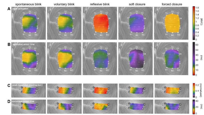
Muscle activation and movement patterns over time across the upper and lower eyelids, shown under different actions. Credit: Anatomical Engineering Group/UCLA

A blink of an eye is vital to protecting the eye by keeping it from drying out. This simple function seems natural and instantaneous, but is it?

Now, a team of UCLA biomechanical engineers and ophthalmologists has uncovered new details about the muscle that controls blinking, offering a pathway toward developing blink-assisting prostheses. Published in PNAS, the study found that the orbicularis oculi – the muscle that controls eyelid movement – contracts in complex patterns that vary by action and move the eyelid in more than just a simple up-and-down motion.

The researchers studied how this muscle behaves differently across various actions including spontaneous blinks, protective rapid closures and squeezed shut-eye motions.

“The eyelid’s motion is both more complex and more precisely controlled by the nervous system than previously understood,” said study corresponding author Tyler Clites, an assistant professor of mechanical and aerospace engineering at the UCLA Samueli School of Engineering. “Different parts of the muscle activate in carefully timed sequences depending on what the eye is doing. This level of muscle control has never been recorded in the human eyelid. Now that we have this information in rich detail, we can move forward in designing neuroprostheses that help restore natural eyelid function.”

In experiments with volunteers, the researchers looked at five different ways the eyes close:

  • Spontaneous blink: An automatic, unconscious blink that occurs regularly to keep the eye lubricated
  • Voluntary blink: An intentional blink, as when someone is asked to blink on command
  • Reflexive blink: A rapid, involuntary blink triggered to protect the eye from a collision
  • Soft closure: A gentle, slow eyelid descent, similar to the beginning of sleep
  • A forced closure: A deliberate squeezing of the eyelids tightly shut

To record activity in the orbicularis oculi with high precision, an ophthalmic surgeon inserted tiny wire electrodes into the eyelid. The researchers then used a motion-capture system to track eyelid movement in ultraslow motion. These tools allowed the team to measure subtle differences in eyelid movement, including speed, direction, and which part of the muscle initiated the action.

Video of spontaneous blink – dynamic muscle activation patterns and eyelid kinematics. Credit: Anatomical Engineering Group/UCLA

“People can lose the ability to blink due to a stroke, tumour, infection or injury. The condition is painful in the short term and can damage the eyes enough to cause vision loss,” said study co-author Dr Daniel Rootman, an associate professor of ophthalmology at the David Geffen School of Medicine at UCLA and director of the UCLA Orbital Disease Center. “We know that a small electric pulse can stimulate the orbicularis oculi muscle to move, but designing one that works well has been elusive. What we now have is a good roadmap to such a device, including where exactly to place electrodes, how to time them, and how strong the pulse should be. These guidelines could help pave the way for the development and clinical testing of such a device, with the ultimate goal of providing real relief for patients.”

With this fundamental knowledge of eyelid biomechanics in hand, the researchers can now work on refining a prototype neuroprosthesis to assist people with blinking.

“Understanding how the eyelid works is crucial to designing an accurate stimulation pattern for a prosthesis, as well as for diagnostic purposes,” said study first author Jinyoung Kim, a UCLA mechanical engineering doctoral student and member of Clites’ research group, the Anatomical Engineering Group at UCLA. “We are more than excited to bridge this gap and move forward to work with patients who have facial paralysis and help improve their lives.”

Source: UCLA Samueli School of Engineering