The protein Dickkopf 3 plays a key role in the development of radiation-induced fibroses – and could be a promising target for novel therapies
Picture by Macrovector on Freepik
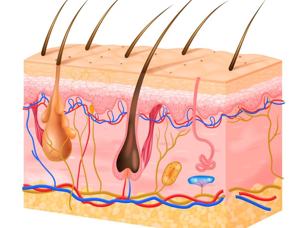
Radiotherapy is one of the main treatment forms for cancer. Among its most common side effects is skin damage, right up to chronic inflammations and fibroses. At present, such long-term damage can only be treated symptomatically and leads to thickened, painful, or sensitive skin for months to years after the radiation treatment. A team led by LMU immunologist Professor Peter Nelson (LMU University Hospital) and Roger Sandhoff and Peter E. Huber from the German Cancer Research Center (DKFZ) has identified a protein called Dickkopf 3 (DKK3) as a main cause of long-term skin damage after radiotherapy – a decisive step for the development of novel, more targeted therapy options.
By investigating mouse models and human cells and tissue samples, the researchers demonstrated that DKK3 is activated after radiotherapy in a certain group of skin cells that are responsible for skin renewal. This activity triggers a chain reaction which promotes inflammations and the formation of scar-like tissue and leads to chronic skin damage. The key findings were driven by the work of LMU students, Li Li and Khuram Shehzad. Their efforts were essential in identifying DKK3 as the critical molecular mediator and in establishing the mechanistic framework presented in the paper. “We also observed similar processes in the kidney,” says Nelson. “This indicates that the activation of DKK3 is a fundamental mechanism that promotes fibrosis in various tissues.”
According to the researchers, these findings underscore that DKK3 represents a promising new treatment target. “Drugs that block DKK3 could one day help prevent or reduce long-term skin damage after radiotherapy and thus improve the quality of life of cancer patients and survivors,” says Nelson. The researchers are currently investigating, moreover, whether this approach could also contribute to the prevention of scar formation in other organs.
The face is privileged when it comes to scarring after injury. A Stanford Medicine study in mice not only discovers why but also finds a drug that helps skin from other sites regenerate.
Tweaking a pattern of wound healing established millions of years ago may enable scar-free injury repair after surgery or trauma, Stanford Medicine researchers have found. If results from their study, which was conducted in mice, translate to humans, it may be possible to avoid or even treat the formation of scars anywhere on or within the body.
Scarring is more than a cosmetic problem. Scars can interfere with normal tissue function and cause chronic pain, disease and even death. It’s estimated that about 45% of deaths in the United States are due to some type of fibrosis – usually of vital organs like the lungs, liver or heart.
Scars on the skin’s surface, while rarely fatal, are stiffer and weaker than normal skin and they lack sweat glands or hair follicles, making it difficult to compensate for temperature changes.
Surgeons have known for decades that facial wounds heal with less scarring than injuries on other parts of the body. This phenomenon makes evolutionary sense: Rapid healing of body wounds prevents death from blood loss, infection or impaired mobility, but healing of the face requires that the skin maintain its ability to function well.
“The face is the prime real estate of the body,” said professor of surgery Michael Longaker, MD. “We need to see and hear and breathe and eat. In contrast, injuries on the body must heal quickly. The resulting scar may not look or function like normal tissue, but you will likely still survive to procreate.”
Exactly how this discrepancy happens has remained a mystery, although there were some clues.
“The face and scalp are developmentally unique,” said professor of surgery Derrick Wan, MD. “Tissue from the neck up is derived from a type of cell in the early embryo called a neural crest cell. In this study we identified specific healing pathways in scar-forming cells called fibroblasts that originate from the neural crest and found that they drive a more regenerative type of healing.”
Activating this pathway in even a subset of fibroblasts around small wounds on the abdomen or backs of mice caused them to heal with much less scarring – similar to untreated facial or scalp wounds.
Longaker, the Deane P. and Louise Mitchell Professor in the School of Medicine, and Wan, the Johnson & Johnson Distinguished Professor in Surgery II, are the senior authors of the study, which was published January 22 in Cell. Plastic surgery resident Michelle Griffin, MD, PhD, and clinical and postdoctoral scholar Dayan Li, MD, PhD, are the lead authors of the research.
“Many of the authors on this paper are fellow physician scientists,” said Li, who is board certified in dermatology. “This project was inspired by what we’ve observed in our patients – facial wounds in general heal with less scarring. We wanted to understand, mechanistically, why this is.”
Proteins determine scarring
Li and his colleagues used laboratory mice to investigate differences in wound healing at various sites on the animals’ bodies. They anesthetised the mice before creating small skin wounds on the face, scalp, back and abdomen. The wounds were stabilised by suturing small plastic rings around them to prevent differences in mechanical forces as the animals moved. Mice were given pain relief during the healing process.
After 14 days, the wounds on the face and scalp expressed lower levels of proteins known to be involved in scar formation as compared with those on the abdomen or back of the animals. The sizes of the scars were also smaller.
The researchers then transplanted skin from the face, scalp, back and abdomen of mice onto the backs of control mice. After the transplants had engrafted, they repeated the experiment on the transplanted skin. As before, wounds in the skin transplanted from the faces of the donor mice expressed lower levels of scarring-associated proteins.
Additionally, Li and his colleagues isolated fibroblasts from skin samples from the four body sites in the donor mice and injected them into the backs of control mice. They observed reduced levels of scarring-associated proteins on the recipient animals’ backs injected with fibroblasts from the donor animals’ faces as compared with fibroblasts from the scalp, back or abdomen.
Now that we understand this pathway and the implications of the differences among fibroblasts that arise from different types of stem cells, we may be able to improve wound healing after surgeries or trauma.”
–Derrick Wan
“We found you don’t need to change or manipulate all fibroblasts within the tissue to have a positive outcome,” Li said. “When we injected fibroblasts that we had genetically altered to more closely resemble facial fibroblasts, we saw that the back incisions healed very much like facial incisions, with reduced scarring, even when the transplanted fibroblasts made up only 10% to 15% of the total number of surrounding fibroblasts. Changing just a few cells can trigger a cascade of events that can cause big changes in healing.”
A less-fibrotic wound healing
Digging deeper, the researchers identified changes in gene expression between facial fibroblasts and those from other parts of the body and followed these clues to identify a signaling pathway involving a protein called ROBO2 that maintains facial fibroblasts in a less-fibrotic state. They also saw something interesting in the genomes of fibroblasts making ROBO2.
“In general, the DNA of the ROBO2-positive cells is less transcriptionally active, or less available for binding by proteins required for gene expression,” Li said. “These fibroblasts more closely resemble their progenitors, the neural crest cells, and they might be more able to become the many cell types required for skin regeneration.”
In contrast, the DNA in fibroblasts from other sites of the body allows free access to genes like collagen that are involved in the creation of scar tissue.
“It seems that, in order to scar, the cells must be able to express these pro-fibrotic genes,” Longaker said. “And this is the default pathway for much of the body.”
ROBO2 doesn’t act alone. It triggers a signalling pathway that results in the inhibition of another protein called EP300 that facilitates gene expression. EP300 plays an important role in some cancers, and clinical trials of a small drug molecule that can inhibit its activity are underway. Li and his colleagues found that using this pre-existing small molecule to block EP300 activity in fibroblasts prone to scarring caused back wounds to heal like facial wounds.
“Now that we understand this pathway and the implications of the differences among fibroblasts that arise from different types of stem cells, we may be able to improve wound healing after surgeries or trauma,” Wan said.
The findings are likely to extend to internal scarring as well, Longaker said. “There’s not a million ways to form a scar,” he said. “This and previous other findings in my lab suggest there are common mechanisms and culprits regardless of the tissue type, and they strongly suggest there is a unifying way to treat or prevent scarring.”
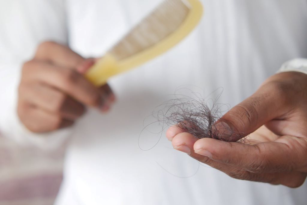
Your guide to safe, effective, and natural hair restoration in 2026
Hair restoration is one of the fastest-growing aesthetic procedures worldwide. The International Society of Hair Restoration Surgery (ISHRS) reports hundreds of thousands of procedures performed globally each year, with demand climbing steadily. As more people seek confidence-boosting solutions to start the new year, South African specialists warn that choosing the wrong clinic can turn a life-changing decision into lasting damage.
Dr Kashmal Kalan, Medical Director at Alvi Armani South Africa, explains: “January brings a sense of renewal. Many people reassess their goals, and hair restoration has become one of the most transformative ways to invest in yourself. It’s no longer just about fitness or weight loss – hair and skin now play a central role in personal confidence.”
A successful hair restoration journey begins long before the procedure and continues well beyond it. At Alvi Armani, every patient undergoes a thorough, personalised consultation. The team evaluates hair loss patterns, donor density, scalp condition, hair type, and personal goals. Advanced AI-assisted microscopic analysis helps ensure patients are suitable and that the procedure is planned for optimal, natural results.
On procedure day, patients enter a calm, controlled environment. Hairline design is finalised, Follicular Unit Extraction (FUE) is performed with precision, and grafts are implanted to follow the natural flow of hair. Recovery is gradual, with initial shedding giving way to new growth. Density and texture refine over 12-18 months, and ongoing check-ins ensure progress stays on track.
Hairline design is the most artistic aspect of the process. Age, facial symmetry, ethnicity, and donor capacity all influence the final outcome. “We aim for perfection within imperfection. The goal is a hairline that complements the face naturally. No one should be able to tell a transplant took place.”
Strategic density planning is equally critical. Every follicle in the donor area is finite, and poor planning can create gaps or thin patches. This can leave permanent aesthetic imbalance. Reputable clinics plan for decades, not just the first few months. Patients should also understand that growth is gradual, and progressive hair loss may require more than one procedure to achieve the desired result.
Alvi Armani ensures every procedure is doctor-led and supported with ongoing care, including stabilisation medications, regenerative therapies, and annual check-ups. Dr Kalan cautions against so-called “dark clinics” offering prices too good to be true, often operating in unhygienic or mobile facilities. “These clinics treat hair restoration as a commodity rather than medicine. They overharvest donor areas, produce unnatural results, and leave patients needing urgent repairs. Repair procedures now make up roughly a quarter of our cases.”
Beyond procedural excellence, Alvi Armani educates patients on lifestyle choices that support lasting results, from nutrition and scalp care to ongoing therapies. While the process requires patience, the rewards – confidence, natural appearance, and the security of a clinic that plans carefully for the future – make it worthwhile.
For anyone considering hair restoration in 2026, the advice is clear: invest in quality from the start. With the right clinic, personalised planning, and medical oversight, patients can achieve safe, natural results that endure for years to come.
According to a new study, lower doses of approved immunotherapy for malignant melanoma can give better results against tumours, while reducing side effects. This is reported by researchers at Karolinska Institutet in the Journal of the National Cancer Institute.
“The results are highly interesting in oncology, as we show that a lower dose of an immunotherapy drug, in addition to causing significantly fewer side effects, actually gives better results against tumours and longer survival,” says last author Hildur Helgadottir, a researcher at the Department of Oncology-Pathology at Karolinska Institutet, who led the study.
The traditional dose of nivolumab and ipilimumab is the one that is approved and established. Due to the extensive side effects, Sweden has increasingly begun to use a treatment regimen with a lower dose of ipilimumab, which is both gentler and cheaper. Ipilimumab is the most expensive part of this immunotherapy and causes the most side effects.
“In Sweden, we have greater freedom to choose doses for patients, while in many other countries, due to reimbursement policies, they are restricted by the doses approved by the drug authorities,” says Hildur Helgadottir.
Lower dose is more effective
The study included nearly 400 patients with advanced, inoperable malignant melanoma, the most serious form of skin cancer. The study shows that the regimen with the lower dose of ipilimumab is more effective, with a higher proportion of patients responding to treatment, 49%, compared to the traditional dose, 37%.
Progression-free survival, the time the patient lives without the disease worsening, was a median of nine months for the lower dose, compared to three months for the traditional dose. Overall survival was also longer, 42 months compared to 14 months.
Serious side effects were seen in 31% of patients in the low-dose group, compared to 51% in the traditional group.
“The new immunotherapies are very valuable and effective, but at the same time they can cause serious side effects that are sometimes life-threatening or chronic. Our results suggest that this lower dosage may enable more patients to continue the treatment for a longer time, which is likely to contribute to the improved results and longer survival,” says Hildur Helgadottir.
There were some differences between the two treatment groups, but even after adjusting for several factors such as age and tumour stage, the better outcome for the lower dose of ipilimumab remained. The study is a retrospective observational study and therefore it is not possible to definitively establish a causal relationship.
When it comes to protecting babies from the sun, many parents wonder if sunscreen is safe and necessary. The truth is, experts advise against using sunscreen on infants under six months old as their skin is thinner and more sensitive, leading to greater absorption of chemicals and a higher risk of irritation and rashes.
Babies under six months have a higher surface-area-to-body-weight ratio, which increases their exposure to sunscreen chemicals. Some chemical ingredients, like oxybenzone, may cause allergic reactions or disrupt hormones. Sunscreen can also impede a baby’s ability to sweat and regulate their body temperature.
Instead, the best protection for young babies is to keep them out of direct sunlight, dress them in lightweight, long-sleeved clothing, and use hats and shade as natural barriers.
For babies over six months, a gentle, broad-spectrum baby sunscreen with at least SPF 30 can be safely applied. However, using sunscreen should complement, not replace, other sun safety measures, which are vital – especially in our sunny South African climate!
Karen Van Rensburg, spokesperson for Sanosan, explains, “Parents often struggle with knowing how much sunscreen to use on their babies. It’s important to understand that while sunscreen is a helpful tool, relying solely on it, especially for very young infants, can be risky. Using physical barriers like shade and protective clothing alongside sunscreen provides the safest approach to sun care for babies.”
To keep babies safe, parents should:
Avoid sun exposure during peak hours (10 a.m. to 4 p.m.)
Use shade and protective clothing as the first defence.
For babies over six months, reapply a suitable sunscreen on a regular basis to maintain protection, especially after going in the water, after drying off or after sweating.
Your baby should not stay in the sun too long even with sunscreen because every sunburn damages the skin and is a serious risk to their health.
This balanced approach highlights that cautious sunscreen use combined with physical protection methods is key to keeping baby skin healthy and safe from sun damage.
Sanosan Baby Sun Cream SPF 50+ is a top-tier sunscreen designed specifically for delicate baby skin including broad range of UVA+UVB protection SPF 50+. With its pleasant texture, this cream absorbs quickly for easy application and delivers 24 hours of nourishing care, making it suitable for babies, children, and adults alike. With its gentle formula, this sun cream helps maintain skin hydration while protecting against sun damage, allowing for worry-free outdoor playtime. Plus, its microplastic-free, and safe for our oceans!
Two global trials show durable improvements in skin clearance, itch, and quality of life by targeting OX40 immune receptor
Source: Unsplash CC0
An international team of investigators led by Emma Guttman-Yassky, MD, PhD, Waldman Professor and System Chair of the Kimberly and Eric J. Waldman Department of Dermatology at the Icahn School of Medicine at Mount Sinai, has reported results from the first phase 3 clinical trials of rocatinlimab, a novel treatment for moderate-to-severe atopic dermatitis (eczema). The landmark findings from the ROCKET-IGNITE and ROCKET-HORIZON studies were published in The Lancet.
Eczema affects hundreds of millions of people worldwide and is notoriously difficult to treat due to its complex and chronic inflammatory pathways. Current biologics focus on blocking “allergy” cytokines but fail to address the memory T cells that sustain disease activity. Rocatinlimab is the first antibody to selectively block the OX40 receptor on effector and memory T cells, rebalancing the immune system and altering the long-term course of disease.
Across the two global, double-blind, placebo-controlled randomised phase 3 clinical trials, nearly 1,500 patients were followed for 24 weeks, and rocatinlimab showed robust and lasting benefits. Patients receiving the treatment were three times more likely to achieve significant improvement in eczema severity, as measured by EASI and vIGA-AD scores, compared to those on placebo. Improvements continued beyond week 24, suggesting that the benefits strengthen over time. The therapy also led to meaningful reductions in itch, pain, and sleep disturbances, enhancing overall quality of life. Importantly, rocatinlimab was well tolerated, with adverse events comparable to placebo, and demonstrated high selectivity by reducing only the OX40R+ CD4+ T cells responsible for eczema’s persistence, without off-target effects.
“These findings represent a major advance for patients living with eczema, who often face years of uncontrolled symptoms and few effective options,” said physician scientist, Dr. Guttman-Yassky, lead author of the study. “By targeting memory T cells through OX40, rocatinlimab not only clears the skin and relieves itch, but continues to improve patients’ lives over time with a strong safety profile. This is the first phase 3 proof that rebalancing these immune cells can transform how we treat atopic dermatitis.”
The results establish OX40 as a validated treatment target in eczema and position rocatinlimab as a potential first-in-class therapy. Patients from the phase 3 trials are now being followed in the ROCKET-ASCEND extension study, which will track outcomes for up to two years. Additional research will explore its role in paediatric patients, in combination with other therapies, and in direct comparisons to existing systemic treatments.
In a new study published in Nature Communications, researchers at the University of Chicago have discovered how prolonged exposure to ultraviolet (UV) radiation can trigger inflammation in skin cells through degradation of a key protein called YTHDF2. This protein acts as a gatekeeper in preventing normal skin cells from becoming cancerous. The finding reveals that YTHDF2 plays a crucial role in regulating RNA metabolism to keep cells in a healthy state and opens the door to developing potential new approaches to skin cancer prevention and treatment.
Uncontrolled inflammation triggers skin cancer
Each year, nearly 5.4 million people in the United States are diagnosed with skin cancer, with more than 90% of cases attributed to excessive UV exposure. UV rays can damage DNA and cause oxidative stress and inflammation in skin cells — leading to redness, pain and blistering, commonly known as sunburn.
“We’re interested in understanding how inflammation caused by UV exposure contributes to the development of skin cancer,” said Yu-Ying He, PhD, Professor of Medicine in the Section of Dermatology at the University of Chicago.
RNA or ribonucleic acid is an essential molecule that helps convert genetic information into proteins. A special class known as non-coding RNAs regulates gene expression without producing proteins. These molecules typically function in either the nucleus, where a cell’s DNA is stored or the cytoplasm, where most cellular activity occurs.
Low levels of YTHDF2 turn normal skin cells cancerous
He’s laboratory studies how environmental stressors, such as UV radiation or arsenic in drinking water, affect molecular pathways and damage cellular systems, leading to cancer. Through screening various enzymes, the researchers found that UV exposure causes a marked decrease in levels of YTHDF2, a “reader” protein that specifically binds to RNA sequences marked with a chemical tag known as N6-methyladenosine (m6A).
“When we removed YTHDF2 from skin cells, we saw that UV-triggered inflammation was much worse,” He said. “This suggests that the YTHDF2 protein plays a key role in suppressing inflammatory responses.”
Although inflammation is essential for fighting off infections, it also plays a major role in causing life-threatening diseases, including cancer. However, the molecular mechanisms that regulate this response, especially after UV damage, are not well understood.
YTHDF2 in regulation of non-coding RNA interactions
Using multi-omics analysis and additional cellular assays, the research team found that YTHDF2 binds to a specific non-coding RNA known as U6, which is modified by m6A and classified as a small nuclear RNA (snRNA). Under UV stress, cancer cells showed increased levels of U6 snRNA, and these modified RNAs were found to interact with toll-like receptor 3 (TLR3), an immune sensor known to activate inflammatory pathways linked to cancer.
Surprisingly, these interactions occurred within endosomes, where cellular compartments are typically involved in recycling materials, not where U6 snRNA is usually located.
“We spent a lot of time figuring out how these non-coding RNAs get to the endosome, since that’s not where they usually reside,” He explained. “For the first time, we showed that a protein called SDT2 transports U6 into the endosome, and YTHDF2 travels with it.”
Once both YTHDF2 and m6A-modified U6 RNA arrive at the endosome, YTHDF2 blocks the RNA from activating TLR3. However, when YTHDF2 is absent – such as after UV damage, the RNA freely binds to TLR3, triggering harmful inflammation.
“Our study uncovers a new layer of biological regulation, a surveillance system through YTHDF2 that helps protect the body from excessive inflammation and inflammatory damage,” He said.
The findings could open the door to new strategies for preventing or treating UV-induced skin cancer by targeting the RNA-protein interactions that regulate inflammation.
The study, “YTHDF2 regulates self non-coding RNA metabolism to control inflammation and tumorigenesis,” was supported by grants from the National Institutes of Health, the University of Chicago Medicine Comprehensive Cancer Center, the ChicAgo Center for Health and EnvironmenT (CACHET), and the University of Chicago Friends of Dermatology Endowment Fund.
In a tiny back room, with outdated instruments and no doctor in sight, a “hair transplant” begins. Weeks later, patients arrive at legitimate clinics with infections that won’t heal, patchy or missing hair, and permanent scarring – some even with necrotic tissue. This isn’t just happening abroad in countries like Turkey and Pakistan; South Africa has its own growing underground hair transplant industry: the so-called “dark clinics”.
These clinics operate everywhere. In mobile setups, or purely online. Many are run by unlicensed practitioners who are far from registered doctors. Many staff aren’t even legally allowed to work in the country. Regulation is patchy. Enforcement is almost impossible given that they operate for a few weeks and then disappear. And the people who pay the price? Everyday men and women desperate to fix hair loss but who are unable to afford quality and safety.
“Most people have no idea what they’re walking into when visiting one of these clinics,” says Dr Kashmal Kalan, Medical Director at Alvi Armani South Africa. “We see patients quite often who come to us after unsatisfactory or botched procedures. And yes, some cases are life-threatening. These clinics operate with no oversight, no hygiene protocols, and zero accountability.”
The harm isn’t just cosmetic. Rogue clinics routinely cut corners. Non-medical staff perform invasive procedures. Sterile instruments? Often non-existent. Infection control? Minimal. Follow-up care? None. Patients are sometimes rushed into procedures without proper consent, unaware of realistic outcomes or potential complications. What starts as a “cheap deal” can quickly spiral into months of medical interventions, emotional trauma, revision surgeries, and financial strain.
Dr Kalan explains the lure: “These clinics play on insecurity, urgency, and the desire for fast results. Social media and online payments make it easy for unqualified operators to reach people. Most patients don’t ask the hard questions. They only learn the truth when it’s too late. One patient described it as the worst mistake of his life – a promise of transformation that turned into months of pain, scarring, and regret.”
Hard truths: Red flags to watch out for
A legitimate clinic is open, accountable, and transparent. Dark clinics thrive on secrecy, pressure tactics, and the promise of cheap shortcuts. Patients must ask:
Is the surgeon fully qualified, licensed, and registered in South Africa?
Do they have a valid Health Professions Council of South Africa (HPCSA) license? Are they legally allowed to practice in South Africa?
Which technique will be used and why?
Follicular Unit Extraction (FUE), Follicular Unit Transplantation (FUT), Direct Hair Implantation (DHI) – what’s suitable for your hair type and hair-loss pattern?
How many grafts will I need, and what can I realistically expect?
Outcomes depend on donor area, hair quality, and pattern of hair loss.
What are the risks, and how are they managed?
Infection control, emergency protocols, and complication plans must be clear.
What aftercare is in place?
Who monitors healing? How frequent are follow-ups? Can you contact someone if there’s a problem?
Can I see genuine before-and-after photos?
Photos must be comparable, real, and relevant to your hair type.
Is the clinic accredited and registered with the relevant health authority?
The facility must be sterile, clean, and transparent.
Regulators such as the HPCSA and The South African Health Products Regulatory Authority (SAHPRA) try to police illegal clinics, but dark clinics operate under the radar – changing locations, staff, and online presence frequently, creating a perfect storm of vulnerability.
The only safe route: choose a qualified, certified, and accountable practitioner. Look for transparency, credentials, consultations, and thorough aftercare. Ask questions.
Dr Kalan warns: “Do your homework. Don’t chase the cheapest option. Cheap shortcuts in cosmetic procedures are rarely worth it. Your hair can be restored safely, but the damage from a dark clinic? That can scar you for life.”
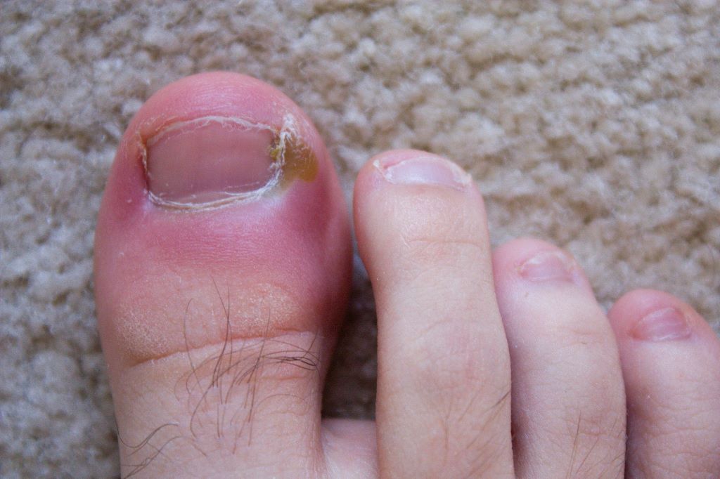
Scientists at Bath and King’s College London have discovered that a common chemical could be used to develop a new treatment for difficult-to-treat nail infections.
Paronychia. Source: Wikimedia Commons CC0
Hydrogen sulphide, the volcanic gas that smells of rotten eggs, could be used in a new treatment for tricky nail infections that acts faster but with fewer side effects, according to scientists at the University of Bath and King’s College London (KCL).
Nail infections are mostly caused by fungi and occasionally by bacteria. They are very common, affecting between 4-10% of the global population, rising to nearly half those aged 70 or over.
These infections can lead to complications, particularly in vulnerable groups such as diabetics and the elderly, but are notoriously difficult to treat.
Current treatments include oral antifungals taken in pill form, and topical treatments which are applied directly to the nail.
Oral antifungals take around 2-4 months to act and are reasonably effective, but they carry risks of side effects, especially in patients with other medical conditions.
Treatments applied directly to the nail are safer, but they often take much longer to work, sometimes taking even years to work, and they frequently relapse or fail.
This is largely because it’s very difficult to get the drug to penetrate through the nail to where the infection resides.
Even the most effective topical treatments have relatively low cure rates, so there is a clear need for new therapeutic approaches that are safe, effective, and capable of reaching microbes embedded deep within the nail.
A team from the University of Bath and King’s College London has now found that hydrogen sulphide (H₂S), a small, naturally occurring gas, could be developed into a promising new treatment.
Previous work has shown that it penetrates the nail plate far more efficiently than existing topical drugs, and now the team has demonstrated that it has strong antimicrobial activity against a wide range of nail pathogens, including fungi that are resistant to common antifungal treatments.
In laboratory tests, the team used a chemical that breaks down to release the H₂S gas and found that it acts in a unique way, disrupting microbial energy production and triggering irreversible damage, ultimately killing the fungi.
Dr Albert Bolhuis, from the University of Bath’s Department of Life Sciences, said: “Thanks to its ability to efficiently reach the site of infection and its novel mode of action, we believe that a topically applied medicine containing hydrogen sulphide could become a highly effective new treatment for nail infections, which avoids the limitations of current therapies.
“Our research lays the foundation for a compelling alternative to existing treatments, with the potential to improve outcomes for patients suffering from persistent and drug-resistant fungal nail infections.”
Hydrogen sulphide is known for its pungent smell of rotten eggs, and has some toxicity, however researchers believe the amounts required are well below toxicity levels and the correct formulation will limit any unpleasant odours.
The research has so far only been done in vitro, but the team hopes to develop a treatment that could be used in patients in the next five years.
Professor Stuart Jones, Director of the Centre for Pharmaceutical Medicine Research at KCL said: “We are looking forward to translating these findings into an innovative topical product that can treat nail infection.”
Science-backed insights and fascinating feats from the world of hair growth
Photo by Natasha Brazil on Unsplash
From a 2.26-metre Afro to hair strong enough to suspend a person mid-air, some of the world’s most jaw-dropping records remind us just how extraordinary human hair can be. While these feats may seem unbelievable, they highlight the biology behind hair’s strength, resilience, and growth potential.
“Exceptional hair growth is the result of discipline, not coincidence,” says Dr Kashmal Kalan, Medical Director at Alvi Armani South Africa. “A healthy scalp and uninterrupted growth cycles form the foundation for strong, resilient hair.”
The Biology Behind Every Strand
Hair grows in three phases: anagen (growth), catagen (transition), and telogen (rest). The anagen phase – lasting anywhere from two to seven years – largely determines how long hair can grow before naturally shedding. People who achieve exceptional lengths often have extended anagen phases, allowing their hair to keep growing far beyond the average.
While genetics set the baseline, lifestyle and environment play a powerful role in influencing growth potential. Nutrition, hormones, stress levels, and scalp health all impact the length and quality of the anagen phase. “We can’t rewrite DNA,” notes Dr Kalan, “but we can influence how genes express themselves.”
Lifestyle Matters
Healthy hair begins long before the styling stage.
Nutrition: Diets rich in protein, healthy fats, and vitamins support follicle strength.
Stress: Elevated stress hormones can shorten the growth phase and trigger shedding.
Sleep: Proper rest gives follicles the recovery time they need.
Scalp care: Gentle exfoliation, oiling, and protective styling can improve circulation and reduce breakage.
Science over hype
Despite bold marketing promises, there are no “miracle” serums that can regrow hair overnight. “Anything claiming dramatic growth in weeks is a red flag,” says Dr Kalan. Scientifically supported treatments – such as platelet-rich plasma (PRP), mesotherapy, and exosome therapy – can optimise follicle function, strengthen roots, and support sustained, natural growth.
“Healthy hair growth is a process grounded in biology, not marketing,” adds Dr Kalan.
Regenerative Approaches for Natural, Lasting Results
At Alvi Armani South Africa, regenerative science is at the core of every treatment. Using advanced techniques such as Follicular Unit Extraction (FUE) alongside PRP and exosome therapy, the clinic works with the body’s own biology to restore growth naturally.
“Our goal is always natural, lasting outcomes – hair that feels and looks strong, resilient, and vibrant,” concludes Dr Kalan.
From strength and endurance to sheer volume and creativity, these hair-related world records showcase just how remarkable human hair can be.
Most golf tees in hair:Anya Bannasch (USA) set a 2024 record with 711 golf tees in her hair – almost double her original goal.
Longest time suspended by hair:Leila Noone, a circus artist, hung from a single knot of her hair for over 25 minutes beneath California’s redwoods in 2025.
Largest afro:Jessica Martinez from New York City is attempting to break the record with an afro measuring about 36 cm high and 51 cm wide, inspiring confidence in natural hair.
Largest ball of human hair:“Hoss”, made entirely from donated hair, continues to grow through global contributions and features in Ripley’s Believe It or Not! exhibitions.
These feats highlight the versatility, strength, and creative expression found in something as simple – and as complex – as human hair.