
When starved of oxygen during a heart attack or stroke, cells unleash a flurry of emergency measures to protect themselves and the body. For decades, scientists have observed that the body’s production of a “three-tailed” lipid molecule consistently surges during this trauma but have puzzled over why. Now, Cornell researchers have uncovered its surprising role in cellular survival: protecting against damage when oxygen runs out.
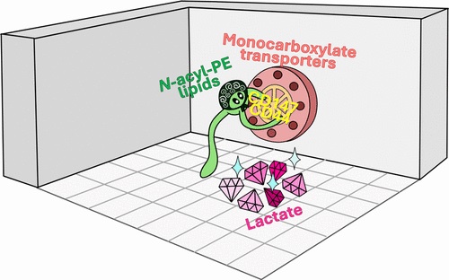
The research shows that the fat molecule, N-acylphosphatidylethanolamine (NAPE), helps cells survive ischaemia by driving lactic acid out of cells. This toxic byproduct builds up during emergency metabolism, and NAPE’s surge appears to be part of the body’s protective response. Though still in an early stage, the findings suggest that boosting or mimicking NAPE could one day help limit tissue damage in heart attack and stroke.
The study, published Sept. 5 in the Journal of the American Chemical Society, was led by graduate student Din-Chi Chiu and Jeremy Baskin, associate professor in the Department of Chemistry and Chemical Biology, and the Weill Institute for Cell and Molecular Biology.
“During heart attack or stroke, when there is an interruption in blood flow, the cells in the affected tissue, whether it is the heart or the brain, have to scramble to be able to continue to produce energy to survive,” Baskin said.
Under normal conditions, cells largely produce energy by a longer and much higher yielding process involving mitochondria.
“However, when energy needs are imminent and oxygen is limited, such as when blood flow is restricted, a metabolic switch occurs to favour glycolysis, which is a quick and dirty way of generating energy,” he said. “But to keep glycolysis going unabated, lactate, or lactic acid, is built up, and because it can be toxic at high levels, cells need to export it to prevent it from building up inside cells to undesirable levels.”
Because NAPE repels water and is short-lived in cells, studying it directly has been nearly impossible. The research team overcame this by designing and synthesising a chemical “look-alike” probe that tagged NAPE’s protein partners under UV light, revealing its interactions.
The researchers observed NAPE latching onto proteins that regulate lactate transport. In particular, it bound to two cell-surface proteins, CD147 and CD44, which control transport proteins that act like gates controlling how lactic acid moves in and out of cells. The team’s experiments showed that when NAPE levels rise, lactate transport ramps up, and blocking those transporters erased the effect.
“The work reframes NAPE as a signaling molecule,” Baskin said. “Our finding that NAPE can stimulate lactate export supports a model in which the role of NAPE in pathological events such as heart attack or stroke is part of a protective response.”
For now, the team is exploring whether different versions of NAPE, with different tail compositions, might fine-tune lactate regulation in unique ways. They are also interested in whether NAPE plays roles in other tissues beyond the heart and brain.
“Future studies in heart and brain tissue will test this hypothesis more directly,” Baskin said. “If confirmed, the work could support the creation of therapies that boost NAPE levels as a way to limit tissue damage in cardiovascular emergencies.”
Source: Cornell University