For older people with irregular heart rhythms who are at high risk of stroke and bleeding, standard care (including the use of blood thinners when indicated) was found to be the better choice compared to a promising, catheter-based procedure, according to a preliminary late-breaking science presentation today at the American Heart Association’s Scientific Sessions 2025.
The trial, Left Atrial Appendage CLOSURE in Patients with Atrial Fibrillation at High Risk of Stroke and Bleeding Compared to Medical Therapy (CLOSURE-AF), compared a catheter-based procedure to medical therapy among patients with atrial fibrillation (AFib), an irregular heart rhythm.
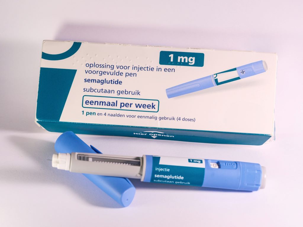
While blood thinners can be highly effective at reducing the risk of stroke among people with AFib, the medication may cause severe bleeding in some people. Due to this risk, researchers are exploring alternative treatments including this catheter-based procedure. The procedure, called a left atrial appendage closure, seals a small pouch in the heart called the left atrial appendage, or LAA, where blood clots can form. If these blood clots enter the bloodstream, it increases the risk of stroke. Closing this pouch reduces the risk of stroke. It also can allow people to stop taking blood thinners for clot prevention.
The CLOSURE AF study compared catheter-based left atrial appendage closure with physician-directed standard medical care (including timely anticoagulant blood thinning when eligible) in patients with atrial fibrillation at high risk for stroke and bleeding. The aim of the study was to demonstrate non-inferiority for catheter-based LAA closure regarding risk of stroke, systemic embolism, cardiovascular/unexplained death or major bleeding. However, this was not reached.
“We expected that catheter-based LAA closure would be comparable to physician-directed standard medical care often using blood thinning anticoagulant medications,” said study lead researcher Ulf Landmesser, MD, chairman of the department of cardiology, angiology and intensive care medicine at Deutsche Herzzentrum Charité and professor of cardiology at Charité University Medicine in Berlin. “However, this was not the case in this trial of older patients at very high risk of bleeding and stroke.
“Our findings indicate that standard physician-directed medical care, including blood thinners for eligible patients, remains a valid management option for those older patients with irregular heartbeat who are at very high risk for stroke and bleeding.”
Landmesser said that the results of the procedure are different for lower-risk patients, and studies investigating this are currently underway. Moreover, ongoing studies are comparing LAA closure in addition to blood thinning in very high-risk patients.
Because medical treatments and LAA closure for AFib remain in development the results of this study may not apply to future research, other techniques or procedures.
Study details, background and design:
More than 900 adults with AFib who were at high risk of stroke and major bleeding participated in this study.
Participants’ average age was 78 years, and 39% were women.
They were enrolled at 42 health care sites in Germany from March 2018 to April 2024, and they were followed for a median of 3 years.
Participants were randomly assigned to one of two treatment groups: standard medical care (including anticoagulant blood thinners, if eligible); or LAA closure.
Researchers compared the frequency of stroke, life-threatening blood clots, cardiovascular/unexplained death and major bleeding between the two treatment groups.
Scientists at NYU are developing a zinc-based treatment for tooth decay that combats bacteria, blocks pain, and avoids staining teeth – all without drilling
Photo by Caroline Lm on Unsplash
Tooth decay is the most common health condition worldwide. While it is preventable and treatable, billions of people are living with cavities and the pain that accompanies them.
Given the massive scale of the problem, there’s a growing movement in dentistry to treat cavities without drilling and filling them. One such approach is applying a clear liquid called silver diamine fluoride to the surface of teeth. Silver diamine fluoride is already FDA-approved to treat tooth sensitivity, and recent NYU research shows that the compound’s antimicrobial properties also make it effective at preventing cavities and stopping small cavities from progressing into larger ones. Because it’s inexpensive and easy to administer, it can be given in schools, in rural areas lacking dentists, or to patients who may have difficulty with dental care, including those with disabilities.
But treatment with silver diamine fluoride comes with one notable drawback: when the silver in it interacts with tooth decay, it turns the treated surface black. While this is not a significant issue for molars at the back of the mouth or baby teeth that fall out, it’s not a great option for teeth seen in a smile.
“Once your teeth are treated with silver diamine fluoride, that stain is permanent, which is a barrier for many people wanting to use the product,” explains Marc Walters, professor of chemistry at NYU.
Walters has long studied silver and other elements used in medicine to carry drugs and imaging contrast agents. Several years ago, he was approached by researchers at NYU College of Dentistry seeking to better understand how silver stains teeth in order to avoid that outcome.
From silver to zinc
Walters had an idea. What if another mineral could be used that was also colourless and antimicrobial but didn’t turn teeth black? This question led him to zinc, an important nutrient found in foods like oysters and beef, as well as in over-the-counter products intended to shorten the duration of colds. Zinc is also used in dentistry, including in toothpaste and mouthwash to fight bacteria and bad breath, as well as in some denture adhesives and cementing agents to affix crowns or temporary fillings.
Walters began studying a zinc phosphate compound to see how it interacts with cavities, and crucially, to determine whether it can permeate deep into teeth. In order to address pain and hypersensitivity, the compound would need to reach the tooth’s dentin, the porous material sandwiched between the hard enamel outer layer and the nerves within. Dentin contains an abundance of microscopic, hollow channels – in fact, 40 000 of these tubules are packed into each square millimetre of dentin.
“We had to develop a solution to give dentists that will be taken up in these very small openings and go deep enough in the tubules so that the material will be retained,” Walters explains.
Walters applied phosphate followed by zinc to slices of a human tooth. Under the microscope, he saw deposits of the compound deep inside the dentin tubules. But while the zinc phosphate successfully permeated the teeth, he knew that a simpler approach that didn’t require applying two treatments would be easier for dentists. “Two steps is one too many,” says Walters.
Drawing inspiration from silver diamine fluoride, Walters developed another zinc-based molecule called zinc tetramine difluoride, which forms a colourless zinc oxide deep inside dentin tubules. The agent starts out as a liquid that is sensitive to concentration and pH. When painted onto a tooth and absorbed, the conditions within dentin tubules prompt a chemical change that quickly turns it into a solid, blocking the tubules and slowly releasing the antimicrobial zinc into the tooth.
His team is continuing to develop several related compounds for the treatment of cavities and has applied for patents of these zinc-based materials in several countries.
Fast and slow
Having both fact-acting and long-lasting properties would offer an ideal combination for fighting cavities and tooth sensitivity, given that many current treatments for sensitive teeth require multiple applications and can take days or weeks to work.
“In one of our studies, two minutes after treatment with our agent, we can see using the electron microscope that the zinc forms long cylinders of mineral that occupy the tubules,” says Walters. “Blocking the dentin tubules cuts off access to the nerves that are much deeper in dentin. It’s like putting a cork in place that shuts off the lower portion of the tubule from the outside environment – and this happens within a minute or two.”
Walters shows an image of a tubule under the microscope that was filled with the zinc compound.
In additional tests, Walters found that zinc oxide persisted in tooth samples for at least one to two months. The goal is to develop a product that lasts for months or even years inside of teeth, stopping hypersensitivity and fighting bacteria on an ongoing basis.
“Not only do you have the analgesic result of having tubules blocked, but you also have a very low solubility agent that can slowly release the zinc into the tubule to prevent the growth of Streptococcus mutans and other bacteria,” Walters adds.
The journey from lab to shelves
With a promising zinc nanocrystal agent in hand, Walters sought out other experts at NYU and beyond. His work caught the attention of Southern Dental Industries (SDI), an Australian company that makes restorative dental materials, including silver diamine fluoride. The company purchased the license for the zinc technology and NYU is working with them to develop it.
Closer to home, Walters began collaborating with Deepak Saxena, professor of molecular pathobiology and director of research innovation and entrepreneurship at NYU College of Dentistry.
Saxena and Walters are collaborating on a new NIH grant to further develop the zinc-based treatment.
As a result of bringing together this diverse expertise Saxena and Walters received a award from NYU, and last month, secured a grant from the NIH.
The NIH grant will fund feasibility studies for Walters’s team to further develop the formulation and confirm its ability to block tubules in a range of dentin samples. It will also fund research through Periomics Care in which Saxena’s team will study the agent’s antimicrobial properties. Specifically, they will look to see if the zinc creates a “zone of inhibition” – preventing the growth of decay-causing bacteria in the vicinity of it or even killing bacteria that comes in contact with it.
“The mouth is full of bacteria. A compound needs to have good antimicrobial activity, which can occur from ionic imbalance, the properties of the zinc, or by the fluoride,” Saxena says. “If a compound does not stain, has good antimicrobial activity, plus it blocks the tubules, then it should be successful in stopping tooth decay and be aesthetically accepted.”
Saxena and Walters are already planning for the next phase of their research, which will include additional studies on the compound’s formulation, effectiveness, toxicity, and shelf life. Ultimately, if these studies go well, the researchers and SDI will approach the FDA for permission to do a clinic trial.
One factor working in their favour: because zinc phosphate has long been used as a dental adhesive, it’s known to be safe and the FDA has already approved it in other forms. These existing products may pave the way for faster research and development of a cavity treatment compared to untested elements, which can take many years to develop.
The future of dentistry
A new non-invasive treatment for cavities could be a game-changer in oral health. “We know that there’s a need – and a market – for a product that stops tooth decay that is effective, cheap, easy to use, and non-staining, given the rise in global numbers of untreated cavities,” Saxena says.
Dentists could use it to treat cavities without needing to scrape or drill out the cavity in preparation. Squirmy kids would need less time in the dentist’s chair. Older adults who get cavities near the roots of their teeth as their gums recede could have a new option for stopping sensitivity and decay in difficult-to-treat areas. If safe and effective, perhaps small quantities could even be available on drugstore shelves and sold directly to consumers.
For Walters and Saxena, their goal is a future with less tooth decay and pain – and if their studies of zinc confirm its potential, silver-stained teeth may be a thing of the past.
Researchers tracked 85 young adults over a four-year period, finding that increases in ultra-processed food consumption were linked with elevated blood sugar and early signs of diabetes risk.
Photo by Jonathan Borba
More than half of calories consumed in the United States come from ultra-processed foods (UPFs), items like fast food and packaged snacks that are often high in sodium, sugar and unhealthy fats. In adults, research has clearly linked these foods to type 2 diabetes and other conditions, but few studies have explored their effects among youth.
Now, researchers from the Keck School of Medicine of USC have completed one of the first studies to examine the link between UPF consumption and how the body processes glucose, which is known to predict diabetes risk. By tracking changes over time, they gained insights into how dietary choices may influence key biological processes.
The researchers studied a group of 85 young adults over a four-year period. They found that an increase in UPF intake was associated with a higher risk for prediabetes, or early-stage high blood sugar that can lead to diabetes. Eating more UPFs was also linked to insulin resistance, where the body becomes less effective at using insulin to control blood sugar. The study, funded in part by the National Institutes of Health, was just published in the journal Nutrition and Metabolism.
“Our findings show that even modest increases in ultra-processed food intake can disrupt glucose regulation in young adults at risk for obesity. These results point to diet as a modifiable driver of early metabolic disease, and an urgent target for prevention strategies among young people,” said senior author Vaia Lida Chatzi, MD, PhD, a professor of population and public health sciences and paediatrics and director of the ShARP Center at the Keck School of Medicine.
Early adulthood is a formative stage where people have reached physical maturity and are building habits that can persist for years. Trading packaged or restaurant meals for whole and raw foods like fruits, vegetables, and whole grains can reduce the likelihood of developing type 2 diabetes later in life.
“Young adulthood is a critical window for shaping long-term health,” Chatzi said. “By focusing on young adults, we have an opportunity to intervene early, before prediabetes and other risk factors become lifelong conditions.”
Signs of prediabetes
The research included 85 young adults from the Metabolic and Asthma Incidence Research (Meta-AIR) study, part of the broader Southern California Children’s Health Study. Participants, aged 17–22, provided data at a baseline visit between 2014 and 2018 and a follow-up visit approximately four years later.
At each visit, participants reported everything they had eaten on one recent weekday and one recent weekend day. Researchers classified foods into two categories: UPFs (such as candy, soda, cereal, packaged spreads, flavored yogurts, and many restaurant foods) and foods that were not ultra-processed. They then calculated what percentage of each participant’s daily caloric intake came from UPFs.
The researchers also collected blood samples from participants before and after they consumed a sugary drink to test how effectively their body responded to blood sugar with insulin. They then conducted a statistical analysis to compare dietary changes with signs of prediabetes, adjusting for differences in age, sex, ethnicity and physical activity levels.
From baseline to follow-up, a 10% increase in UPF consumption was associated with a 64% higher risk for prediabetes and a 56% higher risk for problems with glucose regulation. Participants who reported eating more UPFs at their initial visit were also more likely to have elevated insulin levels at follow-up—an early sign of insulin resistance, where the body must produce more insulin to keep blood sugar in a healthy range.
Limiting ultra-processed foods
The study shows that the risks of UPFs extend to young adults, a group often overlooked in previous research.
“These findings indicate that ultra-processed food consumption increases the risk for pre-diabetes and type 2 diabetes among young adults – and that limiting consumption of those foods can help prevent disease,” said the study’s first author, Yiping Li, a doctoral student in quantitative biomedical sciences at Dartmouth College who previously worked as a researcher at the Keck School of Medicine.
Future studies with larger groups and more detailed diet tracking can help clarify which foods pose the greatest risk for young adults, the researchers said. They also plan to continue investigating the biological mechanisms behind these links, including how specific nutrients in UPFs may influence insulin and blood sugar regulation.
A new University of California San Diego study offers compelling evidence that GLP-1 receptor agonists may do more than regulate blood sugar and weight. In an analysis of more than 6800 colon cancer patients across all University of California Health sites, researchers found that those taking glucagon-like peptide-1 (GLP-1) medications were less than half as likely to die within five years compared to those who weren’t on the drugs (15.5% vs 37.1%).
The study, led by Raphael Cuomo, PhD, used real-world clinical data from the University of California Health Data Warehouse to assess outcomes across the state’s academic medical centres. After adjusting for age, body mass index (BMI), disease severity and other health factors, GLP-1 users still showed significantly lower odds of death, suggesting a strong and independent protective effect.
The survival benefit appeared most pronounced in patients with very high BMI (over 35), hinting that GLP-1 drugs may help counteract the inflammatory and metabolic conditions that worsen colon cancer prognosis. Researchers believe several biological mechanisms could explain the link. Beyond regulating blood sugar, GLP-1 receptor agonists reduce systemic inflammation, improve insulin sensitivity and promote weight loss – all factors that can dampen tumour-promoting pathways. Laboratory studies also suggest that GLP-1 drugs may directly prevent cancer cell growth, trigger cancer cell death and reshape the tumour microenvironment. However, the study authors emphasise that more research is needed to confirm these mechanisms and determine whether the survival benefit observed in this real-world analysis represents a direct anti-cancer effect or an indirect result of improved metabolic health.
Cuomo notes that while these results are observational, they underscore an urgent need for clinical trials to test whether GLP-1 drugs can improve cancer survival rates, especially for patients with obesity-related cancers.
New method for the targeted production of specific cells
Figure 1 Schematic overview of the experimental workflow. MSCs (blue) were singly encapsulated using a microfluidic approach within calcium-crosslinked, RGD-functionalized alginate microgels (pink), followed by a secondary APA and calcium coating to enhance stability. Encapsulated cells were cultured for 21 days and subjected to cyclic hydrostatic pressure in regular cell culture media without any growth factors. Source: İyisan et al., Small Science, 2025.
For the first time, researchers at the Technical University of Munich (TUM) have succeeded in using nanorobots to stimulate stem cells with such precision that they are reliably transformed into bone cells. To achieve this, the robots exert external pressure on specific points in the cell wall. The new method offers opportunities for faster treatments in the future.
Prof Berna Özkale Edelmann’s nanorobots consist of tiny gold rods and plastic chains. Several million of them are contained in a gel cushion measuring just 60 micrometres, together with a few human stem cells. Powered and controlled by laser light, the robots, which look like tiny balls, mechanically stimulate the cells by exerting pressure. “We heat the gel locally and use our system to precisely determine the forces with which the nanorobots press on the cell – thereby stimulating it,” explains the professor of nano- and microrobotics at TUM. This mechanical stimulation triggers biochemical processes in the cell. Ion channels change their properties, and proteins are activated, including one that is particularly important for bone formation.
Heart and cartilage cells: finding the correct stress pattern
If stimulation is carried out at the right rhythm and with the right (low) force, a stem cell can be reliably triggered to develop into a bone cell within three days. This process can be completed within three weeks. “The corresponding stress pattern can also be found for cartilage and heart cells,” asserts Berna Özkale Edelman. “It’s almost like at the gym: we train the cells for a particular area of application. Now we just have to find out which stress pattern suits each cell type,” says the head of the Microbiotic Bioengineering Lab at TUM.
Mechanical forces pave the way for transformation into bone cells
The research team produces bone cells using mesenchymal stem cells. These cells are considered to be the body’s ‘repair cells’. They are approximately 10 to 20 micrometres in size and are generally capable of developing into bone, cartilage or muscle cells, for example. The challenge: The transformation into differentiated cells is complex and has been difficult to control until now. “We have developed a technology that allows forces to be applied to the cell very precisely in a three-dimensional environment,” says TUM scientist Özkale Edelmann. “This represents an unprecedented advance in the field.” The researchers believe that this method can even be used to produce cartilage and heart cells from human stem cells.
Automation is the next step
For treatments, doctors will ultimately need far more differentiated cells – around one million. “That’s why the next step is to automate our production process so that we can produce more cells more quickly,” says Prof Özkale Edelmann.