Day: September 22, 2025

GLP-1 Receptor Agonists Protect the Liver During Alcohol Consumption

Photo by Apolo Photographer on Unsplash

GLP-1 receptor agonists are also promising for the treatment of alcohol use disorder and alcohol-associated liver disease, as growing evidence suggests they reduce the motivation to drink alcohol. Now, surprising new findings reveal that the medications may have direct protective effects on the liver as well.

In a new study, published in npj Metabolism Health and Disease, Yale School of Medicine (YSM) researchers found that in mice, GLP-1RAs reduced an enzyme that metabolises alcohol. That, in turn, decreased the production of toxic alcohol metabolites.

“This is the first time that GLP-1 receptor agonists have been shown to regulate alcohol metabolism in the liver,” says principal investigator Wajahat Mehal, MD, professor of medicine (digestive diseases) at YSM. “If you’re taking semaglutide, then your body will likely handle alcohol differently.”

However, because these drugs slowed the metabolism of alcohol, the mice also had higher blood alcohol levels, the researchers found.

“GLP-1 receptor agonists seem to have very similar effects in mice and humans,” says Mehal. “If these results are also reproduced in humans, people using GLP-1 receptor agonists might be drinking an amount of alcohol that does not normally put them above the legal blood alcohol level, but because they are taking this drug, it does.”

Further studies in humans are needed to understand these impacts of GLP-1 receptor agonists more fully, he stresses.

GLP-1 receptor agonists decrease toxic alcohol metabolites

In the study, researchers gave mice either a GLP-1 receptor agonist or a placebo. They observed that mice receiving the medication had decreased levels of a liver enzyme known as Cyp2e1, which breaks down alcohol into a toxic metabolite called acetaldehyde.

“This is significant because alcohol itself is actually not the most toxic molecule to the liver,” explains Mehal. Rather, acetaldehyde is one of the major drivers of alcohol-related harm to the liver. “These drugs are resulting in less acetaldehyde.”

The findings suggest that not only might GLP-1 receptor agonists help the liver by acting on the brain to reduce alcohol consumption, but also through slowing metabolism of alcohol in the liver, and in turn reducing the levels of toxic metabolites.

Ongoing clinical trials are currently testing the benefits of semaglutide for people living with alcohol-induced liver disease. The study suggests that GLP-1 receptor agonists may offer greater benefits to the liver than previously thought, and that the drug may still help patients who are not abstaining from alcohol.

“Even if some individuals don’t reduce their alcohol intake while they’re on a GLP-1 receptor agonist, they will probably still have hepatic protection, because fewer toxic metabolites will be produced in the liver,” Mehal says.

GLP-1 receptor agonists increase blood alcohol concentration

In another experiment, the researchers measured blood alcohol concentrations of mice 30 minutes after giving them alcohol. They found that mice who had received a GLP-1 receptor agonist had higher blood alcohol concentrations compared to controls, and that these levels took longer to drop.

More research is needed to better understand the consequences of elevated blood alcohol levels on the rest of the body, Mehal says.

“If the liver is not metabolizing alcohol as quickly, the alcohol load could be shifted to other organs,” he poses. “Then, not only might people have a high blood alcohol level, but may also experience more cognitive effects like discoordination.”

The number of people taking these drugs is rapidly increasing—as many as one in eight adults in the U.S. have used or are currently using a GLP-1 receptor agonist. Meanwhile, about half of U.S. adults drink alcohol and 6% report drinking heavily.

As the use of these medications for a range of conditions continues to rise, it is increasingly important to study the interactions between these medications and alcohol, Mehal says.

“There already are large numbers of people who are taking GLP-1 receptor agonists and are drinking either social amounts or excess amounts of alcohol,” says Mehal. “We need to know the effects of these drugs in that setting.

Source: Yale School of Medicine

Landmark Study Finds Perispinal Etanercept of No Benefit to Stroke Trial Participants

A treatment for stroke patients was no more effective than an inactive drug

Source: CC0

The first international trial of an unproven stroke treatment available in the US has concluded that, while harmless, perispinal etanercept is no more effective than an inactive dummy drug, or placebo.

Survivors of stroke have travelled at considerable expense to private clinics in the US to be treated with the arthritis drug etanercept.

In the clinics, the drug is injected into the cervical spinal area, and the patient is then tilted head-down in the belief that this allows the drug to enter the brain.

Stroke is a leading cause of disability throughout the world, affecting more than 7 million people a year. Despite advances, treatments for impairment after stroke remain limited. Some patients call perispinal etanercept a “miracle cure”.

Florey leading stroke researcher, neurologist Professor Vincent Thijs led the Perispinal Etanercept to improve STroke Outcomes – or “PESTO” – trial to investigate this further, supported by funding from the Australian Government.

“We understand why people living with the long-term effects of stroke seek hope and new options,” Professor Thijs said. “With support from the Stroke Foundation and the Medical Research Future Fund, we put this treatment to the test using the gold standard of clinical research – a double-blind randomised trial.”

Half of the PESTO participants were treated with the drug, and half were treated with an inactive dummy drug, with patients and doctors “blind” to who was getting which.

This type of trial eliminates biases because neither doctors nor patients knew who was getting etanercept and who was getting the placebo. Because the results for the 2 patient groups were so similar, we concluded that while the drug did not cause harm, we found no evidence that it led to improved quality of life compared to placebo.

Professor Thijs, who leads the Young Stroke Service at The Florey, said improvements could be due to the placebo effect, a well-established medical phenomenon where some patients in a trial may notice an improvement, despite only receiving a dummy treatment.

Key PESTO trial results, published in Neurology:

  • 126 people from Australia and New Zealand participated in PESTO.
  • 63 received the treatment, 63 the placebo.
  • Their stroke symptoms were measured before the trial and 28 days after.
  • There were no adverse side effects.
  • Among participants who received perispinal etanercept, 52 per cent (33 out of 63) felt better.
  • Among participants who received the placebo, 57 per cent (36 out of 63) felt better.
  • The difference in results between the 2 groups is deemed statistically insignificant.

“It’s important for doctors and the stroke survivor community in Australia and around the world to know that we found no evidence that perispinal etanercept improved quality of life,” Professor Thijs said.

Kelvin Hill, Executive Director of Stroke Programs, Research and Innovation at Stroke Foundation said: “Every Australian stroke patient should have access to the best, evidence-based treatment. Findings of the PESTO study underscore the critical importance of robust research and clinical trials in discovering if treatments work or not.

“Australians experience around 46 000 stroke events every year (one every 11 minutes), and there are now over 440 000 survivors of stroke living in Australia. Stroke Foundation will continue to advocate for more research funding to unlock new effective treatments for stroke; and ensure that advice provided in the Living Clinical Guidelines for Stroke Management enables clinicians to provide the best stroke care possible,” Mr Hill added.

Source: Florey Institute of Neuroscience and Mental Health

Study Affirms Efficacy of Nicotinamide for Skin Cancer Prevention 

Overall, there was a 14% reduction in skin cancer risk. When nicotinamide was taken after a first skin cancer, the risk reduction rose to 54%.

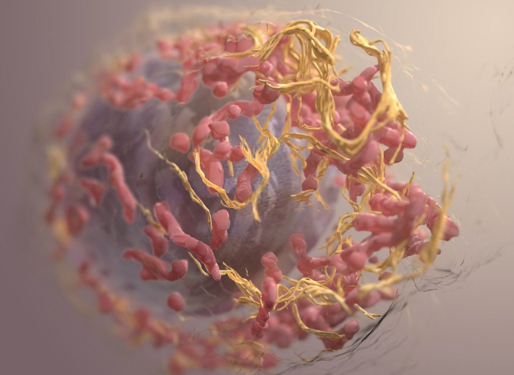
3D structure of a melanoma cell derived by ion abrasion scanning electron microscopy. Credit: Sriram Subramaniam/ National Cancer Institute

The dietary supplement nicotinamide has been recommended by dermatologists for people with a history of skin cancer since 2015, when a clinical study with 386 participants showed that those who took the vitamin B3 derivative developed fewer new occurrences. 

But data to validate those findings in a larger study group has been lacking because nicotinamide can be purchased over the counter without being entered into patients’ medical records.

In a new study published in JAMA Dermatology, researchers found a way to get that data by analysing records from the Veterans Affairs Corporate Data Warehouse. Nicotinamide is on the VA’s official formulary, so the researchers checked the outcomes of 33 833 patients for their next skin cancer diagnosis following baseline treatment with 500 milligrams of nicotinamide twice daily for longer than 30 days. They looked for occurrences of basal cell carcinoma and cutaneous squamous cell carcinoma. 

The researchers compared 12 287 patients who received the treatment with 21 479 who did not. Overall, there was a 14% reduction in skin cancer risk. When nicotinamide was taken after a first skin cancer, the risk reduction rose to 54%, but the benefit declined with treatment initiation following subsequent skin cancers. The risk reduction was much larger for squamous cell carcinoma.  

“There are no guidelines for when to start treatment with nicotinamide for skin cancer prevention in the general population. These results would really shift our practice from starting it once patients have developed numerous skin cancers to starting it earlier. We still need to do a better job of identifying who will actually benefit, as roughly only half of patients will develop multiple skin cancers,” said the study’s corresponding author, Lee Wheless, MD, PhD, assistant professor of Dermatology and Medicine at Vanderbilt University Medical Center and a staff physician at VA Tennessee Valley Healthcare System. 

The researchers were also able to ascertain the outcomes of 1,334 patients who were immunocompromised due to having received solid organ transplants. Among solid organ transplant recipients, no overall significant risk reduction was observed, although early nicotinamide use was associated with reduced occurrences of cutaneous squamous cell carcinoma. 

Source: Vanderbilt University Medical Center

Magnesium Inhibits Colorectal Cancer Development – Mostly in Females

The findings from the current study suggest that magnesium also increases the gut synthesis of vitamin D, which does not go to the blood and takes effect locally.

Photo by Danilo Alvesd on Unsplash

Researchers from Vanderbilt University Medical Center have demonstrated in a precision-based clinical trial that a magnesium supplement increases gut bacteria in humans that have been shown to synthesise vitamin D and inhibit colorectal cancer carcinogenesis.

However, the effect was observed primarily in females – an outcome that the researchers surmised may be attributable to the role that oestrogen plays in shifting magnesium from circulation into cellular uptake.

Intestinal microbiome data and colonoscopy results were analysed from participants who were randomised by whether they had the TRPM7 genotype, which plays a crucial role in regulating magnesium and calcium uptake.

Previously, the investigators showed in the same randomised trial that magnesium enhances the synthesis of vitamin D and increases the blood levels of vitamin D. The findings from the current study suggest that magnesium also increases the gut synthesis of vitamin D, which does not go to the blood and takes effect locally.

These results from the trial were published in The American Journal of Clinical Nutrition.

“Our previous study showed magnesium supplementation increased blood levels of vitamin D when vitamin D levels were low,” said Qi Dai, MD, PhD, professor of Medicine. “The current study reveals that magnesium supplementation also increases the gut microbes which have been shown to synthesise vitamin D in the gut without sunlight and locally inhibit colorectal cancer development.”

The participants were divided into two arms, one that received the magnesium supplement and another that received a placebo. Their gut microbiome was analysed from stools, rectal swabs and rectal tissues. Among participants with adequate TRPM7 function, the magnesium supplement increased Carnobacterium maltaromaticum and Faecalibacterium prausnitzii, which were previously found to work synergistically to increase vitamin D and decrease colorectal carcinogenesis. Among those with inadequate TRPM7 function, the magnesium supplement reduced the abundance of F. prausnitzii in rectal mucosa.

Among 236 participants who all had a history of colorectal polyps, 124 underwent colonoscopies after completing the trial with a 3.5-year median follow-up time. A higher abundance of F. prausnitzii in rectal mucosa was associated with an almost threefold increase in developing additional polyps.

Source: Vanderbilt University Medical Center

Study Assesses Cancers in Children Exposed to Medical Imaging

Study of nearly 4 million children and adolescents finds that 10% of paediatric blood and bone marrow cancers may have stemmed from radiation exposure.

Credit: Pixabay CC0

A study led by UC San Francisco and UC Davis has concluded that radiation from medical imaging is associated with a higher risk of blood cancers in children.

For the study, which appears in NEJM, the researchers examined data from nearly 4 million children and estimated that 1 in 10 blood cancers – some 3000 cancers in all – may be attributable to radiation exposure from medical imaging. The risk increased proportionally based on the cumulative amount of radiation the children received.

The investigation is the first comprehensive assessment using data from children and adolescents in North America that quantifies the association between radiation exposure from medical imaging and blood and bone marrow cancers, such as leukaemia and lymphoma, which are the most common forms of cancer in children and adolescents.

Medical imaging saves lives by enabling timely diagnosis and effective treatment, but it also exposes patients to ionizing radiation, a known carcinogen, particularly through computed tomography (CT).

The authors caution that doctors and parents should avoid excessive radiation doses and minimize exposure when clinically feasible.

“Children are particularly vulnerable to radiation-induced cancer due to their heightened radiosensitivity and longer life expectancy,” said Rebecca Smith-Bindman, MD, a radiologist and professor of Epidemiology and Biostatistics, as well as Obstetrics, Gynecology and Reproductive Sciences at UCSF and the first author of the paper.

“While medical imaging can be lifesaving, our findings underscore the critical need to carefully evaluate and minimise radiation exposure during paediatric imaging to safeguard children’s long-term health,” said Smith-Bindman, who is also a member of the Philip R. Lee Institute for Health Policy Studies. “This involves ensuring that imaging is performed only when it provides essential information for the child’s care and, in cases such as CT scans, using the lowest possible radiation doses.”

Documenting risks in children

The study uses a retrospective cohort design, looking back at the complete imaging histories of 3.7 million children who were born between 1996 and 2016. The children were treated at six health care systems in the U.S. and Ontario, Canada. Investigators found a significant relationship between cumulative radiation dose and the risk of a hematologic malignancy, which includes tumours affecting the blood, bone marrow, lymph, and lymphatic system.

The risk of developing cancer varied significantly by imaging modality. CT, which is used to detect many abnormalities such as tumours, heart disease, and injuries of the spinal cord and brain, entails significant radiation exposure. But radiographs, which are used to diagnose both broken bones and pneumonia, expose children to much lower doses.

Among all the forms of medical imaging, the study found that chest radiography was the most common imaging exam that doctors performed. The most common form of CT was of the head and brain.

For children who underwent a head CT, the researchers attributed about a quarter of the children’s subsequent hematologic malignancies to radiation exposure. For those who had radiographs, by contrast, they estimated that only a small fraction of the children’s subsequent cancers were associated with radiation exposure.

Getting one or two head CTs was associated with a 1.8-fold increased risk of a cancer diagnosis, and this rose to 3.5 times for children who received more scans and were therefore exposed to more radiation.

Altogether, 2961 haematologic malignancies were diagnosed during the study period. Lymphoid malignancies accounted for 79.3%, while myeloid malignancies and acute leukaemia together accounted for 15.5%. About 58% of cancers occurred in males, and about half were diagnosed in children under 5.

The authors said that up to 10% of haematologic malignancies in children and adolescents could be prevented by reducing unnecessary imaging and optimising radiation doses. In many cases, the authors said, substituting non-ionising imaging modalities like ultrasound or MRI may be feasible without compromising diagnostic accuracy.

Benefits vs risks

The authors emphasised that while medical imaging remains an invaluable tool in paediatric care, their findings highlight the need to carefully balance its diagnostic benefits with potential long-term risks.

“This study provides robust, directly observed evidence of a clear dose–response relationship between radiation from medical imaging and hematologic malignancy risk in children and adolescents,” said Diana Miglioretti, PhD, UC Davis Health professor and chief of the Division of Biostatistics.

“Our findings align with international research highlighting that children are especially radiosensitive,” Miglioretti added. “It’s crucial for clinicians to weigh the immediate benefits of imaging against potential long-term health risks and to optimise imaging protocols to minimize radiation exposure.”

Source: University of California – San Francisco

New Study Reveals a Hidden Risk After Cervical Cancer

Cervical cancer. Credit: Scientific Animations CC4.0

For women who’ve overcome cervical cancer, new research from MUSC Hollings Cancer Center points to another health risk that may not be on their radar: anal cancer.

Led by Hollings researchers Haluk Damgacioglu, Ph.D., and Ashish Deshmukh, PhD, co-leader of the Cancer Prevention and Control Research Program, the study sheds light on an under-recognised risk facing women with a history of cervical cancer – and highlights the need for updated screening guidelines. The paper was published in JAMA Network Open.

While cervical cancer is one of the most preventable cancers, with a survival rate of over 90% when found early, clinical guidelines have not addressed what comes next for these patients, who may be at high risk for a related type of cancer.

“We’ve known for a long time that both cervical and anal cancers are caused by HPV, the human papillomavirus,” Deshmukh said. “But what hasn’t been well-understood is how that shared risk might connect the two diseases over a woman’s lifetime.”

Currently, anal cancer screening is recommended for certain high-risk groups, such as people living with HIV, organ transplant recipients and women with a history of vulvar cancer. But there are no clear screening guidelines for women with cervical cancer.

One problem has been a lack of long-term data on their risk and how that risk changes with age and over time. This study helps to fill that gap using high-quality, population-based data.

The researchers turned to the National Cancer Institute’s SEER (Surveillance, Epidemiology and End Results) program – a comprehensive set of registries that tracks cancer diagnoses across the US. They analysed data from more than 85 000 women diagnosed with cervical cancer, tracking them over two decades to see how many went on to develop anal cancer and when those diagnoses occurred.

What they found was striking. Compared with the general population, women with a history of cervical cancer had nearly twice the risk of developing anal cancer.

Anal cancer rates increased with age and over time, with the most diagnoses found in women ages 65 to 74 who were more than 15 years out from their original diagnosis. For women in this age group, the rate of anal cancer diagnoses surpassed a widely accepted threshold for recommending routine screening.

“Our study shows that the risk doesn’t go away – it actually increases with age and over time,” Damgacioglu said.

Why the delay? HPV-related cancers often take years, sometimes decades, to develop. In some cases, the virus may linger undetected or have spread from another part of the body.

“It’s a slow process,” Deshmukh said, “and that’s part of why it’s been so hard to detect. By the time symptoms show up, the cancer is often advanced.”

While anal cancer screening is not as routine as screening for other cancers, reliable methods do exist, including anal cytology (a kind of Pap screen) and anoscopy. Unfortunately, access to specialised screening remains limited. In South Carolina, for example, there is currently only one provider trained to perform high-resolution anoscopy.

That is why it is so important to identify and prioritise the highest-risk groups.

“These results tell us that women who had cervical cancer years ago should be considered for routine anal cancer screening,” Damgacioglu said. “Right now, that’s not happening.”

Deshmukh and his team are now working on a project to determine when and how often screening should happen.

Source: Medical University of South Carolina