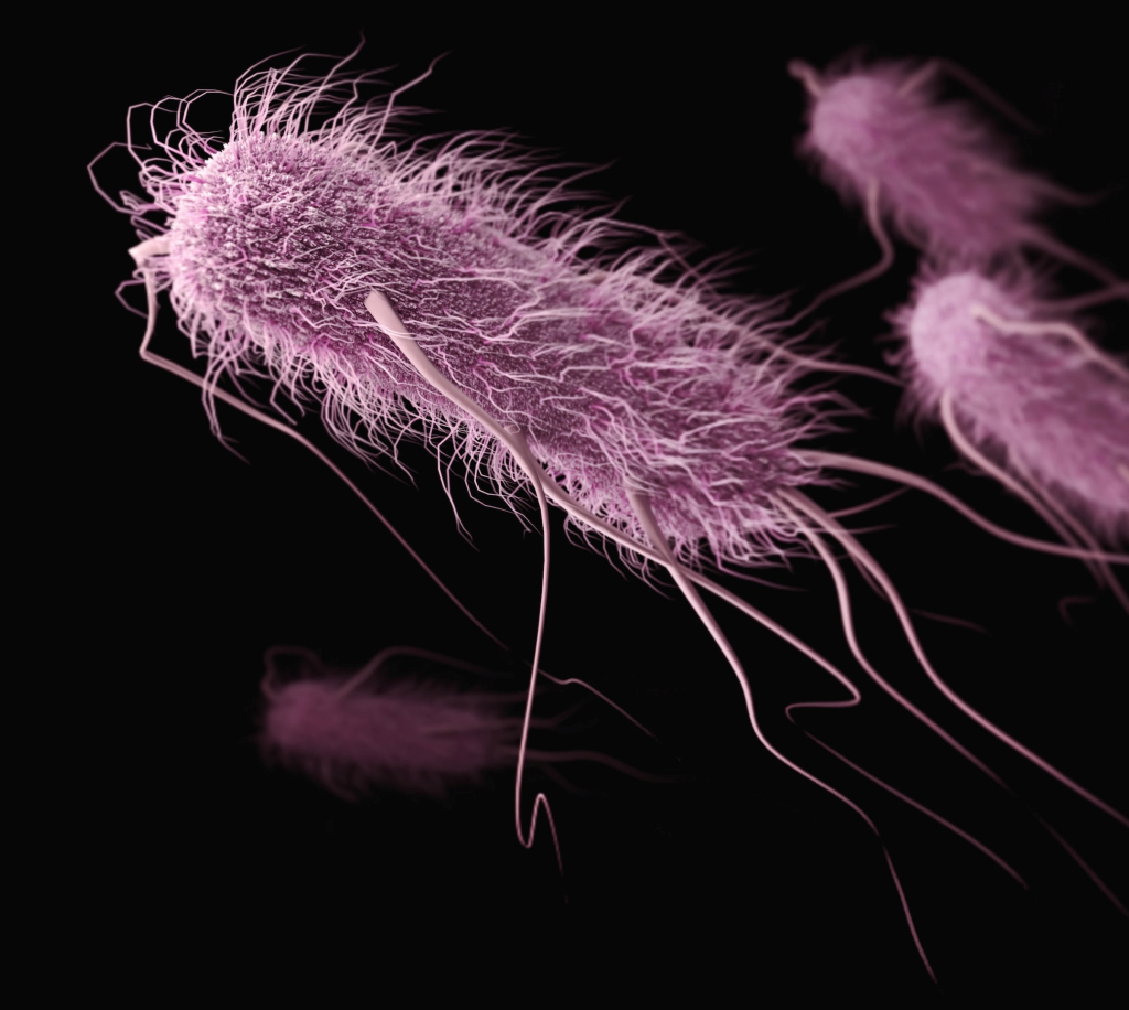
Researchers have identified how pathogenic genes in some Providencia spp., which have gained attention as causes of food poisoning as well as enterohaemorrhagic Escherichia coli. O157 and Salmonella, are transferred within bacterial cells. Their findings are expected to provide new insights into the identification of infection routes of Providencia spp. and the establishment of preventive methods for food poisoning.
Recently, Providencia spp. which have been detected in patients with gastroenteritis, and similar to enterohemorrhagic Escherichia coli. O157 and Salmonella spp., have been attracting attention as causative agents of food poisoning. For children with low immunity, food poisoning can be lethal as it causes severe symptoms such as diarrhoea and dehydration, so clarifying the source of infection and pathogenic factors of Providencia spp., and establishing preventive methods are urgent issues worldwide.
A joint research group led by Professor Shinji Yamasaki, Dr Sharda Prasad Awasthi, a Specially Appointed Lecturer, and graduate student Jayedul Hassan from the Graduate School of Veterinary Science, Osaka Metropolitan University, determined how the pathogenic genes in some Providencia spp. such as Providencia alcalifaciens and Providencia rustigianii are transferred within bacterial cells of genus Providencia. The group has also elucidated that the pathogenic genes of Providencia rustigianii are also transferred to other bacterial cells belonging to Enterobacteriaceae.
Professor Yamasaki concluded, “This achievement is expected to provide new insights into the identification of infection routes of Providencia spp. and the establishment of preventive methods for food poisoning.”
Despite stringent infection-control efforts around the world, hospital-acquired infections (HAIs)keep on popping up from new strains of bacteria. In Science Translational Medicine, researchers report evidence pointing to an unexpected source of such bacteria: the hospitalised patients themselves.
From experiments with mice, researchers at Washington University School of Medicine in St. Louis discovered that urinary tract infections (UTIs) can arise after sterile tubes, called catheters, are inserted into the urinary tract, even when no bacteria are detectable in the bladder beforehand. Such tubes are commonly used in hospitals to empty the bladders of people undergoing surgery. In the mice, inserting the tubes activated dormant Acinetobacter baumannii bacteria hidden in bladder cells, triggering them to emerge, multiply and cause UTIs, the researchers said.
The findings suggest that screening patients for hidden reservoirs of dangerous bacteria could supplement infection-control efforts and help prevent deadly HAIs.
“You could sterilise the whole hospital, and you would still have new strains of A. baumannii popping up,” said co-senior author Mario Feldman, PhD, a professor of molecular microbiology. “Cleaning is just not enough, and nobody really knows why. This study shows that patients may be unwittingly carrying the bacteria into the hospital themselves, and that has implications for infection control. If someone has a planned surgery and is going to be catheterised, we could try to determine whether the patient is carrying the bacteria and cure that person of it before the surgery. Ideally, that would reduce the chances of developing one of these life-threatening infections.”
The notoriously multidrug-resistant A. baumannii is a major threat to patients, causing many cases of UTIs in people with urinary catheters, pneumonia in people on ventilators, and bloodstream infections in people with central-line catheters into their veins.
The researchers set out to investigate why so many A. baumannii UTIs develop after people receive catheters.
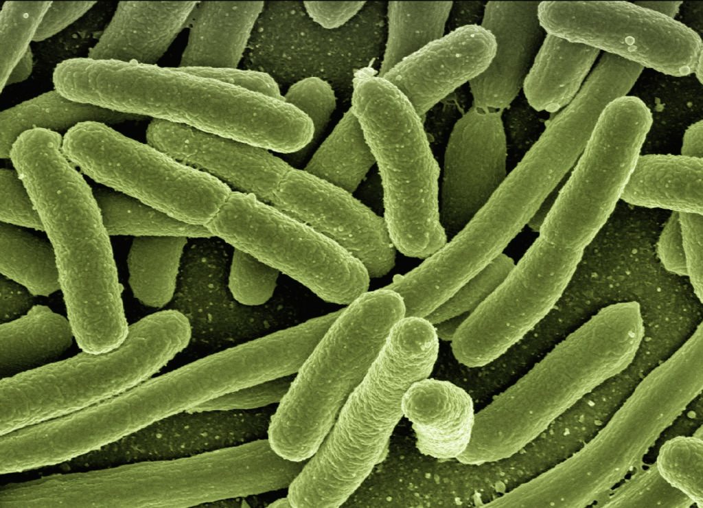
Most UTIs among otherwise healthy people are caused by the bacterium Escherichia coli. Research has shown that E. coli can hide out in bladder cells for months after a UTI seems to have been cured, and then re-emerge to cause another infection.
The researchers investigated whether A. baumannii can hide inside cells like E. coli can. They studied mice with UTIs caused by A. baumannii. They used mice with weakened immune systems because, like people, healthy mice can fight off A. baumannii.
Once the infections had resolved and no bacteria were detected in the mice’s urine for two months, the researchers inserted catheters into the mice’s urinary tracts with a sterile technique. Within 24 hours, about half of the mice developed UTIs caused by the same strain of A. baumannii as the initial infection.
“The bacteria must have been there all along, hiding inside bladder cells until the catheter was introduced,” said co-senior author Scott J. Hultgren, PhD, a professor and expert on UTIs. “Catheterisation induces inflammation, and inflammation causes the reservoir to activate, and the infection blooms.”
Since A. baumannii rarely causes symptoms in otherwise healthy people, many people who carry the bacteria may never know they’re infected, the researchers said. According to the researchers’ literature search, 2% of healthy people carry A. baumannii in their urine.
“I wouldn’t put much weight on the precise percentage, but I think we can say with certainty that some percentage of the population is walking around with A. baumannii,” Feldman said. “As long as they’re basically healthy, it doesn’t cause any problems, but once they’re hospitalised, it’s a different matter. This changes how we think about infection control. We can start considering how to check if patients already have Acinetobacter before they receive certain types of treatment; how we can get rid of it; and if other bacteria that cause deadly outbreaks in hospitals, such as Klebsiella, hide in the body in the same way. That’s what we’re working on figuring out now.”
Scientists have found that common artificial sweeteners can turn previously healthy gut bacteria pathogenic, invading the gut wall and potentially leading to serious health issues.
This study is the first to show the pathogenic effects of some of the most widely used artificial sweeteners (saccharin, sucralose, and aspartame) on two types of gut bacteria, Escherichia coli and Enterococcus faecalis. E. faecalis is capable of crossing the intestinal wall to enter the bloodstream and congregate in the lymph nodes, liver, and spleen, causing a number of infections including septicaemia. To top it off, this commensal bacteria has emerged as a multi-drug resistant pathogen.
Previous studies have shown that artificial sweeteners can affect the composition of gut bacteria, but this new molecular research, led by academics from Anglia Ruskin University (ARU), has shown that sweeteners can also induce pathogenic features in certain bacteria. It found that these pathogenic bacteria can latch onto, invade and kill epithelial Caco-2 cells lining the intestinal wall.
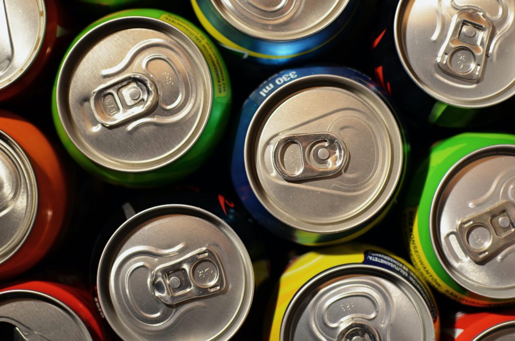
This new study discovered that at a concentration equivalent to two cans of diet soft drink, all three artificial sweeteners significantly increased the adhesion of both E. coli and E. faecalis to intestinal Caco-2 cells, and differentially increased biofilm formation. Bacteria growing in biofilms are less sensitive to antimicrobial resistance treatment and are more likely to secrete toxins and express disease-causing virulence factors.
Additionally, all three sweeteners caused the pathogenic gut bacteria to invade Caco-2 cells found in the wall of the intestine, save for saccharin, which had no significant effect on E. coli invasion.
Senior author Dr Havovi Chichger, Senior Lecturer in Biomedical Science at ARU, said: “There is a lot of concern about the consumption of artificial sweeteners, with some studies showing that sweeteners can affect the layer of bacteria which support the gut, known as the gut microbiota.
“Our study is the first to show that some of the sweeteners most commonly found in food and drink—saccharin, sucralose and aspartame—can make normal and ‘healthy’ gut bacteria become pathogenic. These pathogenic changes include greater formation of biofilms and increased adhesion and invasion of bacteria into human gut cells.
“These changes could lead to our own gut bacteria invading and causing damage to our intestine, which can be linked to infection, sepsis and multiple-organ failure.
“We know that overconsumption of sugar is a major factor in the development of conditions such as obesity and diabetes. Therefore, it is important that we increase our knowledge of sweeteners versus sugars in the diet to better understand the impact on our health.” Source: EurekAlert!
Journal reference: Shil, A & Chichger, H (2021) Artificial Sweeteners Negatively Regulate Pathogenic Characteristics of Two Model Gut Bacteria, E. coli and E. faecalis. International Journal of Molecular Sciences. doi.org/10.3390/ijms22105228.