Johns Hopkins University researchers have grown a novel whole-brain organoid, complete with neural tissues and rudimentary blood vessels, in an advance that could usher in a new era of research into neuropsychiatric disorders such as autism.
“We’ve made the next generation of brain organoids,” said senior author Annie Kathuria, an assistant professor in JHU’s Department of Biomedical Engineering who studies brain development and neuropsychiatric disorders. “Most brain organoids that you see in papers are one brain region, like the cortex or the hindbrain or midbrain. We’ve grown a rudimentary whole-brain organoid; we call it the multi-region brain organoid (MRBO).”
The research, published in Advanced Science, marks one of the first times scientists have been able to generate an organoid with tissues from each region of the brain connected and acting in concert. Having a human cell-based model of the brain will open possibilities for studying schizophrenia, autism, and other neurological diseases that affect the whole brain – work that typically is conducted in animal models.
To generate a whole-brain organoid, Kathuria and members of her team first grew neural cells from the separate regions of the brain and rudimentary forms of blood vessels in separate lab dishes. The researchers then stuck the individual parts together with sticky proteins that act as a biological superglue and allowed the tissues to form connections. As the tissues began to grow together, they started producing electrical activity and responding as a network.
Much smaller compared to a real brain – weighing in at 6 million to 7 million neurons compared with tens of billions in adult brains – these organoids provide a unique platform on which to study whole-brain development.
The researchers also saw the creation of an early blood–brain barrier formation, a layer of cells that surround the brain and control which molecules can pass through.
“We need to study models with human cells if you want to understand neurodevelopmental disorders or neuropsychiatric disorders, but I can’t ask a person to let me take a peek at their brain just to study autism,” Kathuria said. “Whole-brain organoids let us watch disorders develop in real time, see if treatments work, and even tailor therapies to individual patients.”
Using whole-brain organoids to test experimental drugs may also help improve the rate of clinical trial success, researchers said. Roughly 85% to 90% of drugs fail during Phase 1 clinical trials. For neuropsychiatric drugs, the fail rate is closer to 96%. This is because scientists predominantly study animal models during the early stages of drug development. Whole-brain organoids more closely resemble the natural development of a human brain and likely will make better test subjects.
“Diseases such as schizophrenia, autism, and Alzheimer’s affect the whole brain, not just one part of the brain. If you can understand what goes wrong early in development, we may be able to find new targets for drug screening,” Kathuria said. “We can test new drugs or treatments on the organoids and determine whether they’re actually having an impact on the organoids.”
Dr Kashmal Kalan urges patients to prioritise health before surgery – and offers hope to those recovering from illness
Hair loss is often viewed as a cosmetic concern, but emerging clinical insights confirm what many medical professionals have long understood: overall health is one of the most significant contributors to hair loss, and a crucial factor in whether hair restoration procedures succeed.
According to Dr Kashmal Kalan, Medical Director at Alvi Armani South Africa, chronic conditions such as diabetes, hypertension, and high cholesterol are closely linked to diffuse hair thinning, particularly when left undiagnosed or poorly managed.
“These conditions disrupt blood flow, create oxidative stress, and limit nutrient supply to the hair follicles. That directly affects hair growth and viability, especially in patients with a genetic predisposition to balding.”
While pattern baldness is widely understood, lifestyle factors are often overlooked until the condition becomes advanced. “People are often surprised to learn that their smoking, alcohol use, stress levels, or even recreational drug use may be accelerating their hair loss or interfering with their recovery.”
In fact, undisclosed drug use can compromise not only natural regrowth but also post-surgical outcomes. “We’ve seen poor graft uptake and higher complication rates in these cases. That’s why our pre-surgical assessments are so thorough. We need full transparency to ensure patient safety and the best possible results.”
Hair restoration is a medical procedure, not a cosmetic quick fix – and a patient’s internal health matters just as much as surgical precision. At Alvi Armani South Africa, all patients undergo full blood work and health screening before being approved for surgery.
“This is vital not only for safety, but often for diagnosis. Hair loss can sometimes be the first visible symptom of an underlying condition. Through our screenings, we’ve detected cases of unmanaged diabetes, hypertension, and even early autoimmune markers.”
Even once cleared for surgery, long-term success requires commitment from both doctor and patient. “The patient’s role is just as important as the surgeon’s. They need to maintain their health so the body can heal and support strong, sustainable regrowth.”
In July, Alvi Armani South Africa announced a partnership with the Cancer Association of South Africa (CANSA), offering free consultations and personalised advice to cancer survivors – many of whom face permanent scarring or delayed hair regrowth after treatment.
“Hair loss after cancer goes far deeper than appearance,” he notes. “It impacts confidence, identity, and how survivors re-enter everyday life. The good news for survivors is that minimally invasive Follicular Unit Extraction (FUE) techniques can provide an effective pathway to emotional and physical restoration – but only when the body is ready.”
For those in earlier stages of hair loss, early intervention is key. “If the cause is lifestyle-related, healthier habits can help. If it’s genetic, medications or non-surgical treatments may stabilise the loss, sometimes delaying or even eliminating the need for surgery. But ultimately, it’s simple: healthy hair starts with a healthy body.
“We can deliver technically flawless procedures, but healing still depends on the patient. When people approach hair restoration with the same seriousness as any other medical treatment, the results – and their overall wellbeing – are far better,” concludes Dr Kalan.
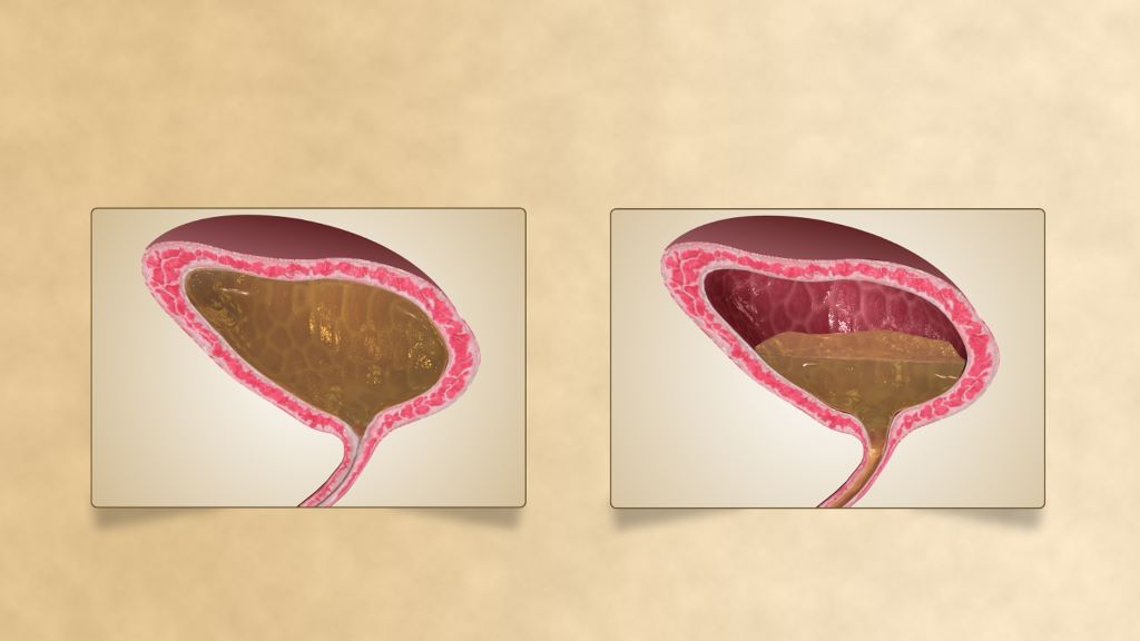
Urinary incontinence is a devastating condition, leading to significant adverse impacts on patients’ mental health and quality of life. Disorders of urination are also a key feature of all neurological disorders.
A USC research team has now made major progress in understanding how the human spinal cord triggers the bladder emptying process. The discovery could lead to exciting new therapies to help patients regain control of this essential function.
In the pioneering study, a team from USC Viterbi School of Engineering and Keck School of Medicine of USC has harnessed functional ultrasound imaging to observe real-time changes in blood flow dynamics in the human spinal cord during bladder filling and emptying.
The work was published in Nature Communications and was led by Charles Liu, the USC Neurorestoration Center director at Keck School of Medicine of USC and professor of biomedical engineering at USC Viterbi, and Vasileios Christopoulos, assistant professor at the Alfred E. Mann Department of Biomedical Engineering.
The spinal cord regulates many essential human functions, including autonomic processes like bladder, bowel, and sexual function. These processes can break down when the spinal cord is damaged or degenerated due to injury, disease, stroke, or aging. However, the spinal cord’s small size and intricate bony enclosure have made it notoriously challenging to study directly in humans.
Unlike in the brain, routine clinical care does not involve invasive electrodes and biopsies in the spinal cord due to the obvious risks of paralysis.
Furthermore, fMRI imaging, which comprises most of human functional neuroimaging, does not exist in practical reality for the spinal cord, especially in the thoracic and lumbar regions where much of the critical function localises.
“The spinal cord is a very undiscovered area,” Christopoulos said. “It’s very surprising to me because when I started doing neuroscience, everybody was talking about the brain. And Dr. Liu and I asked, “What about the spinal cord?”
“For many, it was just a cable that transfers information from the brain to the peripheral system. The truth was that we didn’t know how to go there—how to study the spinal cord in action, visualize its dynamics and truly grasp its role in physiological functions.”
Functional ultrasound imaging: A new window into the spinal cord
To overcome these barriers, the USC team employed functional ultrasound imaging (fUSI), an emerging neuroimaging technology that is minimally invasive. The fUSI process allowed the team to measure where changes in blood volume occur on the spinal cord during the cycle of urination.
However, fUSI requires a “window” through the bone to image the spinal cord. The researchers found a unique opportunity by working with a group of patients undergoing standard-of-care epidural spinal cord stimulation surgery for chronic low back pain.
“During the implantation of the spinal cord stimulator, the window we create in the bone through which we insert the leads gives us a perfect and safe opportunity to image the spinal cord using fUSI with no risk or discomfort to the study volunteers,” said co-first author Darrin Lee, associate director of the USC Neurorestoration Center, who performed the surgeries.
“While the surgical team was preparing the stimulator, we gently filled and emptied the bladder with saline to simulate a full urination cycle under anaesthesia while the research team gathered the fUSI data,” added Evgeniy Kreydin from the Rancho Los Amigos National Rehabilitation Center and the USC Institute of Urology, who was already working closely with Liu to study the brain of stroke patients during micturition using fMRI.
“This is the first study where we’ve shown that there are areas in the spinal cord where activity is correlated with the pressure inside the bladder,” Christopoulos said.
“Nobody had ever shown a network in the spinal cord correlated with bladder pressure. What this means is I can look at the activity of your spinal cord in these specific areas and tell you your stage of the bladder cycle – how full your bladder is and whether you’re about to urinate.”
Christopoulos said the experiments identified that some spinal cord regions showed positive correlation, meaning their activity increased as bladder pressure rose, while others showed negative (anti-correlation), with activity decreasing as pressure increased. This suggests the involvement of both excitatory and inhibitory spinal cord networks in bladder control.
“It was extremely exciting to take data straight from the fUSI scanner in the OR to the lab, where advanced data science techniques quickly revealed results that have never been seen before, even in animal models, let alone in humans,” said co-first author Kofi Agyeman, biomedical engineering postdoc.
New hope for patients
Liu has worked for two decades at the intersection of engineering and medicine to develop transformative strategies to restore function to the nervous system. Christopoulos has spent much of his research career developing neuromodulation techniques to help patients regain motor control.
Together, they noted that for patients, retaining control of the autonomic processes that many of us take for granted is more fundamental than even walking.
“If you ask these patients, the most important function they wanted to restore was not their motor or sensory function. It was things like sexual function and bowel and bladder control,” Christopoulos said, noting that urinary dysfunction often leads to poor mental health. “It’s a very dehumanising problem to deal with.”
Worse still, urinary incontinence leads to more frequent urinary tract infections (UTIs) because patients must often be fitted with a catheter. Due to limited sensory function, they may not be able to feel that they have an infection until it is more severe and has spread to the kidneys, resulting in hospitalisation.
This study offers a tangible path toward addressing this critical need for patients suffering from neurogenic lower urinary tract dysfunction. The ability to decode bladder pressure from spinal cord activity provides proof-of-concept for developing personalised spinal cord interfaces that could warn patients about their bladder state, helping them regain control.
Currently, almost all neuromodulation strategies for disorders of micturition are focused on the lower urinary tract, largely because the neural basis of this critical process remains unclear.
“One has to understand a process before one can rationally improve it,” Liu said.
This latest research marks a significant step forward, opening new avenues for precision medicine interventions that combine invasive and noninvasive neuromodulation with pharmacological therapeutics to make neurorestoration of the genitourinary system a clinical reality for millions worldwide.